
A 35-Year-old lady was brought to the hospital with a history of one-month unresolving fever. She had a dental procedure done two months ago. She was seen by a GP clinic and given two courses of antibiotics. Later she feels reduced effort tolerance and swelling in both lower legs. She has a past medical history of cardiac ailment.
infective endocarditis vegetation echocardiogram mitral valve
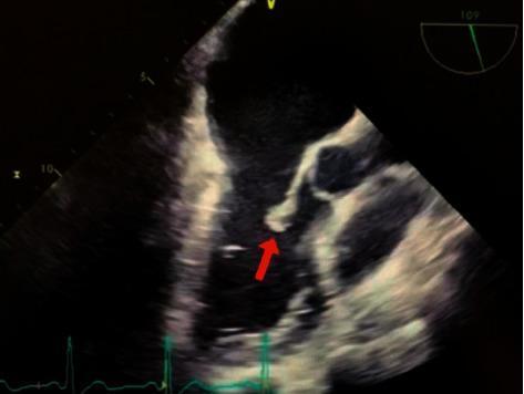
This diagnostic image is a transesophageal echocardiogram (TEE) demonstrating infective endocarditis of the mitral valve. The ultrasound shows a clear, irregular hyperechoic mass, indicated by a red arrow, which represents a vegetation attached to the valve apparatus. Specifically, the vegetation is localized to the lateral posterior (P2) segment of the mitral valve leaflet. Adjacent cardiac structures, including the mitral valve leaflets and chordae tendineae, are visible within the left heart chambers. The image illustrates the classic sonographic presentation of vegetations, which are hallmark findings in bacterial endocarditis (clinically associated here with Staphylococcus hominis). The scan provides critical diagnostic evidence for assessing valve morphology and the risk of embolic events. Educational focus includes the identification of valvular vegetations and understanding their typical location on the atrial side of atrioventricular valves in the context of systemic infection.
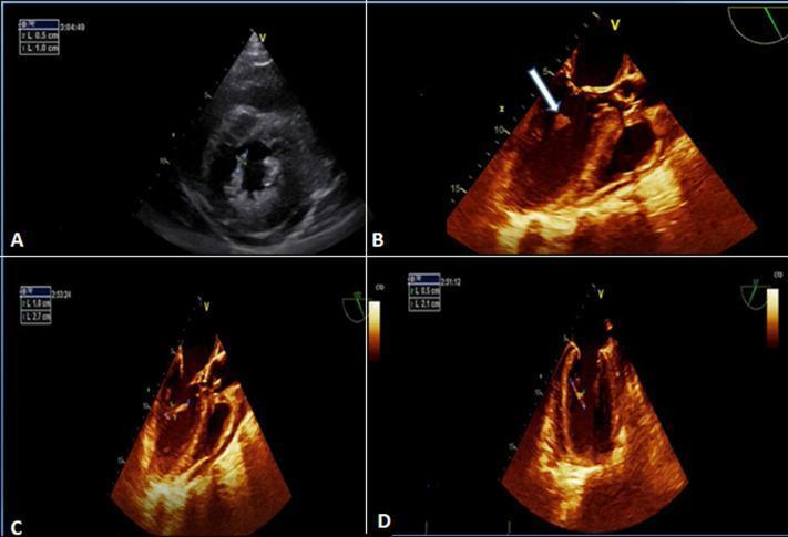
This composite of echocardiographic images demonstrates classic findings of infective endocarditis involving the mitral valve apparatus. Image A is a parasternal short-axis view from a transthoracic echocardiogram (TTE) showing an echogenic, textured mass consistent with vegetation located on the posteromedial mitral valve pillar. Images B, C, and D are transoesophageal echocardiograms (TEE) using a sepia-toned enhancement for improved tissue characterization. These views provide higher resolution of the vegetation, which appears as an irregular, hyper-echogenic, and frond-like mobile structure. In image B, an arrow specifically identifies the vegetation's attachment to the mitral valve apparatus. The diagnostic imaging highlights key features of valvular infection, including vegetation morphology, size assessment (noted in caliber measurements on the screen), and its anatomical relationship to the sub-valvular structures. This material is highly relevant for cardiology and infectious disease training regarding the diagnostic criteria for endocarditis.
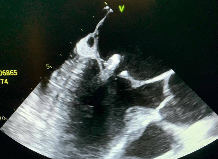
This diagnostic image is a transesophageal echocardiogram (TEE) demonstrating infective endocarditis of the mitral valve. The ultrasound shows a large, pedunculated, and irregularly shaped echogenic mass (vegetation) attached to the posterior mitral valve leaflet. The vegetation appears mobile, extending significantly into the heart chamber. The underlying mitral valve leaflets exhibit areas of increased echogenicity and thickening, consistent with valvular calcification and chronic degenerative changes, which may serve as a nidus for reinfection. This finding is clinically significant for the diagnosis of bacterial endocarditis, often necessitated when transthoracic echocardiography (TTE) is inconclusive. The image highlights the critical role of TEE in visualizing valvular vegetations, assessing their size (approximately 1.3 cm in this case), and identifying comorbid conditions like valve calcification in patients with positive blood cultures or suspected cardiac source of emboli.
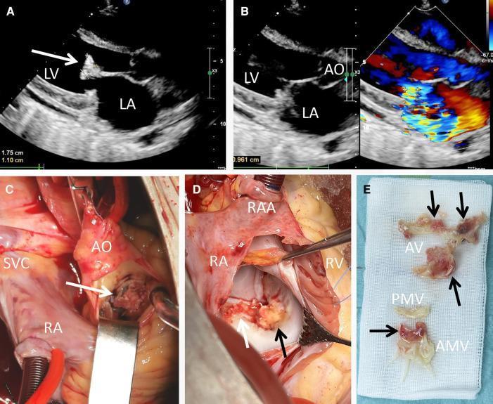
This composite figure presents clinical imaging and surgical findings of infective endocarditis. (A) Transthoracic echocardiogram (TTE) in a long-axis view shows a large, pedunculated echogenic vegetation (white arrow) measuring 1.75 cm x 1.10 cm attached to the anterior mitral valve leaflet. (B) TTE with color Doppler demonstrates a mosaic-pattern jet across the mitral valve, indicating severe mitral regurgitation. (C) Intraoperative view through the aorta (AO) reveals a yellowish vegetation (white arrow) infiltrating the aortic valve cusp. (D) Surgical exposure of the mitral valve via the right atrium (RA) shows extensive friable, yellowish vegetations (arrows) involving the anterior mitral cusp and annulus. (E) Gross specimen photograph displays the excised material on surgical gauze, with arrows pointing to the vegetations associated with the aortic valve (AV) and anterior mitral valve (AMV) tissue. The images collectively demonstrate the progression from diagnostic ultrasonography to surgical management of multi-valvular endocarditis, highlighting the morphology and destructive nature of infective vegetations.
Osler nodes Janeway lesions splinter hemorrhages infective endocarditis skin findings
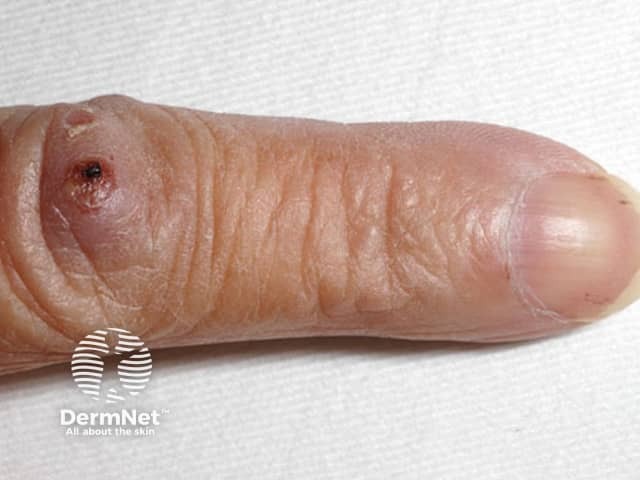
This clinical photograph documents a lateral view of the index finger with visible cutaneous signs of infective endocarditis. The primary subject is dermatologic manifestations: Osler nodes—tender, subcutaneous nodules located at the fingertip/distal phalanx region—alongside splinter hemorrhages beneath the nail plate. The image demonstrates a small, dark lesion near the fingertip base and tiny linear reddish-brown streaks under the nail plate. The surrounding skin shows mild edema and inflammatory changes. These findings are classic for subacute bacterial endocarditis and immune complex–mediated vasculitis. Diagnostic significance: Osler nodes indicate immune-complex deposition in the dermis with concurrent bacteremia and endocardial infection; splinter hemorrhages reflect distal microemboli or microvasculopathy. Differential considerations include Janeway lesions (painless), rheumatoid nodules, vasculitic lesions, or trauma-related changes. Clinical correlation: chest auscultation may reveal a cardiac murmur; blood cultures and inflammatory markers (CRP, ESR) may be positive; treatment decisions rely on echocardiography and microbiology. Imaging modality: clinical photography using a digital camera; technique: macro/close-up; lighting: natural or diffused; magnification: not specified. Anatomical location: distal phalanx of the index finger, fingertip pad; laterality: not specified; anatomical plane: lateral/volar aspect. Use cases: bedside assessment, educational dermatology and internal medicine, documentation of endocarditis signs.
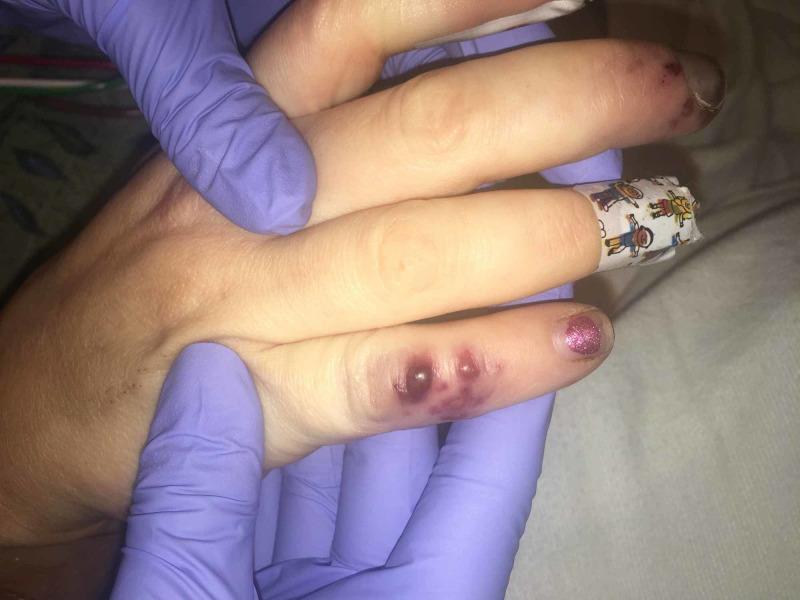
This clinical photograph displays characteristic peripheral stigmata of infective endocarditis on a patient's hand. The primary findings are Osler's nodes, seen as painful, raised, violaceous nodular lesions located on the distal aspect of the fourth digit. These nodules exhibit a deep red-to-purple hue and appear slightly edematous, indicative of immune complex-mediated vasculitis. Additionally, the index finger displays splinter hemorrhages, visible as small, linear, dark reddish-brown streaks beneath the nail bed, representing micro-emboli or capillary damage. The middle finger is partially occluded by a patterned medical adhesive bandage. A healthcare provider in purple nitrile gloves is holding the hand, facilitating the clinical examination. These visual signs are critical diagnostic indicators for bacterial endocarditis, often associated with systemic bacteremia and valvular vegetations. The image serves as an educational tool for identifying classic dermatological manifestations of systemic cardiovascular infection.
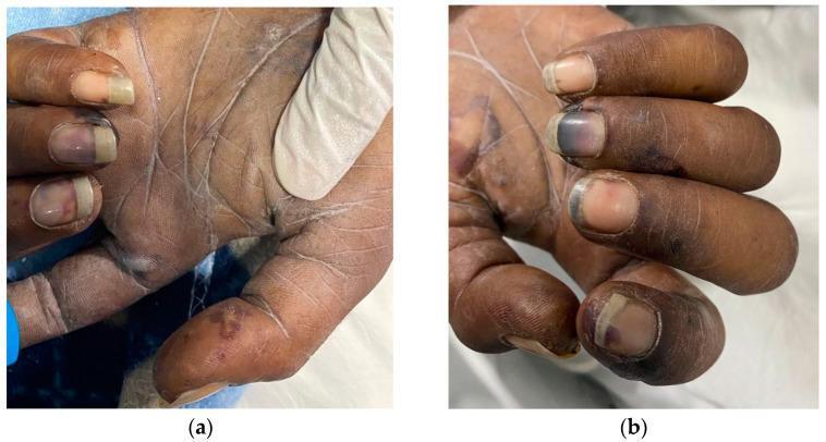
Two clinical photographs (a and b) show the palmar surfaces and fingers of a patient exhibiting peripheral stigmata of infective endocarditis. The palms and fingertips demonstrate multiple Janeway lesions, which appear as non-tender, small, erythematous to violaceous macules and papules. The nail beds of several fingers display splinter hemorrhages, characterized by thin, linear, dark-red to brown streaks running longitudinally under the nail plate. Some digits also show subungual hematomas or dark purplish discoloration near the distal edges, and there is visible bulbous enlargement of the distal phalanges consistent with digital clubbing. The skin texture appears dry with prominent creases and subtle areas of hypopigmentation on the palms. These findings are classic dermatologic manifestations of microembolic or immunologic processes associated with systemic infection, specifically infective endocarditis.
Clinical Case Analysis: Infective Endocarditis (IE)
Diagnosis: Subacute Infective Endocarditis
| Clinical Feature | Significance |
|---|---|
| Dental procedure 2 months ago | Portal of entry for bacteremia (oral flora - viridans streptococci) |
| Pre-existing cardiac disease | Turbulent flow/endothelial damage - ideal surface for vegetation |
| 1-month unresolving fever | Subacute course - insidious onset over weeks |
| Two failed antibiotic courses | Short courses suppress but do not cure endovascular infection |
| Reduced effort tolerance + bilateral leg edema | Heart failure from valve destruction/regurgitation |
Pathogenesis
"IE can develop on previously normal valves, but cardiac abnormalities predispose to such infections; rheumatic heart disease, mitral valve prolapse, bicuspid aortic valves, and calcific valvular stenosis are all common substrates."
- Dental procedure → transient bacteremia with oral flora (most likely Streptococcus viridans)
- Bacteria seed the damaged/abnormal valve (her pre-existing cardiac disease)
- Organisms adhere to sterile platelet-fibrin deposits → form a vegetation (thrombotic debris + organisms)
- Vegetation enlarges over weeks → valve dysfunction → regurgitation → progressive heart failure
- Vegetations can embolise → systemic complications
"In community-acquired IE, 50–60% of cases are caused by Streptococcus viridans, a relatively benign group of normal oral flora. Typically, such infections occur on damaged or deformed valves and present as subacute IE." - Robbins & Kumar
Acute vs. Subacute IE
| Feature | Acute IE | Subacute IE (this patient) |
|---|---|---|
| Onset | Days | Weeks to months |
| Organism | S. aureus | S. viridans, HACEK |
| Valve | Normal or abnormal | Usually pre-existing disease |
| Source | Healthcare/IV drugs | Dental/GI procedures |
| Course | Destructive, high mortality | Insidious, more treatable |
Modified Duke Criteria - Applying to This Case
Definite IE = 2 Major, OR 1 Major + 3 Minor, OR 5 Minor
- Positive blood cultures: ≥2 separate cultures with typical organisms (viridans streptococci, S. aureus, HACEK, enterococci)
- Evidence of endocardial involvement on echo: oscillating vegetation, abscess, new valvular regurgitation, or new partial prosthetic dehiscence
- Predisposing heart condition ✓ (pre-existing cardiac disease)
- Fever >38°C ✓ (1-month history)
- Vascular phenomena - major arterial emboli, Janeway lesions, conjunctival hemorrhages, mycotic aneurysm
- Immunologic phenomena - Osler nodes, Roth spots, glomerulonephritis, rheumatoid factor
- Microbiologic evidence - positive blood culture not meeting major criteria
Peripheral Stigmata of IE to Look For
| Sign | Description | Mechanism |
|---|---|---|
| Osler nodes | Tender subcutaneous nodules on finger/toe pads | Immune complex deposition |
| Janeway lesions | Non-tender erythematous macules on palms/soles | Septic microemboli |
| Roth spots | Retinal hemorrhages with pale center | Immune complex vasculitis |
| Splinter hemorrhages | Linear dark streaks under nails | Microemboli |
| Conjunctival petechiae | Petechiae on conjunctiva | Microemboli |
| Clubbing | Digital clubbing in chronic cases | Chronic hypoxia |
| Murmur | New or changing cardiac murmur | Valve destruction |
Echocardiographic Findings
"Echocardiography should be obtained as soon as possible for any patient with suspected endocarditis. Evidence of vegetation, abscess, prosthetic valve dehiscence, or new valvular regurgitation are major diagnostic criteria."
- TTE (transthoracic echo) - first-line investigation
- TEE (transoesophageal echo) - higher sensitivity, recommended if TTE non-diagnostic or prosthetic valve suspected
Investigations to Order
- Blood cultures x3 (drawn from different sites, >1 hour apart) - before any antibiotics
- Echocardiography (TTE then TEE if needed)
- Full blood count - leukocytosis, normocytic anaemia common
- ESR, CRP - elevated (non-specific markers of infection/inflammation)
- Urinalysis - microscopic haematuria, proteinuria (immune complex glomerulonephritis)
- Rheumatoid factor - elevated in ~50% of subacute IE (minor criterion)
- Renal function, LFTs
- 12-lead ECG - PR prolongation may indicate aortic root abscess/conduction disease
- Chest X-ray - pulmonary oedema, cardiomegaly (from heart failure)
Management
Antibiotic Therapy
| Organism | Antibiotic Regimen |
|---|---|
| Penicillin-susceptible Streptococcus | Penicillin G 12-18 million units IV/day x 4 weeks, OR Ceftriaxone 2g IV once daily x 4 weeks |
| Penicillin-resistant Streptococcus | Vancomycin (if penicillin-allergic or resistant) |
| S. aureus (MRSA) | Vancomycin + Gentamicin |
| Empiric (pending cultures) | Penicillinase-resistant penicillin/cephalosporin for native valve |
Heart Failure Management
- Diuretics for fluid overload (bilateral leg oedema/dyspnoea)
- Monitor for worsening valve regurgitation - may need urgent surgical intervention
Surgical Indications (from Braunwald's)
- Heart failure unresponsive to medical management ← this patient has developing HF
- Large vegetation (>10mm) with high embolic risk
- Perivalvular abscess or fistula
- Persistent bacteremia despite antibiotics
- Fungal endocarditis
Antibiotic Prophylaxis (Why It Failed Here)
- Prosthetic cardiac valves
- Previous IE
- Congenital heart disease (unrepaired cyanotic, or within 6 months of repair)
- Cardiac transplant with valvulopathy
Complications to Monitor For
- Embolic events - stroke, splenic/renal infarcts, septic pulmonary emboli (right-sided IE)
- Perivalvular abscess - seen on TEE; PR prolongation on ECG is a warning sign
- Progressive valve destruction - worsening regurgitation, heart failure
- Mycotic aneurysms - intracranial → subarachnoid haemorrhage
- Immune complex glomerulonephritis - renal failure
- Metastatic infection - osteomyelitis, septic arthritis
Summary
- Admit to hospital (IE requires inpatient management with a multidisciplinary team)
- Draw 3 sets of blood cultures before antibiotics
- Echocardiography (TTE/TEE) urgently
- IV antibiotics (empiric, then targeted based on culture)
- Cardiothoracic surgery review given emerging heart failure
"Every patient with IE should be managed in the inpatient setting of a medical center with experienced medical and surgical specialists... this 'team' approach has resulted in improved outcomes." - Braunwald's Heart Disease
- Braunwald's Heart Disease (Cardiovascular Medicine), Chapter on IE
- Robbins & Kumar Basic Pathology, p.368
- Fuster and Hurst's The Heart, 15th Edition
- The Washington Manual of Medical Therapeutics
Explain the pathogenesis of infective endocarditis, including the relationship between transient bacteremia following invasive dental procedures, pre-existing cardiac lesions, endothelial injury, and vegetation formation.
infective endocarditis pathogenesis vegetation formation diagram mechanism
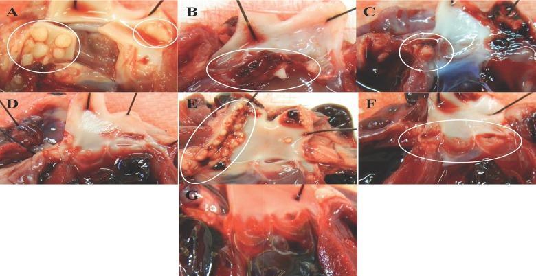
A composite clinical photograph showing seven panels (A-G) of gross anatomical specimens depicting experimental infective endocarditis (IE) in a rabbit model. The images focus on the aortic valve leaflets to demonstrate vegetation formation induced by Staphylococcus aureus enterotoxin gene cluster (egc) proteins. Panels A, B, C, E, and F show varying degrees of pathological vegetations (circled in white), appearing as nodular, yellowish-white masses (SElO, SElU) or reddish-brown thrombotic clusters (SElM, SEI). Morphology ranges from clustered, multifocal nodules to linear arrangements along the valve margins. In contrast, panels D (SEG) and G (Vector control) represent comparative groups where the valve leaflets remain relatively smooth, intact, and free of significant vegetation or inflammatory mass. The series illustrates how specific superantigens (SAgs) contribute to the pathogenesis and severity of valvular lesions in endocarditis, providing a visual comparison of lesion distribution, size, and color across different bacterial protein expressions.
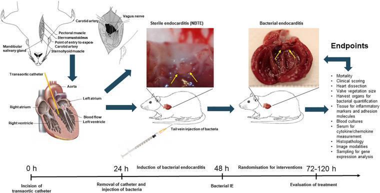
A scientific infographic and flow diagram depicting an in vivo experimental model of rat infective endocarditis (IE). The visual combines anatomical diagrams, clinical photographs, and a timeline. The process starts with a transaortic catheterization (0h) shown in a schematic of the rat neck and heart, where a catheter enters the aorta and reaches the left ventricle to induce mechanical endothelial damage. At 24h, the catheter is removed, and bacteria are administered via tail vein injection. Two clinical macro photographs compare heart valve pathology: 'Sterile endocarditis (NBTE)' shows small, discrete, pale thrombotic vegetations (indicated by yellow arrows), while 'Bacterial endocarditis' shows bulky, irregular, dark-colored masses and extensive vegetational growth on the valve leaflets. The timeline progresses through the induction of bacterial IE (48h) and evaluation of treatments (72-120h). Listed endpoints for the model include mortality, valve vegetation size, bacterial quantification, inflammatory markers, and histopathology. This diagram illustrates the pathogenesis from non-bacterial thrombotic endocarditis to active infection and is intended for medical research and educational contexts.
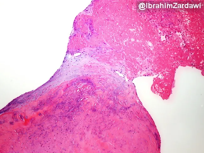
Infective endocarditis histology image demonstrating a valve leaflet vegetation attached to the endocardial surface. The lesion is a friable thrombus-rich mass composed predominantly of fibrin and platelets with an inflammatory cell infiltrate. Bacterial colonies are not evident on routine hematoxylin and eosin staining, which can occur in culture-negative endocarditis or after prior antibiotic exposure; additional stains or culture may reveal organisms. The vegetation is anchored to the valve leaflet, illustrating the pathological substrate of infection and its interface with native tissue. This pattern supports a diagnosis of infective endocarditis in the clinical context and helps distinguish from noninfectious vegetations such as marantic endocarditis or Libman-Sacks endocarditis. Clinically, the histology guides antimicrobial strategy, duration, and need for surgical evaluation, given the embolic potential of friable vegetations. Educationally, this image is valuable for cardiac pathology and infectious disease training, highlighting thrombus-dominated composition, inflammatory infiltration, and the frequent absence of visible bacteria on standard histology.
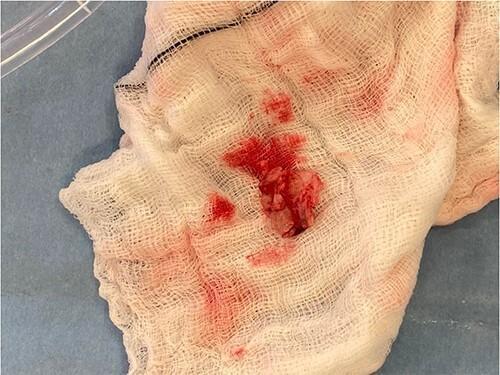
This intraoperative clinical photograph displays a macro-level view of excised cardiac vegetation, typical of infective endocarditis, resting on a white, sterile surgical gauze swab. The vegetation appears as an irregular, friable, and multi-lobulated tissue mass with a reddish-pink hue, indicating significant vascularization or associated blood clot formation. The specimen's texture is heterogeneous and rough, consistent with a collection of fibrin, platelets, and microorganisms typically found on heart valve surfaces during active infection. The surrounding white gauze shows several focal areas of bright red serosanguinous staining where the vegetation has been placed. This image is medically significant for illustrating the gross pathological morphology of a mitral valve vegetation following surgical excision and valve debridement. It serves as a visual record of the endovascular disease process, highlighting the size and friability of the mass that contributed to valvular regurgitation and potential embolic risk.
nonbacterial thrombotic endocarditis NBTE sterile vegetation valve histology
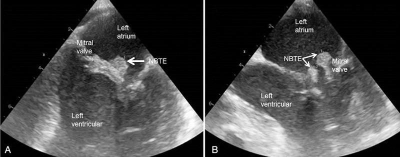
This diagnostic image consists of two panels (A and B) showing transesophageal echocardiography (TEE) in a long-axis view. The images depict the left atrium, left ventricle, and the separating mitral valve. In both panels, prominent sterile vegetations characteristic of nonbacterial thrombotic endocarditis (NBTE), also known as Marantic endocarditis, are visible as highly echogenic masses attached to the mitral valve leaflets. These vegetations protrude into the left atrial space. Panel A displays a single large irregular vegetation, while Panel B reveals the fragmented or multi-lobulated nature of the thrombi. The clinical context involves a patient with pancreatic cancer, illustrating a paraneoplastic manifestation of malignancy-associated hypercoagulability. This image is used to teach the identification of embolic sources in patients with cancer-related ischemic stroke and highlights the importance of TEE in diagnosing valvular vegetations that may be missed on transthoracic echocardiography.
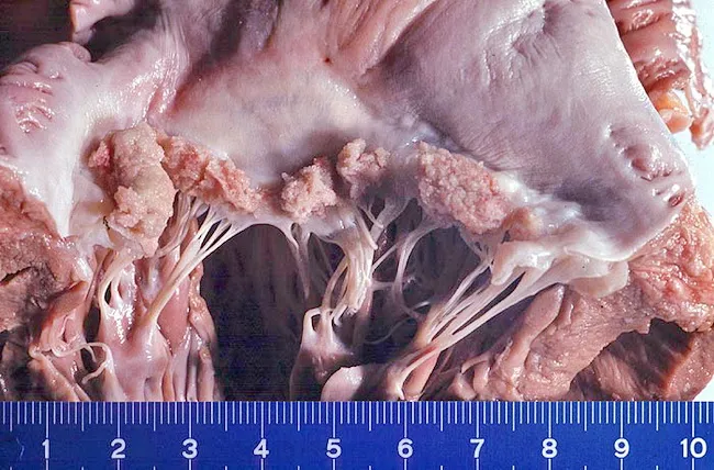
Gross pathology photograph of a heart valve with sterile, friable vegetations consistent with non-bacterial thrombotic endocarditis (NBTE, also called marantic endocarditis). The specimen shows the valvular leaflets and adjacent endocardium with pale, translucent to pinkish nodular thickenings along the lines of valve closure and on the atrial surface in this left-sided valve representation, most commonly the mitral valve. These vegetations are composed primarily of fibrin and platelets with little or no inflammatory cells and lack detectable organisms on routine assessment. The context of an advanced mucin-producing carcinoma—here endometrial carcinoma—supports a hypercoagulable state that predisposes to NBTE and embolic events. On gross inspection, the vegetations are usually small to moderate in size, friable, and may appear as irregular, spongy masses that can detach and embolize to the systemic circulation. The absence of vegetation-associated suppuration or ulceration helps distinguish NBTE from infectious endocarditis. Clinically, NBTE carries diagnostic significance by prompting evaluation for occult malignancy or systemic hypercoagulability, and management centers on anticoagulation rather than antibiotic therapy alone. This image underscores the macroscopic appearance used in pathology and cardiology to correlate echocardiographic findings with tissue-level pathology and to guide multidisciplinary care in oncology-associated thromboembolic syndromes.
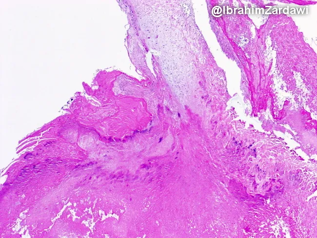
Histology of a cardiac valve vegetation from an intravenous drug user with infective endocarditis. The specimen was processed for light microscopy and stained with hematoxylin and eosin (H&E). The vegetative mass adheres to the valvular surface and shows a dominant thrombus composed of densely packed fibrin and platelets, with admixed bacterial colonies appearing as dark purple clusters within the eosinophilic matrix. The core is friable and irregular, with a leafy, chain-like fringe along the valve edge. The bacterial colonies are variably distributed, forming focal microcolonies that disrupt surrounding tissue architecture. There is limited or variable inflammatory cell infiltrate; neutrophils may be present at the margins, consistent with acute infective endocarditis. The overall pattern is consistent with septic endocarditis in an intravenous drug user, where bacteria colonize a pre-existing thrombus on the valve surface, producing vegetations that may embolize. Clinically, histology complements microbiology and imaging to confirm diagnosis and guide antibiotic therapy. Diagnostic significance includes identification of infectious vegetations on valves, distinguishing from sterile thrombi, and informing prognosis. Potential differential diagnoses include nonbacterial thrombotic endocarditis and Libman-Sacks endocarditis in alternative clinical settings. This image is valuable for education on endocarditis pathology, valve destruction, and the histopathology of septic vegetations.
Pathogenesis of Infective Endocarditis
Step 1 - The Susceptible Substrate: Pre-existing Cardiac Lesions
"The natural cardiac endothelium is resistant to infection. In animal models of endocarditis, damage to the endothelium is required to establish infections, despite a large inoculum of bacteria." - Fuster and Hurst's The Heart, 15th ed.
"The undamaged endothelium is resistant to infection by most bacteria." - Harrison's Principles of Internal Medicine, 22nd ed.
| Cardiac Lesion | Mechanism of Endothelial Risk |
|---|---|
| Mitral regurgitation | Regurgitant jet traumatises atrial surface of mitral leaflet and atrial wall |
| Aortic stenosis / regurgitation | High-velocity jets across stenotic orifice damage downstream endothelium |
| Ventricular septal defect | Low-pressure right side of defect exposed to traumatic shear forces |
| Bicuspid aortic valve | Turbulent, disorganised flow causes leaflet endothelial activation |
| Mitral valve prolapse | Mechanical stress on leaflets; leading pre-existing risk factor today |
| Rheumatic heart disease | Scarred, deformed leaflets - historically the most common predisposing lesion |
| Prosthetic valves | Foreign surface; accounts for 10-20% of all IE cases |
"High velocity jets may traumatize the endothelium, as occurs in the left ventricular outflow tract in hypertrophic obstructive cardiomyopathy. In other situations, risk is related to turbulent, disorganized flow downstream from an orifice where flow velocities first increase and then decelerate in a lower pressure zone. The disorganization of flow that results increases regional platelet and endothelial activation, and the slower eddy zones in proximity to the endothelium, increase the potential for bacterial interaction with the wall." - Fuster and Hurst's The Heart
Step 2 - Endothelial Injury and NBTE Formation
- Collagen exposure activates circulating platelets
- Platelets adhere and aggregate at the injury site
- The coagulation cascade is triggered locally
- Fibrin is deposited over the platelet aggregate
"Endothelial injury... allows either direct infection by virulent organisms or the development of a platelet-fibrin thrombus - a condition called nonbacterial thrombotic endocarditis (NBTE). This thrombus serves as a site of bacterial attachment during transient bacteremia." - Harrison's Principles of Internal Medicine, 22nd ed.
"The resultant endothelial damage promotes platelet/fibrin deposition and the formation of sterile vegetations (nonbacterial thrombotic endocarditis)." - Tintinalli's Emergency Medicine
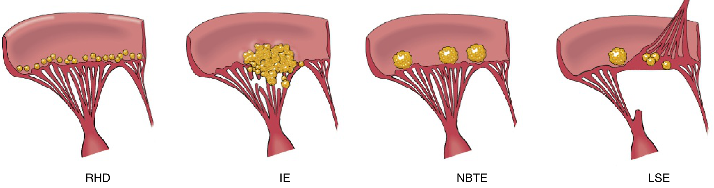
Step 3 - Transient Bacteremia from the Dental Procedure
"The periodontal tissues of the mouth are commonly replete with organisms capable of causing endocarditis, and bacteremia often follows mechanical trauma to the oral mucosa from eating, brushing, flossing, or dental procedures." - Fuster and Hurst's The Heart
"Transient bacteremia (as little as 10 organisms per milliliter of blood for <30 minutes) may colonize and convert to infective endocarditis." - Tintinalli's Emergency Medicine
Step 4 - Bacterial Adherence via MSCRAMMs
"The organisms that commonly cause IE have surface adhesin molecules, collectively called microbial surface components recognizing adhesin matrix molecules (MSCRAMMs) that mediate adherence to NBTE sites or injured endothelium." - Harrison's Principles of Internal Medicine, 22nd ed.
| Organism | Adhesin Molecule | Target |
|---|---|---|
| S. aureus | Clumping factor (ClfA) | Fibrinogen, fibrin |
| S. aureus | Fibronectin-binding proteins (FnBPA/B) | Fibronectin |
| S. aureus | von Willebrand factor binding | Intact or injured endothelium |
| E. faecalis | Fss2 (fibrinogen-binding protein) | Fibrinogen |
| E. faecalis | Ace (collagen-binding adhesin) | Collagen |
| E. faecalis | Ebp pili | Platelet adherence |
| Viridans streptococci | Dextran (cell wall polysaccharide) | Platelet matrix |
| All gram-positive organisms | Fibronectin-binding proteins | Fibronectin in NBTE |
Step 5 - Vegetation Growth and the "Protected Niche"
- Bacteria replicate within the thrombus
- They stimulate further platelet activation and fibrin deposition (by activating the extrinsic coagulation cascade and causing monocytes to release cytokines)
- Activated endothelial cells deposit additional fibronectin, providing more adhesion points for more bacteria
- The vegetation grows progressively larger - up to several centimetres - and becomes increasingly friable
"Bacteria circulating in the bloodstream... may then bind to and replicate in this microthrombus, amplifying the extrinsic clotting pathway, monocyte release of cytokines, and local deposition of fibronectin by activated endothelial cells. The result is a macroscopic infective mass that promotes further deposition of platelets and fibrin, resulting in increasing vegetation size and fragility." - Fuster and Hurst's The Heart
"Adherent organisms stimulate further deposition of platelets and fibrin, leading to generation of a 'protected site' that phagocytic cells cannot easily penetrate." - Tintinalli's Emergency Medicine
"Infected vegetations are avascular and contain bacteria in great density, often with 10⁹ to 10¹⁰ colony forming units per gram. Neutrophils and host defense molecules have limited penetration into these lesions, allowing them to grow relatively unchecked and resulting in continuous high-grade bacteremia." - Fuster and Hurst's The Heart
- Why short antibiotic courses fail: oral antibiotics at standard doses cannot achieve bactericidal concentrations inside the avascular vegetation
- Why IE requires 4-6 weeks of IV antibiotics: only sustained, high-concentration bactericidal therapy can sterilise the vegetation
Biofilm Formation
"Some bacteria, including streptococci, generally form sessile, layered communities of cells irreversibly attached to cardiac surfaces that are enclosed in a protective matrix of exopolymeric products called biofilm. Biofilms are up to 1000-fold more resistant to antibiotic therapy than planktonic cells." - Fuster and Hurst's The Heart
Step 6 - S. aureus - The Exception That Bypasses NBTE
"Some highly virulent organisms such as Staphylococcus aureus may not require underlying trauma to establish infection." - Fuster and Hurst's The Heart
Step 7 - Consequences of Vegetation
A. Local Cardiac Destruction
- Valve leaflet perforation → acute regurgitation → acute heart failure
- Chordae rupture → flail leaflet
- Perivalvular abscess (ring abscess) → extends into annulus/myocardium
- Conduction system invasion → heart block (PR prolongation on ECG)
B. Embolisation
- Fragments break off the friable vegetation → septic emboli
- Left-sided IE: brain (stroke/abscess), spleen, kidneys, coronary arteries
- Right-sided IE (IV drug users - tricuspid): pulmonary septic emboli
"Shedding of emboli is common because of the friable nature of the vegetations. Since the fragmented vegetations contain large numbers of organisms, abscesses often develop at the sites where emboli lodge, leading to development of septic infarcts and aneurysms resulting from bacterial infection of the arterial wall (mycotic aneurysms)." - Robbins & Kumar Basic Pathology
C. Immune Complex Deposition (Immunologic Phenomena)
- Sustained bacteremia → chronic antigen exposure → antigen-antibody complex formation
- Complexes deposit in: glomeruli (focal/diffuse glomerulonephritis), skin, retinal vessels
- Produces: microscopic haematuria, Osler nodes, Roth spots, elevated rheumatoid factor
Pathogenesis Summary - Sequential Model
PRE-EXISTING CARDIAC LESION
(mitral regurgitation, rheumatic disease, bicuspid valve, MVP)
          ↓
TURBULENT / HIGH-VELOCITY BLOOD FLOW
          ↓
ENDOTHELIAL INJURY
(collagen + matrix molecules exposed)
          ↓
PLATELET ADHESION + FIBRIN DEPOSITION
          ↓
NBTE (Sterile platelet-fibrin vegetation)
          ↓
TRANSIENT BACTEREMIA
(dental procedure → viridans streptococci enter bloodstream)
          ↓
BACTERIAL ADHERENCE via MSCRAMMs
(fibronectin-binding proteins, dextran → fibrin-platelet matrix)
          ↓
BACTERIAL REPLICATION within thrombus
          ↓
AMPLIFIED PLATELET + FIBRIN DEPOSITION
(monocyte cytokines, extrinsic coagulation, fibronectin by endothelial cells)
          ↓
GROWING INFECTED VEGETATION
(avascular, 10⁹-10¹⁰ CFU/g, protected from phagocytes + antibiotics)
          ↓
┌─────────────────────────────────────────────────┐
│ Local destruction │ Embolisation │ Immune complexes│
│ (valve regurgitation,│ (stroke, mycotic│ (GN, Osler,    │
│ abscess, CHF)       │ aneurysm)       │ Roth, RF+)     │
└─────────────────────────────────────────────────┘
Why Did the Antibiotic Courses Fail?
- They were short courses - insufficient to sterilise the avascular, densely packed vegetation
- Oral route - oral antibiotics may not achieve bactericidal concentrations within the fibrin-encased vegetation
- Biofilm - sessile bacteria within biofilm are up to 1000x more resistant
- Vegetation size grows - early suppression of bacteremia may temporarily reduce fever while the vegetation continues to enlarge structurally
- Harrison's Principles of Internal Medicine, 22nd ed. - Chapter on IE Pathogenesis
- Fuster and Hurst's The Heart, 15th ed. - PATHOGENESIS section
- Robbins & Kumar Basic Pathology - Infective Endocarditis, Morphology
- Tintinalli's Emergency Medicine - Pathophysiology of IE
What is GN,Osler,Roth,RF+ ??
GN, Osler, Roth, RF+ Explained
🔴 GN = Glomerulonephritis
- What it is: Inflammation of the kidney's filtering units (glomeruli)
- Mechanism: Circulating antigen-antibody complexes (formed during prolonged bacteremia) deposit in the glomerular basement membrane → activates complement → inflammatory damage
- Clinical signs: Microscopic haematuria (blood in urine), proteinuria, rising creatinine
- Type: Focal or diffuse proliferative glomerulonephritis; can rarely cause renal failure
🔴 Osler Nodes
- What they are: Painful, tender, raised subcutaneous nodules
- Location: Pads of the fingers and toes (pulp of digits)
- Colour: Reddish-purple, slightly raised, 2-15mm
- Mechanism: Immune complex deposition in the skin → local vasculitis (NOT septic emboli)
- Key feature: They are PAINFUL - this distinguishes them from Janeway lesions
🔴 Roth Spots
- What they are: Retinal haemorrhages with a pale (white) centre
- Location: Seen on fundoscopy (back of the eye)
- Mechanism: Immune complex-mediated retinal vasculitis → haemorrhage; pale centre = fibrin/platelet plug or ischaemic retinal oedema
- Clinical significance: A minor Duke criterion; seen in ~2% of IE cases but highly specific when present
🔴 RF+ = Rheumatoid Factor Positive
- What it is: A circulating autoantibody (IgM against the Fc portion of IgG)
- Why it appears in IE: Prolonged antigenic stimulation from chronic bacteremia drives polyclonal B-cell activation → RF is produced as a non-specific immunological byproduct
- Frequency: Positive in ~50% of subacute IE cases
- Why it matters: It is a Minor Duke Criterion for IE diagnosis; also explains why some IE patients are misdiagnosed as rheumatoid arthritis early in the illness
Summary Table
| Finding | Full Name | Mechanism | Location | Key Feature |
|---|---|---|---|---|
| GN | Glomerulonephritis | Immune complex deposition in kidney | Glomeruli | Haematuria, proteinuria |
| Osler | Osler nodes | Immune complex vasculitis | Fingertip/toe pads | Painful nodules |
| Roth | Roth spots | Immune complex retinal vasculitis | Retina (on fundoscopy) | White-centred haemorrhage |
| RF+ | Rheumatoid Factor positive | Polyclonal B-cell stimulation | Blood test | 50% of subacute IE |
All 4 Are Caused by the Same Root Process
Prolonged bacteremia (weeks)
        ↓
Continuous antigen load in bloodstream
        ↓
Antigen-Antibody complex formation
        ↓
Complex deposition in small vessels + kidneys
        ↓
Complement activation → Inflammation
        ↓
┌──────────────┬─────────────┬──────────────┬──────────────┐
│   Kidney     │    Skin     │    Eye       │   Blood      │
│ (GN)         │ (Osler      │ (Roth        │ (RF+)        │
│              │  nodes)     │  spots)      │              │
└──────────────┴─────────────┴──────────────┴──────────────┘