I now have all the content needed. Let me update the todos and write the comprehensive answer.> Note: Davidson's Principles and Practice of Medicine is not available in the textbook library. The following comprehensive summary is drawn from the three best equivalent sources available: Harrison's Principles of Internal Medicine (22nd Ed, 2025), Goldman-Cecil Medicine, and Robbins & Cotran Pathologic Basis of Disease - which together cover everything Davidson's would cover on this topic and more.
Hodgkin Lymphoma - Complete Guide
1. Definition
Hodgkin lymphoma (HL) is a malignancy of mature B lymphocytes characterized by the presence of neoplastic giant cells called Reed-Sternberg (RS) cells within a reactive inflammatory background of lymphocytes, macrophages, eosinophils, and plasma cells. It consists of two major types:
- Classical Hodgkin Lymphoma (cHL) - ~90% of cases
- Nodular Lymphocyte-Predominant Hodgkin Lymphoma (NLPHL) - ~10% of cases
It was the first human cancer to be successfully cured with radiation therapy and chemotherapy.
- Goldman-Cecil Medicine, Chapter 172
2. Epidemiology
| Feature | Detail |
|---|
| Incidence | ~8,830-9,000 new cases/year in the US; ~30,000 annually in North America + Europe |
| Age distribution | Bimodal - peak in young adults (25-30 yrs) AND late adulthood (>50-55 yrs) |
| Sex | Slightly more common in males |
| Race | More common in Whites than Blacks; rare in Asian populations |
| Incidence rate | ~2.7 per 100,000 (age-adjusted) |
| Mortality rate | ~0.5 per 100,000 (age-adjusted) |
| Geography | Highest in USA, Canada, Switzerland, northern Europe; lowest in eastern Asia |
- In the Indian subcontinent, the age distribution is strongly shifted toward childhood (unlike the Western bimodal pattern)
- In the developing world, EBV infection occurs in childhood; in developed countries it is often delayed to teenage years
- HIV infection increases HL risk 5- to 10-fold
- Goldman-Cecil Medicine, p.1638-1640; Harrison's, p.2635
3. Etiology and Pathobiology
Epstein-Barr Virus (EBV)
- EBV is the leading suspect but definitive proof is lacking
- A 2- to 3-fold elevated risk of HL follows infectious mononucleosis
- EBV detectable in 20-40% of non-HIV HL cases; in nearly 100% of HIV-associated HL cases
- RS cells in HIV-associated HL express LMP-1 (EBV latent membrane protein-1), a transforming oncoprotein
- EBV genomes from multiple disease sites are clonal, suggesting early lymphomagenesis involvement
Genetic Factors
- Siblings of affected individuals have a 3- to 7-fold increased risk
- Identical twins have a much higher concordance rate than non-identical twins
- Certain HLA alleles confer susceptibility or protection
The Reed-Sternberg Cell - Origin
Through elegant molecular studies of single isolated RS cells:
- Clonal IGH gene rearrangements and signs of somatic hypermutation establish their origin from germinal center or post-germinal center B cells
- Despite B-cell origin, RS cells have silenced most B-cell gene expression due to aberrant transcription factor activity (loss of PAX5, OCT2, BOB.1, PU.1 expression)
- RS cells aberrantly activate NF-kB signaling, which promotes their survival and proliferation
- Reactive cells recruited by RS cells (T-cells, eosinophils, macrophages) typically comprise >90% of the tumor cellularity
- Robbins & Cotran, p.3813-3815
4. Classification (WHO)
The WHO recognizes 5 subtypes of HL:
A. Classical HL (cHL) - 4 subtypes
All share the same RS cell immunophenotype:
- CD15+, CD30+ (in 85-99% of cases)
- PAX-5 positive (weak/dim)
- CD19-, CD20- (or weak) - B-cell antigens silenced
- CD45- (LCA negative)
1. Nodular Sclerosis (NSCHL)
- Most common subtype (~60-70% of cHL in Western countries)
- Predominates in young adults, especially females
- Typical presentation: mediastinal involvement
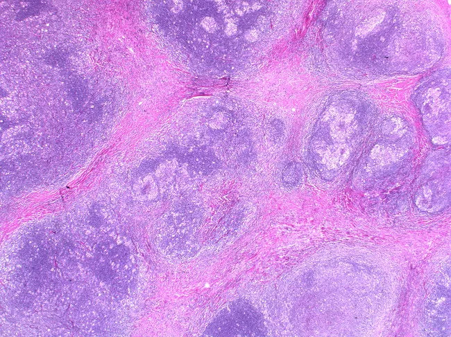
- Histology: dense collagen bands divide the lymph node into nodules; RS cells are lacunar variants (cytoplasm retraction artifact in formalin)
- EBV association: ~25%
2. Mixed Cellularity (MCCHL)
- Second most common (~20-30%)
- More common in older adults, HIV-infected individuals, developing countries
- Histology: diffuse or vaguely nodular infiltrate of RS cells in a rich reactive background without fibrosis
- EBV association: ~75%
3. Lymphocyte-Rich
- Rare (~5%)
- Best prognosis among cHL
- Nodular or diffuse pattern with abundant lymphocytes, few RS cells
4. Lymphocyte-Depleted
- Rarest and most aggressive cHL subtype
- Diffuse fibrosis OR abundant RS cells with few lymphocytes
- More common in elderly, HIV-infected, developing world
- EBV association: ~100% in HIV-associated cases
- Most present at advanced stage with systemic symptoms
B. Nodular Lymphocyte-Predominant HL (NLPHL)
- ~5-10% of all HL
- Different biology from cHL - more related to indolent B-cell NHL
- Histology: nodules of small lymphocytes; RS cells are replaced by "popcorn cells" (L&H variants) with multilobed nuclei
- Immunophenotype DIFFERS from cHL: L&H cells are CD20+, BCL6+, CD15-, CD30-
- Ongoing somatic hypermutation = true germinal center B-cell origin
- EBV: rarely associated
- Demographics: mostly males <35 years, cervical or axillary LN involvement; mediastinal and bone marrow involvement rare
- 3-5% risk of transformation to diffuse large B-cell lymphoma
- Prognosis: excellent (may be more likely to recur than classic subtypes but still very good)
- Robbins & Cotran, p.3810-3905; Harrison's, p.2635-2637
5. Histopathology - Reed-Sternberg Cells
The RS cell is the pathognomonic cell of cHL.
Classic RS cell:
- Large cell with abundant pale cytoplasm
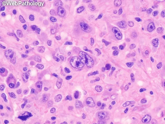
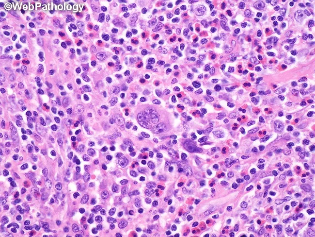
- Bilobed or multilobed nucleus with prominent eosinophilic nucleoli - the classic "owl's eye" appearance
- Mirror-image nuclei when binucleated
Variants:
- Lacunar cells (nodular sclerosis) - retraction of cytoplasm in formalin gives a "lacunar" appearance
- Mononuclear (Hodgkin cells) - single nucleus with large nucleolus
- "Popcorn cells" / L&H cells (NLPHL) - multilobed nuclei resembling popcorn kernels
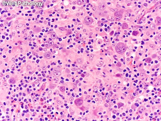
Classic RS cells with prominent eosinophilic "owl's eye" nucleoli in mixed cellularity cHL
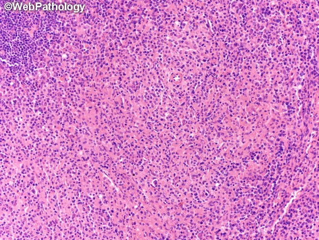
Nodular sclerosis cHL: fibrous collagen bands separating nodules with lacunar RS cells
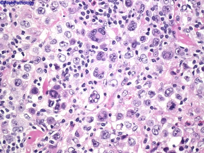
Lymphocyte-depleted cHL: sheets of RS cells with sparse lymphocytes
6. Clinical Features
Lymphadenopathy
- Most patients present with painless, nontender lymphadenopathy
- Common sites: cervical > supraclavicular > axillary (most frequent in the upper body)
- Mediastinal adenopathy in >50% of patients at diagnosis - sometimes the initial manifestation
- Spread is orderly by contiguity (unlike NHL which spreads non-contiguously)
- Mesenteric nodes and Waldeyer's ring: rarely involved (unlike NHL)
- Subdiaphragmatic presentation: unusual, more common in older males
Constitutional "B" Symptoms
Approximately one-third of patients present with B symptoms:
- Fever >38°C (unexplained, persistent or recurrent)
- Night sweats (drenching)
- Weight loss >10% of body weight over 6 months
Special/Unusual Manifestations
- Pel-Ebstein fever: cyclical fever pattern - days to weeks of fever followed by afebrile intervals (more common in mixed-cellularity, abdominal involvement, older patients)
- Severe pruritus (itching) - unexplained
- Alcohol-induced lymph node pain - characteristic though rare
- Paraneoplastic cerebellar degeneration
- Erythema nodosum, ichthyosiform skin changes
- Nephrotic syndrome
- Immune hemolytic anemia and thrombocytopenia
- Hypercalcemia
- Cutaneous anergy (depressed cell-mediated immunity) due to IL-10 secretion by RS cells suppressing Th1 responses
- Harrison's, p.2664-2665; Robbins & Cotran, p.3912-3915
7. Ann Arbor Staging System
| Stage | Definition |
|---|
| I | Single lymph node region OR one extralymphatic site (Ie) |
| II | Two or more lymph node regions on the same side of the diaphragm; IIe = with local extralymphatic extension |
| III | Lymph node regions on both sides of the diaphragm; IIIe with extralymphatic extension |
| - III₁ | Subdiaphragmatic: spleen, splenic hilar, celiac, or portal nodes only |
| - III₂ | Subdiaphragmatic: paraaortic, iliac, or mesenteric nodes |
| IV | Diffuse involvement of extranodal organ(s); liver or bone marrow involvement |
Suffix modifiers:
- A = No B symptoms
- B = Presence of fever >38°C, drenching night sweats, or weight loss >10%
- E = Contiguous extranodal extension from a nodal site
- S = Splenic involvement
- X = Bulky disease (mediastinal mass >1/3 of maximum chest diameter, or any mass >10 cm)
Tumor stage is the most important prognostic variable (more than histologic subtype with current treatment)
- Harrison's Table 114-3; Goldman-Cecil Table 172-3
8. International Prognostic Score (IPS) for Advanced HL
The IPS uses 7 factors, each scoring 1 point (score 0-7):
- Serum albumin <4 g/dL
- Hemoglobin <10.5 g/dL
- Male sex
- Age ≥45 years
- Stage IV disease
- White blood cell count ≥15,000/µL
- Lymphocyte count <600/µL OR <8% of WBC
- Score 0-1: ~80% 5-year freedom from progression
- Score ≥5: ~55% 5-year freedom from progression
9. Investigations
Blood Tests
- CBC with differential (may show lymphopenia, eosinophilia, neutrophilia, anemia)
- Erythrocyte sedimentation rate (ESR) - elevated, prognostic value
- Serum chemistry: LDH, albumin, alkaline phosphatase, bilirubin
- Serum creatinine and protein electrophoresis
- HIV serology (mandatory - risk factor, affects management)
- Hepatitis B and C serology (before rituximab/chemotherapy)
Imaging
- Chest X-ray (PA + lateral) - identifies mediastinal involvement
- CT scan of neck, thorax, abdomen, and pelvis (contrast-enhanced, slices ≤1 cm)
- FDG-PET/CT - now mandatory for:
- Initial staging (replaces bone marrow biopsy for BM assessment)
- Mid-treatment response assessment (interim PET)
- End-of-treatment response assessment
- More sensitive and specific than CT or gallium scanning
- MRI - when precise bone or soft tissue extent needed, or IV contrast contraindicated
Histology (Essential)
- Excisional lymph node biopsy is the gold standard
- Fine-needle aspiration is inadequate - excisional or core biopsy required
- Immunohistochemistry panel: CD15, CD30, CD20, PAX5, CD45, EBV (LMP-1)
Bone Marrow Biopsy
- Largely replaced by PET/CT for staging in cHL
- Still performed in select cases (e.g., unexplained cytopenias)
- Goldman-Cecil, p.1750-1756; Harrison's, p.2668
10. Differential Diagnosis
| Condition | Key Distinguishing Features |
|---|
| Non-Hodgkin lymphoma | Non-contiguous spread, extranodal common, different immunophenotype |
| Infectious mononucleosis | EBV monospot/serology, lymphocytosis, heterophile antibodies |
| Cat-scratch disease | History of animal contact, Bartonella serology |
| Sarcoidosis | Bilateral hilar adenopathy, ACE elevated, non-caseating granulomas, no RS cells |
| Mediastinal germ cell tumor | AFP/hCG elevated, young males, anterior mediastinum |
| Primary mediastinal large B-cell lymphoma | CD20+, CD15-, CD30+/-, different clinical course |
11. Treatment
Early Stage (Stage I-II) - Favorable
Standard: Combined modality therapy
- ABVD x 2 cycles (Adriamycin/doxorubicin, Bleomycin, Vinblastine, Dacarbazine)
- Followed by Involved-Field Radiation Therapy (IFRT) or Involved-Node Radiation Therapy (INRT) at 20-30 Gy
- PET-adapted approach: if interim PET is negative after 2 cycles ABVD, radiation may be omitted
Early Stage (Stage I-II) - Unfavorable (Bulky disease, B symptoms, ESR >50)
- ABVD x 4 cycles + radiation therapy
Advanced Stage (Stage III-IV)
First-line options:
- ABVD x 6 cycles (most common worldwide)
- BV-AVD (Brentuximab vedotin + AVD - A+AVD): favored in stage III-IV; replaces bleomycin with brentuximab (anti-CD30 antibody-drug conjugate); superior PFS to ABVD in ECHELON-1 trial
- eBEACOPP (escalated Bleomycin, Etoposide, Adriamycin, Cyclophosphamide, Oncovin, Procarbazine, Prednisone): higher response rates but more toxic; used in very high-risk/bulky advanced disease
ABVD Regimen Details
| Drug | Mechanism |
|---|
| Doxorubicin (Adriamycin) | Topoisomerase II inhibitor, DNA intercalation |
| Bleomycin | DNA strand breaks (lung toxicity: 5-10%) |
| Vinblastine | Vinca alkaloid, microtubule inhibitor |
| Dacarbazine | Alkylating agent |
Given every 28 days; 2-6 cycles depending on stage.
Response-Adapted/PET-Guided Therapy
- Interim PET (after 2 cycles) is used to guide escalation or de-escalation
- Negative interim PET = excellent prognosis; radiation may be omitted
- Positive interim PET = treatment intensification considered
- Goldman-Cecil, p.1800-1830; Harrison's, p.2687-2691
12. Relapsed/Refractory Disease
- Salvage chemotherapy (e.g., ICE, DHAP, ESHAP) to achieve remission
- Followed by high-dose chemotherapy + autologous hematopoietic stem cell transplantation (ASCT) - potentially curative in ~50% of relapsed cases
- Brentuximab vedotin (BV) - anti-CD30 antibody-drug conjugate:
- Active in relapsed/refractory cHL post-ASCT
- BV maintenance post-ASCT reduces relapse in high-risk patients
- Checkpoint inhibitors (anti-PD-1 agents):
- Nivolumab and Pembrolizumab - highly active in relapsed/refractory HL
- HL RS cells overexpress PD-L1 (due to chromosome 9p24.1 amplification) making them highly susceptible to PD-1 blockade
- ORR ~65-80% in heavily pretreated patients
- Allogeneic HSCT - considered in some relapsed cases post-autologous transplant
13. Late Complications of Treatment
This is a major clinical challenge - cured patients may suffer long-term treatment toxicity:
| Complication | Causative Agent | Risk |
|---|
| Secondary malignancies | Radiation, alkylating agents, etoposide | Lung cancer, breast cancer, melanoma, AML/MDS |
| Cardiovascular disease | Doxorubicin (cardiomyopathy), radiation (coronary artery disease, valvular disease) | Significantly elevated vs. general population |
| Pulmonary toxicity | Bleomycin (pneumonitis, fibrosis) | 5-10% clinically significant |
| Hypothyroidism | Neck radiation | Common post-neck RT |
| Infertility | Alkylating agents (BEACOPP > ABVD) | Reduced with ABVD vs. BEACOPP |
| Peripheral neuropathy | Vinblastine, brentuximab vedotin | Dose-dependent |
| Avascular necrosis | Steroids | Less common with ABVD |
ABVD is preferred over BEACOPP for most patients partly because of lower gonadotoxicity and secondary malignancy risk.
14. Special Situations
HL in Pregnancy
- Complete staging minimizing radiation (abdominal ultrasound preferred)
- Can often continue pregnancy to term without treatment
- If symptomatic/progressive: ABVD is safe in 2nd and 3rd trimester (low fetal risk)
- Alternative: single-agent vinblastine (lowest effective dose) to control symptoms until delivery, then full chemotherapy post-delivery
HL in HIV
- Incidence increased 5- to 10-fold
- Almost always EBV-associated
- Histology: usually mixed cellularity or lymphocyte-depleted
-
80% present with advanced stage + B symptoms
- Extranodal involvement common, especially bone marrow
- Treatment: HAART + ABVD with vigorous supportive care (G-CSF, prophylactic antimicrobials)
- With appropriate care, cure rates now comparable to HIV-negative patients
HL in the Elderly (>65 years)
- 5-year OS falls from ~80% in younger patients to <50% in those >65 years
- Worse outcome due to comorbidities and inability to tolerate full doses
- When full staging and full-dose chemotherapy are given, outcomes equivalent to younger patients
- May need to reduce bleomycin (pulmonary disease) or omit doxorubicin (cardiac disease) in >70 years
- Goldman-Cecil, p.1891-1906
15. Prognosis
| Scenario | Outcome |
|---|
| Overall cure rate | >85% |
| Stage I-IIA | Cure rate ~90% |
| Stage IV A/B | Disease-free survival at 5 years: 60-70% |
| Post-ASCT relapse | ~50% can still be cured |
Favorable prognostic factors:
- Early stage (I or II)
- No B symptoms (A designation)
- No bulky disease
- Female sex
- Low IPS score
- Negative interim PET after 2 cycles
Unfavorable prognostic factors:
- Advanced stage (III or IV)
- B symptoms
- Bulky disease
- Elevated ESR
- High IPS score
- Lymphocyte-depleted subtype
- Mixed cellularity in older/HIV patients
16. Comparison: HL vs. NHL
| Feature | Hodgkin Lymphoma | Non-Hodgkin Lymphoma |
|---|
| Node involvement | Often localized, single axial group (cervical, mediastinal, para-aortic) | Frequently multiple peripheral nodes |
| Spread pattern | Orderly, contiguous | Non-contiguous, unpredictable |
| Mesenteric/Waldeyer's ring | Rarely involved | Commonly involved |
| Extranodal presentation | Rare | Common |
| Hallmark cell | Reed-Sternberg cell | None (varies by subtype) |
| Age peak | Bimodal (young adults + elderly) | Increases with age (most >50 yrs) |
| Curability | >85% cure rate | Variable (some indolent, some aggressive) |
- Robbins & Cotran, Table 13.7
Summary
Hodgkin lymphoma is a curable malignancy of germinal center B-cell origin, defined by pathognomonic Reed-Sternberg cells with their "owl's eye" morphology and a distinctive CD30+/CD15+/CD20- immunophenotype. It presents as painless lymphadenopathy (predominantly upper body), often with mediastinal involvement, and may be accompanied by B symptoms (fever, night sweats, weight loss). Treatment with ABVD-based chemotherapy, guided by PET/CT response assessment, cures >85% of patients. The current challenge is minimizing long-term treatment toxicity (secondary malignancies, cardiovascular disease) while maintaining these excellent cure rates.
Sources: Harrison's Principles of Internal Medicine 22nd Ed (2025) - Chapter 114; Goldman-Cecil Medicine - Chapter 172; Robbins & Cotran Pathologic Basis of Disease - Chapter 13.> Important note on the source: Davidson's Principles and Practice of Medicine is not in the textbook library. The content above is sourced from the three most authoritative equivalent references available: Harrison's Principles of Internal Medicine (22nd Ed, 2025), Goldman-Cecil Medicine, and Robbins & Cotran Pathologic Basis of Disease. These texts cover Hodgkin lymphoma at the same depth or greater than Davidson's, and the clinical content is virtually identical.
Hodgkin Lymphoma - Complete Reference
1. Definition
Hodgkin lymphoma (HL) is a malignancy of mature B lymphocytes characterized by the presence of pathognomonic neoplastic giant cells called Reed-Sternberg (RS) cells within a reactive inflammatory background (lymphocytes, eosinophils, macrophages, plasma cells - comprising >90% of tumor cellularity).
Two major types:
- Classical Hodgkin Lymphoma (cHL) - ~90% of cases
- Nodular Lymphocyte-Predominant Hodgkin Lymphoma (NLPHL) - ~10%
It was the first human cancer to be successfully cured with radiation and chemotherapy.
2. Epidemiology
| Feature | Detail |
|---|
| Incidence (USA) | ~8,830-9,000 new cases/year |
| Age distribution | Bimodal - peak 1: young adults (25-30 yrs); peak 2: adults >50-55 yrs |
| Sex | Slightly more common in males |
| Race | More common in Whites than Blacks; rare in Asians |
| Geography | Highest: USA, Canada, Switzerland, northern Europe; Lowest: East Asia |
| India | Age distribution shifted strongly into childhood |
| HIV | Increases HL risk 5-10 fold |
| Cure rate | >85% overall |
3. Etiology and Pathobiology
Epstein-Barr Virus (EBV)
- Leading etiologic suspect - no definitive proof yet
- 2-3 fold elevated risk follows infectious mononucleosis
- EBV in RS cells: 20-40% of non-HIV HL; ~100% of HIV-associated HL
- RS cells in HIV-HL express LMP-1 (EBV latent membrane protein-1 - transforming oncoprotein)
- Clonal EBV genomes at multiple disease sites = direct early lymphomagenesis role
Genetic Factors
- Siblings: 3-7 fold increased risk
- Identical twins: high concordance
- HLA alleles confer susceptibility/protection
Reed-Sternberg Cell - Molecular Origin
- Clonal IGH gene rearrangements + somatic hypermutation signs
- Origin: germinal center or post-germinal center B cells
- Most B-cell gene expression is silenced (loss of PAX5, OCT2, BOB.1, PU.1)
- NF-kB aberrantly activated - promotes RS cell survival and proliferation
- RS cells secrete cytokines (IL-10, IL-5, CCL5, etc.) that recruit the reactive milieu and suppress anti-tumor immunity
4. WHO Classification
Classical HL (cHL) - all share RS cell immunophenotype: CD15+, CD30+, PAX5 weak+, CD20-, CD45-
| Subtype | Frequency | Key Features | EBV Association |
|---|
| Nodular Sclerosis | 60-70% | Young adults, female preponderance, mediastinal mass, collagen bands, lacunar RS cells | ~25% |
| Mixed Cellularity | 20-30% | Older adults, HIV, developing countries, diffuse infiltrate, classic RS cells | ~75% |
| Lymphocyte-Rich | ~5% | Best prognosis, few RS cells, abundant lymphocytes | ~40% |
| Lymphocyte-Depleted | Rarest | Elderly, HIV, advanced stage, sheets of RS cells or diffuse fibrosis | ~100% (HIV-assoc) |
NLPHL (Nodular Lymphocyte-Predominant)
- Popcorn cells (L&H cells): multilobed nuclei like popcorn kernels
- Immunophenotype: CD20+, BCL6+, CD15-, CD30- (opposite of cHL!)
- Ongoing somatic hypermutation = true germinal center B cells
- Males <35 years, cervical/axillary LN; mediastinal + BM involvement rare
- EBV: rarely associated
- 3-5% risk transformation to diffuse large B-cell lymphoma
- Prognosis: excellent
5. Histopathology - Reed-Sternberg Cells
Classic RS Cell (cHL):
- Large cell, abundant pale cytoplasm
- Bilobed/multilobed nucleus with prominent eosinophilic "owl's eye" nucleoli
- Mirror-image nuclei when binucleated - the hallmark
Variants:
- Lacunar cells (nodular sclerosis) - cytoplasm retraction artifact in formalin
- Mononuclear Hodgkin cells - single large nucleus/nucleolus
- Popcorn/L&H cells (NLPHL) - multilobed, popcorn-like nuclei
Classic RS cells with eosinophilic "owl's eye" nucleoli in mixed cellularity cHL
Nodular sclerosis cHL: thick collagen bands creating discrete nodules with lacunar RS cells
6. Clinical Features
Lymphadenopathy
- Painless, nontender enlarged lymph nodes
- Distribution: cervical > supraclavicular > axillary (upper body predominance)
- Mediastinal adenopathy in >50% at diagnosis (may be the presenting feature)
- Spread: orderly and contiguous (unlike NHL's non-contiguous spread)
- Rarely involves mesenteric nodes or Waldeyer's ring (unlike NHL)
- Subdiaphragmatic presentation: unusual, more common in older males
B Symptoms (~1/3 of patients)
- Fever >38°C (unexplained, persistent, or recurrent)
- Drenching night sweats
- Weight loss >10% of body weight over 6 months
Special Features
- Pel-Ebstein fever: cyclical pattern of days-weeks of fever alternating with afebrile intervals (classic for HL)
- Alcohol-induced lymph node pain - characteristic (rare but highly specific)
- Severe unexplained pruritus
- Cutaneous anergy (depressed cell-mediated immunity due to IL-10 from RS cells)
- Paraneoplastic cerebellar degeneration
- Nephrotic syndrome (minimal change disease association)
- Immune hemolytic anemia / thrombocytopenia
- Hypercalcemia
- Erythema nodosum, ichthyosiform skin changes
7. Ann Arbor Staging System
| Stage | Definition |
|---|
| I | Single lymph node region OR one extralymphatic site (Ie) |
| II | ≥2 node regions, same side of diaphragm; IIe = + local extralymphatic extension |
| III | Node regions on both sides of diaphragm; III₁ = spleen/celiac/portal; III₂ = paraaortic/iliac/mesenteric |
| IV | Diffuse extranodal organ involvement; liver or bone marrow involvement |
| A | No B symptoms |
| B | Fever >38°C, night sweats, or weight loss >10% |
| E | Contiguous extranodal extension from a nodal site |
| X | Bulky disease (mediastinal mass >1/3 chest diameter, or any mass >10 cm) |
Stage is the most important prognostic variable in current practice.
8. Investigations
Mandatory Workup
- History + physical exam (all lymph node groups, liver, spleen)
- CBC with differential (lymphopenia, eosinophilia, anemia possible)
- ESR (elevated; prognostic value)
- Serum chemistry: LDH, albumin, alkaline phosphatase, bilirubin, creatinine
- Serum protein electrophoresis
- HIV serology (mandatory)
- Hepatitis B + C serology
- Chest X-ray (PA + lateral)
- Contrast CT of neck, thorax, abdomen, pelvis (slices ≤1 cm)
- FDG-PET/CT - now mandatory for staging, mid-treatment assessment, end-of-treatment assessment
Tissue Diagnosis
- Excisional lymph node biopsy (gold standard)
- FNA alone is inadequate - core or excisional biopsy required
- IHC panel: CD15, CD30, CD20, CD45, PAX5, EBV (LMP-1)
Bone Marrow Biopsy
- Largely replaced by PET/CT in cHL (BM involvement tends to be patchy, may be missed on unilateral biopsy)
- Still considered in unexplained cytopenias
9. International Prognostic Score (IPS) - Advanced HL
7 adverse factors (1 point each):
- Serum albumin <4 g/dL
- Hemoglobin <10.5 g/dL
- Male sex
- Age ≥45 years
- Stage IV disease
- WBC ≥15,000/µL
- Lymphocytes <600/µL OR <8% of WBC
- Score 0-1 → ~80% 5-year freedom from progression
- Score ≥5 → ~55% 5-year freedom from progression
10. Treatment
ABVD Regimen (Cornerstone)
| Drug | Class |
|---|
| A - Doxorubicin (Adriamycin) | Anthracycline - topoisomerase II inhibitor |
| B - Bleomycin | DNA strand break agent (lung toxicity risk) |
| V - Vinblastine | Vinca alkaloid - microtubule inhibitor |
| D - Dacarbazine | Alkylating agent |
Given every 28 days.
Stage-Based Treatment Summary
| Stage | Treatment |
|---|
| Early favorable (I-II, no bulk, no B symptoms) | ABVD x2 + IFRT 20-30 Gy; or ABVD x2-4 (if interim PET negative, RT may be omitted) |
| Early unfavorable (I-II with bulk or B symptoms) | ABVD x4 + IFRT 30 Gy |
| Advanced (III-IV) | ABVD x6 OR BV-AVD (A+AVD) x6 - superior PFS by ECHELON-1 trial |
| High-risk advanced | Escalated BEACOPP (more toxic, reserved for very high-risk) |
Brentuximab Vedotin (BV)
- Anti-CD30 antibody-drug conjugate (ADC) delivering monomethyl auristatin E (MMAE)
- Replaces bleomycin in A+AVD (BV-AVD) regimen
- Improved PFS vs. ABVD in advanced HL (ECHELON-1 trial)
- Also used as consolidation post-ASCT in high-risk relapsed/refractory disease
PET-Guided Therapy
- Interim PET after cycle 2 - guides escalation or de-escalation
- Negative interim PET = excellent prognosis; radiation may be safely omitted in early favorable disease
- Positive interim PET = consider treatment intensification
11. Relapsed/Refractory Disease
- Salvage chemotherapy (ICE, DHAP, ESHAP, GDP)
- Autologous HSCT (high-dose chemoradiation + stem cell rescue) - potentially curative in ~50%
- Brentuximab vedotin - maintenance post-ASCT in high-risk cases
- Anti-PD-1 checkpoint inhibitors:
- Nivolumab and Pembrolizumab - ORR 65-80%
- HL RS cells overexpress PD-L1 (due to 9p24.1 amplification + JAK2 amplification) = highly susceptible to PD-1 blockade
- Allogeneic HSCT - for select relapsed cases post-autologous transplant
12. Late Treatment Complications
| Complication | Agent | Notes |
|---|
| Secondary malignancies | Radiation + alkylating agents | Breast cancer (young women), lung cancer, AML/MDS |
| Cardiovascular disease | Doxorubicin + chest RT | Cardiomyopathy, premature CAD, valve disease |
| Pulmonary fibrosis | Bleomycin | 5-10%; minimized by PFT monitoring and dose limits |
| Hypothyroidism | Neck radiation | Common; requires lifelong monitoring |
| Infertility | Alkylating agents | BEACOPP >> ABVD for gonadotoxicity |
| Peripheral neuropathy | Vinblastine, BV | Dose-dependent; dose reduction if needed |
The challenge now is curing HL while minimizing these late toxicities - hence the shift toward less radiation, PET-guided therapy, and replacing bleomycin with BV.
13. Special Situations
HL in Pregnancy
- Minimize radiation imaging; use ultrasound + single PA chest X-ray with shielding
- Often can continue pregnancy to term without treatment
- If treatment required: ABVD safe in 2nd and 3rd trimester
- Alternative: single-agent vinblastine until delivery, then full chemotherapy
HL in HIV
- 5-10x increased risk; virtually always EBV-associated
- Usually mixed cellularity or lymphocyte-depleted histology
-
80% present with advanced stage + B symptoms + extranodal involvement
- Treatment: HAART + ABVD + G-CSF + antimicrobial prophylaxis
- With proper support, cure rates now comparable to HIV-negative patients
HL in the Elderly (>65 years)
- 5-year OS <50% vs. ~80% in younger patients
- Worse outcomes due to comorbidities, inability to receive full chemotherapy doses
- Target: full staging + full-dose chemotherapy (with G-CSF support)
- In >70 years or pre-existing pulmonary/cardiac disease: reduce bleomycin dose or omit doxorubicin
14. Prognosis
| Scenario | Outcome |
|---|
| Overall cure rate | >85% |
| Stage I-IIA | Cure ~90% |
| Stage IVA/IVB (5-yr DFS) | 60-70% |
| Relapsed post-ASCT | ~50% still curable |
Favorable factors: early stage, no B symptoms, no bulky disease, female sex, low IPS, negative interim PET
Unfavorable factors: advanced stage, B symptoms, bulky disease, high IPS, lymphocyte-depleted subtype
15. HL vs. NHL Quick Comparison
| Feature | Hodgkin Lymphoma | Non-Hodgkin Lymphoma |
|---|
| Node pattern | Single axial group, contiguous spread | Multiple peripheral nodes, non-contiguous |
| Mediastinal involvement | Very common | Less common (except some subtypes) |
| Mesenteric/Waldeyer's ring | Rarely involved | Commonly involved |
| Extranodal disease | Rare at presentation | Common |
| Hallmark cell | Reed-Sternberg cell (CD15+, CD30+) | Varies by subtype |
| Prognosis | Excellent, >85% curable | Highly variable |
Sources: Harrison's Principles of Internal Medicine 22nd Ed (2025), Ch. 114 | Goldman-Cecil Medicine, Ch. 172 | Robbins & Cotran Pathologic Basis of Disease, Ch. 13