

Screenshot_2026-08-12-19-02-24-586_com.google.android.apps.docs.jpg
Answer all questions according to mbbs first year
Reading File
This is a comprehensive AIIMS Rajkot 1st MBBS Physiology exam paper. Let me search the medical library for all the key topics simultaneously.
Finding Sources
Finding Sources
Finding Sources
Finding Sources
Good results found. Now let me read the key sections in parallel:
Reading File
Reading File
Finding Sources
Finding Sources
Excellent data collected. Now let me read insulin actions and get remaining topics:
Reading File
Finding Sources
Finding Sources
Finding Sources
I now have comprehensive data from the textbooks. Let me compile all the answers:Here are complete answers to all questions from the AIIMS Rajkot 1st MBBS Physiology Paper I, based on standard textbooks (Costanzo, Ganong, Guyton):
AIIMS Rajkot - 1st MBBS Physiology Paper I - Complete Answers
SECTION A
QUESTION 1 (10 Marks)
Hypoglycemic Hormone = Insulin
Name & Introduction
Insulin is secreted by Beta (β) cells of the Islets of Langerhans in the pancreas. It is the principal hypoglycemic (blood glucose-lowering) hormone of the body.
Actions of Insulin
Insulin is the "hormone of abundance" - when nutrients exceed demands, insulin directs storage.
A. Carbohydrate Metabolism
| Action | Result |
|---|---|
| Increases glucose uptake into cells (via GLUT-4) | Decreases blood glucose |
| Increases glycogen synthesis (glycogenesis) | Glucose stored as glycogen |
| Decreases glycogenolysis | Less glucose released from liver |
| Decreases gluconeogenesis | Less new glucose formed |
B. Fat Metabolism
| Action | Result |
|---|---|
| Increases lipogenesis (fat synthesis) | Decreases blood fatty acids |
| Decreases lipolysis | Decreases blood fatty acids |
| Decreases ketone body formation | Prevents ketoacidosis |
C. Protein Metabolism
| Action | Result |
|---|---|
| Increases amino acid uptake into cells | Decreases blood amino acids |
| Increases protein synthesis | Anabolic effect |
| Decreases protein catabolism | Muscle building |
D. Other Actions
- Promotes K+ uptake into cells (hypokalemia with excess insulin)
- Promotes cell growth and proliferation
- Stimulates gene transcription
Mechanism of Action
Insulin binds to a tetrameric receptor (2α + 2β subunits). The β subunits have intrinsic tyrosine kinase activity that autophosphorylates and activates downstream proteins - this is unlike most hormones which use second messengers.
Physiological Basis for Symptoms of Deficiency (Diabetes Mellitus)
| Deficiency Effect | Symptom |
|---|---|
| No glucose uptake into cells → hyperglycemia | Osmotic diuresis → Polyuria, Polydipsia, Dehydration |
| No glucose entry into cells (starvation in abundance) | Polyphagia (increased hunger) |
| Increased lipolysis → excess free fatty acids → ketone bodies | Diabetic ketoacidosis (DKA) - nausea, vomiting, Kussmaul breathing, coma |
| Increased protein catabolism | Weight loss, muscle wasting |
| Decreased wound healing, decreased WBC function | Susceptibility to infections |
| Osmotic damage to lens | Cataracts |
| Vascular changes from chronic hyperglycemia | Retinopathy, nephropathy, neuropathy |
- Costanzo Physiology 7th Ed., p.447-450
QUESTION 2 - Short Notes (4 × 5 marks)
2a) Cushing Syndrome - Causes and Features (5 marks)
Definition
Cushing syndrome is the clinical condition resulting from chronic excess of glucocorticoids (cortisol).
Causes
- Exogenous (most common): Prolonged pharmacologic use of corticosteroids
- Cushing's Disease: Pituitary adenoma secreting excess ACTH → drives adrenal cortex to overproduce cortisol
- Primary adrenal tumor: Adrenal adenoma/carcinoma independently overproduces cortisol (ACTH is LOW due to negative feedback)
- Ectopic ACTH syndrome: ACTH-secreting tumors outside pituitary (e.g., small cell lung carcinoma)
Clinical Features (due to excess cortisol and androgens)
Fat redistribution:
- Central (truncal) obesity
- Moon face (round face)
- Buffalo hump (supraclavicular and dorsocervical fat)
- Thin extremities (muscle wasting)
Metabolic:
- Hyperglycemia (steroid diabetes)
- Hypertension (cortisol has weak mineralocorticoid activity + sensitizes vessels to catecholamines)
- Osteoporosis
Protein catabolism:
- Muscle weakness and wasting
- Purple/violaceous striae (stretch marks from loss of connective tissue)
- Poor wound healing
- Thin skin
Androgen excess (from adrenal):
- Hirsutism and virilization in females
- Menstrual irregularities
Biochemical distinction:
-
Cushing's disease (pituitary): ACTH elevated; suppressed by HIGH-dose dexamethasone
-
Cushing's syndrome (adrenal/ectopic): ACTH low; not suppressed by dexamethasone
-
Costanzo Physiology 7th Ed., p.442
2b) Types of Contraceptives and Mechanism of Action (5 marks)
Hormonal Contraceptives
1. Combined Oral Contraceptive Pill (COC)
- Contains: Estrogen + Progestin
- Mechanism:
- Inhibit GnRH → suppress FSH and LH surge → prevent ovulation (primary mechanism)
- Thicken cervical mucus → prevent sperm penetration
- Alter endometrium → prevent implantation
2. Progestin-only Pills ("Mini-pill")
- Mechanism:
- Thicken cervical mucus
- Suppress LH surge (less reliable ovulation suppression)
- Thin endometrium
3. Depot Medroxyprogesterone (DMPA injection)
- IM injection every 3 months
- Mechanism: Strong suppression of LH surge → no ovulation
4. Subdermal Implants (e.g., Implanon)
- Contains etonogestrel; releases continuously
- Mechanism: Same as progestin-only
5. Emergency Contraception (Morning-after pill)
- High-dose levonorgestrel within 72 hours
- Mechanism: Delays or inhibits ovulation; may impair implantation
Non-Hormonal Contraceptives
6. Intrauterine Device (IUD)
- Copper IUD: Copper ions are spermicidal (inhibit sperm motility)
- Hormonal IUD (Mirena): releases levonorgestrel locally
7. Barrier Methods
- Condoms, diaphragm: Physical block to sperm
8. Surgical
- Tubectomy (tubal ligation): Blocks passage of egg
- Vasectomy: Blocks sperm release
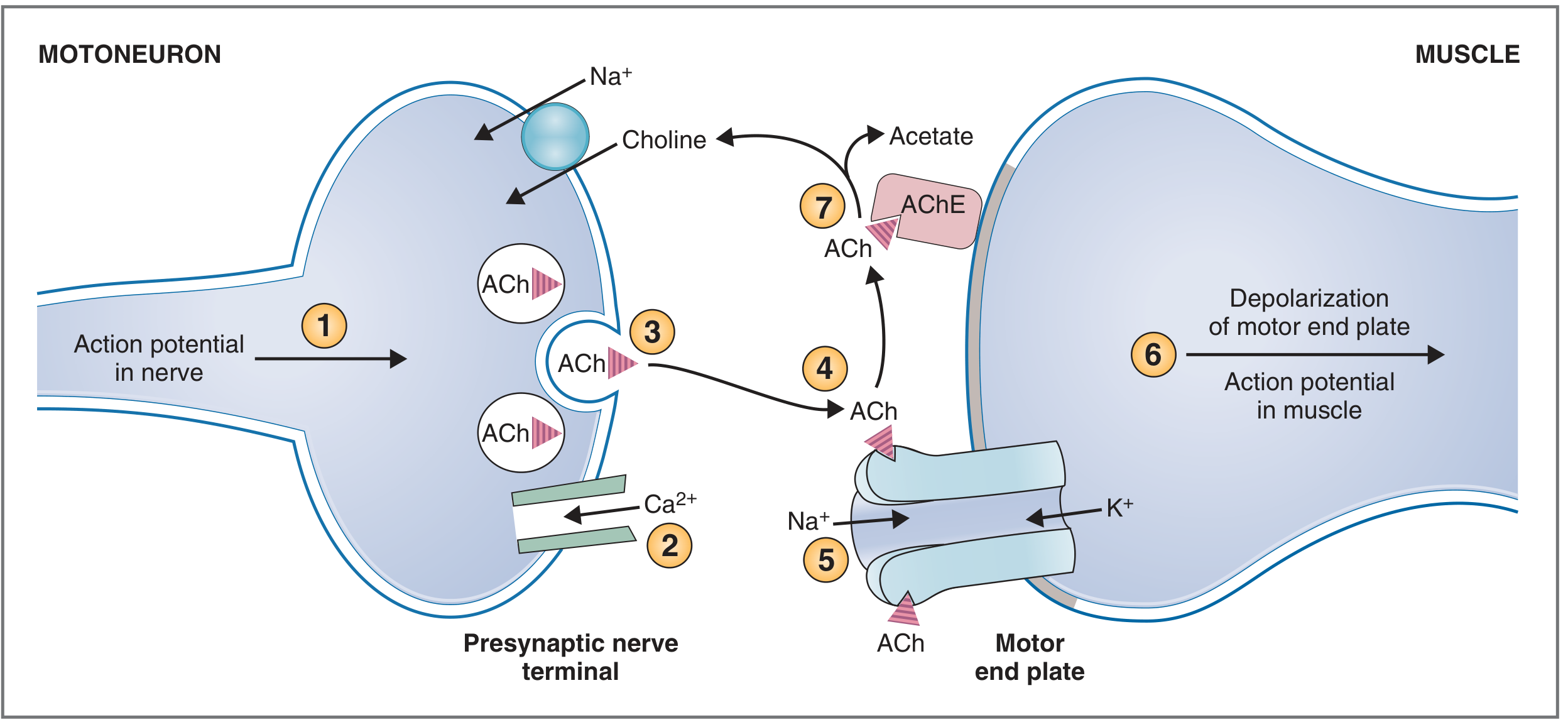
2c) Sequence of Events in Neuromuscular Transmission (5 marks)
The neuromuscular junction (NMJ) is the synapse between a motor neuron (motoneuron) and a skeletal muscle fiber.
Step-by-Step Sequence:
1. Action potential propagates down the motor neuron to the presynaptic terminal (by sequential depolarization of Na+ channels).
2. Depolarization of the presynaptic terminal opens voltage-gated Ca²+ channels → Ca²+ flows into the terminal down its electrochemical gradient.
3. Ca²+ entry triggers fusion of synaptic vesicles containing acetylcholine (ACh) with the plasma membrane → ACh is released by exocytosis (quantal release - smallest unit = one quantum/vesicle).
- ACh is synthesized from Acetyl CoA + Choline by choline acetyltransferase.
4. ACh diffuses across the synaptic cleft and binds to nicotinic ACh receptors (NnACh-R) on the motor end plate of the muscle.
5. Binding opens ligand-gated channels permeable to both Na+ and K+ (but Na+ influx dominates) → membrane depolarizes → generates an End Plate Potential (EPP).
6. The EPP depolarizes adjacent muscle membrane above threshold → generates a muscle action potential that propagates along the sarcolemma and down T-tubules → triggers excitation-contraction coupling (Ca²+ release from SR → cross-bridge cycling → contraction).
7. ACh is rapidly degraded in the synaptic cleft by acetylcholinesterase (AChE) into choline + acetate.
- Choline is taken back into the presynaptic terminal by an Na+-choline cotransporter (recycling).
Mnemonic: AP → Ca²+ → ACh release → Binds NnR → EPP → Muscle AP → Contraction → AChE terminates
- Costanzo Physiology 7th Ed., p.34
2d) Functional Differences Between Skeletal and Smooth Muscle (5 marks)
| Feature | Skeletal Muscle | Smooth Muscle |
|---|---|---|
| Innervation | Somatic (voluntary) motor neuron | Autonomic nervous system (involuntary) |
| Control | Voluntary | Involuntary |
| Striation | Present (actin-myosin organized in sarcomeres) | Absent (no sarcomeres, dense bodies instead) |
| Nuclei | Multiple, peripheral | Single, central |
| Speed of contraction | Fast (ms range) | Slow (seconds range) |
| Fatigue | Fatigues rapidly | Resistant to fatigue |
| Myosin light chain kinase | Troponin-Tropomyosin system for regulation | Calmodulin-MLCK system (no troponin) |
| Ca²+ source | SR (sarcoplasmic reticulum) | Both SR and extracellular Ca²+ |
| Pacemaker activity | None (requires nerve stimulation) | Present in some (e.g., gut, ureter) |
| Plasticity | Fixed resting length | Stress relaxation - can contract over wide range of lengths |
| Tone | No resting tone | Maintains resting tone |
| Gap junctions | Absent (functionally) | Present (single-unit smooth muscle acts as syncytium) |
| Examples | Biceps, quadriceps | Gut, bladder, blood vessels, uterus |
QUESTION 3 - Short Answer Questions (4 × 2 marks)
3a) Myasthenia Gravis (2 marks)
Myasthenia Gravis (MG) is an autoimmune disease of the neuromuscular junction.
- Pathophysiology: Autoantibodies against nicotinic ACh receptors (NnAChR) on the motor end plate → receptor destruction and complement-mediated damage → reduced EPP → failure to trigger muscle AP
- Also: Anti-MuSK (muscle-specific kinase) antibodies in seronegative cases
- Features: Fluctuating muscle weakness, worse with activity (fatigable weakness), improves with rest; ptosis, diplopia, dysphagia, respiratory muscle weakness
- Diagnosis: Edrophonium (Tensilon) test - AChE inhibitor rapidly reverses weakness
- Association: Thymoma/thymic hyperplasia
- Treatment: AChE inhibitors (neostigmine, pyridostigmine), immunosuppressants, thymectomy
3b) LH Surge (2 marks)
The LH surge is a massive, brief rise in LH secretion from the anterior pituitary that occurs in the mid-cycle (around day 13-14 of a 28-day cycle).
- Trigger: Rising estrogen levels from the dominant follicle cause a positive feedback on the anterior pituitary (switch from negative to positive feedback above a threshold ~200 pg/mL for >36 hours)
- Result: LH surge triggers:
- Ovulation - final maturation and rupture of the Graafian follicle (~36-40 hours after LH surge peak)
- Resumption of meiosis I in the oocyte
- Luteinization - conversion of ruptured follicle into corpus luteum
- FSH also surges simultaneously (smaller)
- Clinical use: LH surge detection is used to time ovulation in fertility treatments
3c) Major Role of Parathormone (PTH) (2 marks)
PTH is secreted by chief cells of parathyroid glands in response to low blood Ca²+ (hypocalcemia).
Major role: Raise blood calcium (hypercalcemic hormone)
Mechanisms:
- Bone: Activates osteoclasts → bone resorption → releases Ca²+ and PO₄³- into blood
- Kidney (proximal tubule): Increases Ca²+ reabsorption; increases PO₄³- excretion (phosphaturia)
- Kidney (activates 1α-hydroxylase): Converts 25-OH-D₃ to active 1,25-(OH)₂-D₃ (calcitriol) → which increases intestinal Ca²+ absorption
- Net effect: Blood Ca²+ ↑, Blood PO₄³- ↓ (phosphate is excreted)
- Receptor: PTH binds to Gs-protein-coupled receptor → activates adenylyl cyclase → ↑cAMP
3d) Role of Gap Junctions in Cellular Transport (2 marks)
Gap junctions are protein channels made of connexins (6 connexin proteins form one connexon; two connexons align across adjacent cells to form a gap junction channel).
Roles in cellular transport:
- Ion transport: Allow free passage of small ions (Na+, K+, Ca²+) - enabling electrical coupling between cells (e.g., cardiac myocytes, smooth muscle)
- Small molecule transfer: Pass molecules <1000 Da - cAMP, IP₃, ATP, glucose, amino acids
- Electrical syncytium: In cardiac muscle and single-unit smooth muscle, gap junctions allow action potentials to spread directly cell-to-cell without neurohumoral transmission
- Metabolic coupling: Share nutrients and signaling molecules between adjacent cells
- Embryonic development: Coordinate cell differentiation via chemical signaling
- NOT permeable to: Large proteins, nucleic acids, organelles
SECTION B
QUESTION 4 (Case-Based: Hemiplegia with UMN lesion)
52-year-old man with sudden left-sided loss of voluntary movement, left-sided hemiplegia with upper motor neuron type facial palsy on left side
4a) Pathway Affected and Diagram (4 marks)
The pathway affected is the Corticospinal Tract (Pyramidal Tract) on the right side (contralateral to the left-sided weakness).
Pathway (draw and label):
Motor Cortex (Primary - Precentral gyrus, Area 4)
         ↓
Internal Capsule (Posterior limb, Genu)
         ↓
Cerebral Peduncle (Midbrain)
         ↓
Pons (Corticobulbar fibers diverge to cranial nerve nuclei here)
         ↓
Pyramids of Medulla
         ↓
DECUSSATION of Pyramids (at medullary-spinal cord junction)
         ↓
Lateral Corticospinal Tract (contralateral spinal cord)
         ↓
Anterior Horn Cells (Lower Motor Neurons)
         ↓
Muscle
Note: For upper motor neuron facial palsy on the same (left) side as the hemiplegia, the lesion must be above the pons (where corticobulbar fibers decussate to reach the contralateral facial nucleus). The right-sided lesion causes left-sided hemiplegia AND left-sided UMN facial palsy.
Most likely lesion site: Right internal capsule or right cerebral hemisphere (cortex/corona radiata)
4b) Functions of the Corticospinal Tract (2 marks)
- Voluntary movement: Initiates and controls precise, skilled voluntary movements, especially of distal limb muscles (fine finger movements)
- Speed and dexterity: Fast-conducting pathway for fractionated (individual finger) movements
- Tone: Carries inhibitory signals to reduce tone in antigravity muscles (damage leads to spasticity)
- Reflexes: Modulates spinal cord reflex arcs; damage leads to hyperreflexia (release of inhibition)
- Somatosensory: Some fibers end in dorsal horn and modify sensory processing
4c) Differences: UMN Lesion vs LMN Lesion (2 marks)
| Feature | UMN Lesion | LMN Lesion |
|---|---|---|
| Site | Above anterior horn cell (cortex, corticospinal tract) | Anterior horn cell, nerve root, peripheral nerve, NMJ |
| Weakness | Present (paresis/plegia) | Present (paresis/plegia) |
| Tone | Increased (spasticity) - clasp-knife | Decreased (flaccidity) |
| Reflexes | Exaggerated (hyperreflexia) | Absent or diminished (hyporeflexia) |
| Babinski sign | Present (extensor plantar) | Absent |
| Atrophy | Mild (disuse atrophy only) | Severe (denervation atrophy) |
| Fasciculations | Absent | Present |
| Clonus | Present | Absent |
| Distribution | Hemiplegic / paraplegic (large groups) | Individual muscles / nerve distribution |
4d) Commonest Site of Lesion Causing Hemiplegia (1 mark)
The commonest site is the Internal Capsule (posterior limb and genu).
- Here all corticospinal and corticobulbar fibers are tightly packed together
- A small lesion (e.g., lacunar infarct) causes complete contralateral hemiplegia with facial involvement
- This is the site affected in the most common type of stroke (hypertensive small vessel disease)
QUESTION 5 - Short Notes (4 × 5 marks)
5a) Auditory Pathway (5 marks)
The auditory pathway carries sound information from the cochlea to the auditory cortex.
Components (in order):
- Cochlea (receptor) - Hair cells of the Organ of Corti transduce sound vibrations to electrical signals
- Cochlear nerve (CN VIII - vestibulocochlear nerve) - 1st order neurons (cell bodies in Spiral Ganglion)
- Cochlear nucleus (dorsal and ventral) - in pons/medulla junction (1st synapse)
- Trapezoid body - Major decussation point; most fibers cross to the contralateral side but bilateral representation begins here
- Superior olivary nucleus - 2nd synapse (sound localization, binaural integration)
- Lateral lemniscus - Ascends through pons
- Inferior colliculus (midbrain) - Important relay for auditory reflexes (e.g., startle reflex)
- Medial Geniculate Nucleus (MGN) of thalamus - Final relay before cortex
- Primary Auditory Cortex (A1) - Located in Heschl's gyri (transverse temporal gyri), Brodmann's areas 41 and 42, in the superior temporal gyrus
Key features:
- Bilateral representation at all levels above the cochlear nucleus (so unilateral central lesions rarely cause complete deafness)
- Tonotopic organization maintained throughout - each frequency has a spatial map
- The crossed pathway (contralateral ear) is dominant
5b) Types of Aphasia (5 marks)
Aphasia is an acquired disorder of language due to brain damage (usually left hemisphere).
| Type | Lesion Site | Comprehension | Fluency | Repetition | Features |
|---|---|---|---|---|---|
| Broca's (Motor/Expressive) | Broca's area (Area 44,45) - inferior frontal gyrus | Intact | Non-fluent | Impaired | Speaks in short, effortful phrases; telegraphic speech; aware of deficit; frustration |
| Wernicke's (Sensory/Receptive) | Wernicke's area (Area 22) - posterior superior temporal gyrus | Impaired | Fluent | Impaired | Fluent but meaningless speech; neologisms, paraphasias; unaware of deficit |
| Conduction Aphasia | Arcuate fasciculus (connects Broca to Wernicke) | Intact | Fluent | Severely impaired | Cannot repeat; can understand and produce spontaneous speech |
| Global Aphasia | Large perisylvian lesion | Impaired | Non-fluent | Impaired | Severe loss of all language functions |
| Anomic Aphasia | Angular gyrus or diffuse | Relatively intact | Fluent | Intact | Word-finding difficulty; inability to name objects (anomia) |
| Transcortical Motor | Anterior to Broca | Intact | Non-fluent | Intact | Similar to Broca but repetition preserved |
| Transcortical Sensory | Posterior to Wernicke | Impaired | Fluent | Intact | Similar to Wernicke but repetition preserved |
5c) Role of Hypothalamus in Thermoregulation (5 marks)
The hypothalamus is the master thermostat of the body, maintaining core body temperature at 37°C (set point).
Structure
- Anterior hypothalamus / Preoptic area: Contains heat-sensitive neurons; controls heat LOSS mechanisms
- Posterior hypothalamus: Controls heat PRODUCTION/conservation mechanisms
Sensors
- Central thermoreceptors: Neurons in preoptic area respond to blood temperature changes
- Peripheral thermoreceptors: Warm and cold receptors in skin send afferent signals
Responses to HIGH temperature (heat)
- Cutaneous vasodilation - shunts blood to skin → heat loss by radiation/conduction
- Sweating - evaporative heat loss (most effective mechanism in humans)
- Decreased metabolic rate - reduce heat production
- Behavioral responses - seeking cool environment, removing clothing
Responses to LOW temperature (cold)
- Cutaneous vasoconstriction - reduces heat loss from skin
- Shivering - involuntary skeletal muscle contractions → heat production
- Non-shivering thermogenesis - catecholamines and thyroid hormone increase metabolic rate; brown adipose tissue activation
- Piloerection - minimal in humans
- Behavioral - huddling, adding clothing
Fever
- Pyrogens (bacteria, LPS) stimulate macrophages to release interleukin-1 (IL-1), IL-6, TNF-α
- These trigger prostaglandin E₂ (PGE₂) synthesis in hypothalamus
- PGE₂ raises the set point → body feels cold → shivering, vasoconstriction until new set point reached
- Aspirin/NSAIDs: Inhibit cyclooxygenase → block PGE₂ → reduce fever
5d) Refractive Errors of the Eye and Their Correction (5 marks)
Refractive errors occur when the eye cannot focus light precisely on the retina.
1. Myopia (Near-sightedness)
- Defect: Eyeball too long or corneal/lens curvature excessive → light focuses in front of retina
- Vision: Near objects clear; distant objects blurred
- Correction: Concave (diverging) lens - diverges light rays before entering eye
2. Hypermetropia/Hyperopia (Far-sightedness)
- Defect: Eyeball too short or insufficient refractive power → light focuses behind the retina
- Vision: Distant objects clearer; near objects blurred (requires constant accommodation effort)
- Correction: Convex (converging) lens - converges light before entering eye
3. Astigmatism
- Defect: Unequal curvature in different meridians of cornea (non-spherical cornea) → light focuses at two different points instead of one
- Vision: Blurring and distortion at all distances; may cause headaches
- Correction: Cylindrical lens - corrects in one meridian only
4. Presbyopia
- Defect: Age-related loss of elasticity of lens → reduced accommodation (cannot focus on near objects)
- Typically begins after age 40
- Correction: Convex (reading) glasses for near work; bifocals
5. Aphakia
- Defect: Absence of lens (post-cataract surgery)
- Strong convex lens needed
QUESTION 6 - Short Answer Questions (4 × 2 marks)
6a) Referred Pain - Definition, Examples, Mechanism (2 marks)
Definition: Referred pain is pain that is felt at a location different from the actual site of the painful stimulus (the damaged/diseased organ). The pain is referred to the skin or deep structures that share the same dermatomal innervation as the viscus.
Examples:
- Cardiac ischemia (heart attack): Pain felt in left chest, left arm, jaw, left shoulder (T1-T4 segments)
- Diaphragmatic irritation: Pain referred to the shoulder tip (C3,4,5 - phrenic nerve)
- Appendicitis: Initial pain around umbilicus (T10 dermatome), later shifts to right iliac fossa
- Ureteric colic: Flank pain radiating to groin/inner thigh (L1-L2)
- Gallbladder: Right shoulder tip and right scapula
Mechanism - Convergence-Projection Theory (most accepted):
- Afferent pain fibers from viscera and from skin converge on the SAME 2nd order neuron in the dorsal horn (same spinal segment)
- The brain misinterprets the source of pain as coming from the skin (because somatic pain is far more common and has been experienced before)
- The cortex "projects" the pain to the familiar (somatic/skin) location
6b) Graded Potential (2 marks)
A graded potential is a local, non-propagated change in membrane potential that is proportional in amplitude to the stimulus intensity.
Characteristics:
| Feature | Graded Potential | Action Potential |
|---|---|---|
| Amplitude | Proportional to stimulus (graded) | All-or-none (fixed) |
| Propagation | Decremental (dies with distance) | Propagated without decrement |
| Summation | Can summate (temporal & spatial) | Cannot summate |
| Threshold | Not required | Required (all-or-none) |
| Duration | Variable | Fixed (1-2 ms) |
Examples:
- End plate potential (EPP) at NMJ
- Receptor potential (generator potential)
- EPSP and IPSP at synapses
- Slow waves in smooth muscle
Importance: Graded potentials act as the input signals of neurons; if they summate sufficiently to reach threshold at the axon hillock, they trigger an action potential.
6c) Effect of Autonomic Stimulation on Intestinal Motility (2 marks)
| Feature | Sympathetic Stimulation | Parasympathetic Stimulation |
|---|---|---|
| Neurotransmitter | Norepinephrine | Acetylcholine |
| Receptor | α and β adrenergic | Muscarinic (M₂, M₃) |
| Motility (peristalsis) | Decreased (inhibits) | Increased (stimulates) |
| Tone | Decreased | Increased |
| Sphincters | Contracts (closes - ileocecal, internal anal) | Relaxes (opens) |
| Secretion | Decreased | Increased |
Sympathetic: "Fight or flight" → diverts blood away from gut, inhibits digestion
Parasympathetic (vagus nerve for small intestine; pelvic nerve for large intestine): "Rest and digest" → promotes digestion and propulsion
Note: The gut has its own intrinsic nervous system (Enteric Nervous System - Meissner's and Auerbach's plexuses) that can function independently of the ANS.
6d) Abnormal Taste Sensations (2 marks)
| Term | Definition | Cause |
|---|---|---|
| Ageusia | Complete loss of taste sensation | CN VII (chorda tympani), CN IX lesion; zinc deficiency; drugs |
| Hypogeusia | Decreased taste sensitivity | Aging, radiation, chemotherapy, COVID-19 |
| Dysgeusia | Distorted or perverted taste (a normal substance tastes abnormal) | Liver disease, diabetes, pregnancy, drugs (metronidazole - metallic taste), zinc deficiency |
| Parageusia | Abnormal taste in the absence of any stimulus (taste hallucination) | Partial seizures, psychiatric disorders |
| Cacogeusia | Perception of bad/unpleasant taste | Dental infections, psychiatric conditions |
| Phantogeusia | Taste without any stimulus | Temporal lobe lesions |
Common clinical examples:
- COVID-19: Ageusia/hypogeusia due to damage to taste receptor cells or olfactory neurons
- Zinc deficiency: Hypogeusia (zinc required for taste receptor turnover)
- Chorda tympani damage (during middle ear surgery): Ipsilateral taste loss on anterior 2/3 of tongue
- Glossopharyngeal nerve (CN IX) damage: Taste loss from posterior 1/3 of tongue
All answers based on Costanzo Physiology 7th Ed., Ganong's Review of Medical Physiology 26th Ed., Guyton & Hall Medical Physiology, and Adams & Victor's Neurology - appropriate for MBBS 1st year standard.
This is a shared conversation. Sign in to Orris to start your own chat.