
Pivd plus hemangioma in d5 vertibrae further management
vertebral hemangioma management treatment
thoracic disc herniation PIVD management
vertebral hemangioma MRI spine thoracic
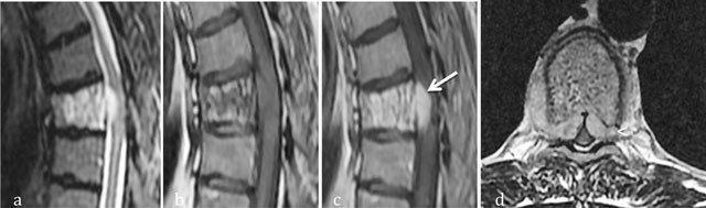
This composite diagnostic image features four MRI views of the thoracic spine illustrating an aggressive hemangioma at the T6 vertebral level. (a) Sagittal T2-weighted image shows high signal intensity within the vertebral body, indicating hypervascularity or edema. (b) Sagittal T1-weighted image shows a corresponding area of intermediate signal. (c) Post-contrast sagittal T1-weighted image demonstrates intense enhancement of the vertebral body with clear extension into the anterior epidural space (arrow) and anterior soft tissues (arrowhead), causing narrowing of the spinal canal. (d) Axial T1-weighted post-contrast image confirms circumferential involvement of the T6 vertebral body and bilateral soft tissue extension into the epidural space, resulting in mild cord compression. The presence of extra-osseous extension and cortical breakthrough characterizes this as an aggressive variant of a vertebral hemangioma, a key differentiator from typical latent hemangiomas. These features are critical for diagnosing potential spinal cord compression in the context of musculoskeletal oncology and neuroradiology.
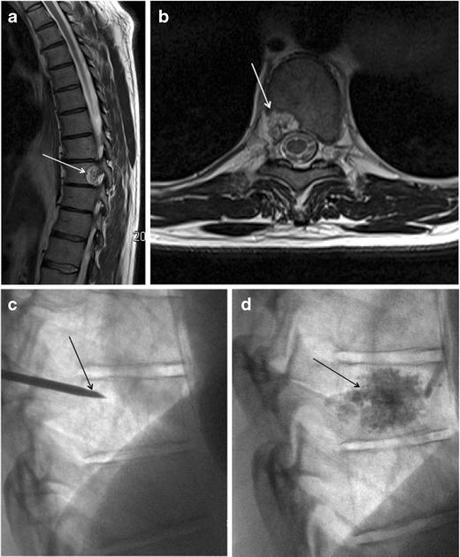
This composite figure demonstrates the diagnostic imaging and interventional management of a spinal hemangioma in the thoracic spine.
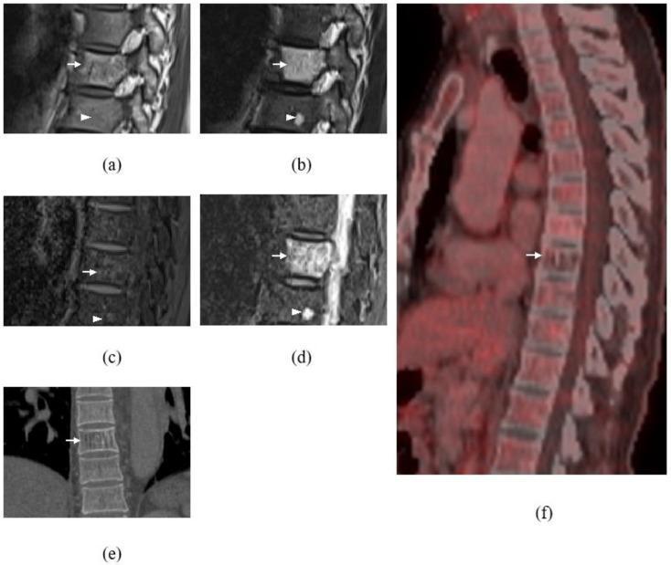
This composite educational image features multi-modal diagnostic imaging of the thoracic spine, focusing on vertebral body lesions at T9 and T10.
PIVD at D5 + Vertebral Hemangioma at D5: Further Management
- Determine if the hemangioma is incidental or aggressive/symptomatic
- Plan management for the disc prolapse without ignoring hemangioma risks (especially intraoperative hemorrhage)
- Address both lesions in an integrated manner
Step 1: Characterize the Hemangioma (Critical First Step)
MRI Assessment (Gold Standard)
- Typical/latent hemangioma: High signal on BOTH T1 and T2 (fat + vascular) - usually incidental, no treatment needed
- Aggressive hemangioma: Hypointense on T1, hyperintense on T2 - indicates a large vascular component; treat independently
Six Radiographic Criteria for Compressive Risk (Campbell's 2026)
- Thoracic location T3-T9 (D5 falls within this range - YES, this criterion is met)
- Entire vertebral body involvement
- Neural arch/pedicle involvement
- Irregular "honeycomb" appearance
- Expanded, poorly defined cortex
- Soft tissue swelling
Step 2: Clinical Classification
| Presentation | Likely Source | Action |
|---|---|---|
| Thoracic myelopathy (spasticity, weakness, Babinski +ve) | Disc OR hemangioma compressing cord | MRI urgently, may need combined surgery |
| Radiculopathy (band-like chest/abdominal pain) | Disc at D4-5 level | Conservative first, then disc surgery |
| Pure axial back pain | Could be hemangioma | Hemangioma workup, exclude disc as cause |
| Progressive neurologic deficit | Aggressive hemangioma or large disc | Surgery, embolization pre-op |
Step 3: Management Algorithm
A. If Hemangioma is Incidental (typical MRI features, no symptoms from it)
- Conservative (6-8 weeks first): NSAIDs, physiotherapy, analgesics, activity modification
- If conservative fails or neurologic compromise exists: Surgical discectomy
- Thoracic discectomy is rare (<2% of all discectomies) - approach depends on disc type:
- Soft disc prolapse: Thoracoscopic approach preferred
- Hard/calcified disc: Thoracotomy or costotransversectomy
- Surgical caution: The co-existing hemangioma at D5 increases intraoperative bleeding risk - even if "incidental," the surgeon must be prepared for vascular ooze from the vertebral body
- Thoracic discectomy is rare (<2% of all discectomies) - approach depends on disc type:
- The hemangioma can be observed with serial MRI every 6-12 months
B. If Hemangioma is Symptomatic / Aggressive
- Effective in 50-80% of symptomatic hemangiomas
- Dose: 30-40 Gy in fractionated doses
- Useful when surgery is not feasible or lesion is moderately symptomatic
- As per the 2023 review (PMID: 37122908), radiation remains the preferred non-surgical option
- Bone cement (PMMA) injection stabilizes the vertebral body and obliterates vascular spaces
- Effective for aggressive hemangiomas with pain and vertebral instability
- Contraindicated if posterior vertebral wall is breached (cement may leak into spinal canal)
- Can be done percutaneously; inflatable bone tamps (kyphoplasty) show similar good results
- Direct intralesional alcohol injection obliterates vascular spaces
- CT angiography required beforehand to map functional vascular spaces
- Less than 1 mL of ethanol used per session to avoid neurological damage
- Indicated when surgery is planned AND hemangioma has significant vascular component
- Reduces intraoperative blood loss substantially
- Best done 24-48 hours before surgery
- Embolization alone may give temporary pain relief but neurologic improvement is variable
- Indications: progressive neurologic deficit, spinal instability, failed conservative/radiation
- Anterior resection of hemangioma + disc + reconstruction
- If large posterior element involvement: posterior decompression + instrumented fusion
- T9-L1 or appropriate segment fusion after decompression (as shown in the Campbell's surgical case at T11)
Step 4: Integrated Surgical Planning (If Both Need Surgery)
- Pre-op CT angiography - map hemangioma vascularity and disc-cord relationship
- Pre-op embolization - 24-48 hrs before surgery to reduce bleeding
- Single-stage surgery preferred:
- Anterior approach: corpectomy/discectomy + vertebral body reconstruction + cage/strut graft
- Posterior stabilization: pedicle screw fixation for stability
- Blood bank preparation - crossmatch at minimum 4 units
- Neuromonitoring: Intraoperative SSEP/MEP is mandatory for thoracic cord surgery
Key Investigations Before Finalizing Management
| Investigation | Purpose |
|---|---|
| MRI spine (whole) with contrast | Characterize hemangioma (T1/T2 signal), cord compression, disc extent |
| CT spine with bone windows | Cortical integrity, pedicle involvement, posterior wall continuity |
| CT angiography | Vascular mapping of hemangioma if surgery/ethanol planned |
| X-ray standing (AP/Lateral) | Assess alignment, corduroy pattern |
| Neurological exam (complete) | Myelopathy grading (JOA score), sensory level, reflexes |
| Bone scan (optional) | Activity of hemangioma - though unreliable (can be hot or cold) |
Summary
| Scenario | Primary Action |
|---|---|
| Incidental hemangioma + disc causing symptoms | Treat disc (conservative → surgical), monitor hemangioma |
| Both hemangioma and disc causing symptoms | Workup for aggressive features, embolize, combined surgery |
| Myelopathy with progressive deficit | Urgent MRI, pre-op embolization, anterior decompression + fusion |
| Hemangioma symptomatic, disc incidental | Radiation or vertebroplasty, observe disc |
- Campbell's Operative Orthopaedics 15th Ed 2026, pp. 2582-2583
- Bailey and Love's Short Practice of Surgery 28th Ed, p. 534
- Recent surgical management review: Kasliwal 2024, PMID: 37992532
- Vertebral hemangioma review: Kato et al. 2024, PMID: 38789994
There is band like feel in chest with radiculopathy pain
T5 dermatome band chest thoracic radiculopathy nerve root
thoracic dermatomes map T5 chest wall
Band-like Chest Sensation + Radiculopathy at D5 Level - Management
Why This Symptom Occurs
- A tight, band-like or constricting feeling encircling the chest
- Burning, aching, or electric pain radiating along the T5 dermatomal strip
- Numbness or paresthesia (tingling, pins and needles) along the same band
- This is intercostal neuralgia from thoracic nerve root origin
Immediate Differential Diagnoses to Rule Out
| Differential | How to distinguish |
|---|---|
| Cardiac angina | ECG, troponin - pain typically not dermatomal, aggravated by exertion |
| Herpes zoster (shingles) | Look for vesicular rash in the same band; preceded by burning |
| Aortic dissection | Sudden onset, tearing pain, BP difference between arms - urgent |
| Pleuritis / pneumothorax | Pleuritic quality, breathing-related change, CXR |
| Diabetic truncal neuropathy | Multiple dermatomal bands, diabetic history, no disc on MRI |
| Intramedullary cord lesion | Upper motor neuron signs below the level |
Management for D5 PIVD with Radiculopathy (Band-like Pain)
Phase 1: Conservative Management (First 6-8 Weeks) - Try This First
Medications
| Drug | Role | Dose guidance |
|---|---|---|
| NSAIDs (e.g., diclofenac, etoricoxib) | First-line analgesic, anti-inflammatory | Standard anti-inflammatory doses with food |
| Gabapentin / Pregabalin | Neuropathic pain (the burning, band-like sensation) | Gabapentin 300mg TDS, titrate; Pregabalin 75mg BD |
| Tricyclic antidepressants (amitriptyline) | Neuropathic pain modulation, sleep | 10-25mg at night |
| Short-term oral steroids (methylprednisolone) | Reduce acute nerve root inflammation | 6-day taper pack |
| Muscle relaxants (methocarbamol, baclofen) | Paraspinal spasm associated with disc | Short course only |
| Tramadol / weak opioids | Rescue pain management | Only if above insufficient |
Physical Measures
- Relative rest (not absolute bed rest) for 1-2 weeks during acute phase
- Thoracic spine bracing/corset: Reduces motion at D4-5 level, offloads disc pressure
- Hot fomentation over paravertebral muscles for spasm relief
- Physiotherapy (after acute phase subsides):
- Postural correction (thoracic kyphosis worsens disc pressure)
- Core strengthening - deep paraspinal and abdominal muscles
- Extension exercises (McKenzie-based for thoracic disc)
- Traction - limited role in thoracic spine but may help
Interventional (Pain Clinic)
- Thoracic epidural steroid injection (TESI): Delivers corticosteroid (e.g., methylprednisolone 80mg + local anaesthetic) directly to the epidural space at D4-5 - very effective for nerve root pain
- Selective nerve root block (SNRB) at T5: Fluoroscopy/CT guided injection at the T5 foramen - both diagnostic (confirms T5 as the symptomatic level) AND therapeutic
- Intercostal nerve block: Along the T5 intercostal groove - gives temporary but good relief of the band-like pain
Important note on hemangioma: Epidural injections must be done with imaging guidance and the injectionist must be aware of the D5 hemangioma - avoid inadvertent needle trauma to the vascular lesion
Phase 2: Re-evaluate at 6-8 Weeks
Phase 3: Surgical Indications (When to Operate)
- Progressive myelopathy - worsening weakness in legs, gait ataxia, Babinski positive
- Bowel or bladder dysfunction - suggests cord compression, URGENT surgery
- Persistent unremitting radicular pain not responding to 6-8 weeks of conservative treatment
- Neurological deterioration on examination
- Hemangioma becomes aggressive (soft tissue component, cord compression on MRI)
Surgical Options for D5 PIVD (if needed)
| Approach | Best for | Notes |
|---|---|---|
| Anterior transthoracic discectomy + fusion | Central / midline herniation | Gold standard; requires thoracotomy |
| Thoracoscopic (VATS) discectomy | Soft disc, lateral/posterolateral | Less morbid, technically demanding |
| Transpedicular / lateral extracavitary | Lateral HNP, posterolateral disc | Posterior approach, avoids thoracotomy |
| Costotransversectomy | Lateral disc, calcified disc | Good access, avoids full thoracotomy |
- Pre-operative CT angiography and embolization of hemangioma 24-48 hours before surgery
- Anterior approach gives simultaneous access to disc AND vertebral body hemangioma
- Bone cement (vertebroplasty) can be done at same setting if vertebral body is unstable
- Instrumented fusion (pedicle screws T3-T7) likely needed if corpectomy performed
Red Flag Symptoms - Act Urgently If These Appear
| Red Flag | Meaning | Action |
|---|---|---|
| Leg weakness progressing | Cord compression from disc or hemangioma | Emergency MRI, surgical planning within 24-48 hrs |
| Difficulty walking, scissor gait | Myelopathy | Urgent neurosurgical referral |
| Urinary retention or incontinence | Cord or cauda compression | Emergency surgery |
| Bilateral band pain + clonus | Significant myelopathy | Do not delay |
Summary Flow
Band-like chest pain + radiculopathy at D5
        ↓
Confirm T5 dermatome involvement clinically
Rule out cardiac, aortic, herpes causes
        ↓
MRI (already done) - confirm disc is compressing T5 root
Check hemangioma MRI signal (T1/T2)
        ↓
NO myelopathy signs?
  → Conservative 6-8 weeks (NSAIDs + gabapentin + physiotherapy)
  → + Epidural/selective nerve root block if pain severe
        ↓
Persistent or worsening?
  → Surgery (anterior transthoracic or thoracoscopic)
  → With pre-op embolization of hemangioma
- Miller's Review of Orthopaedics 9th Ed, pp. 767-768
- Bailey & Love's Short Practice of Surgery 28th Ed, p. 534
- Campbell's Operative Orthopaedics 15th Ed 2026, p. 2583
And for hemangioma?
vertebral hemangioma treatment vertebroplasty embolization radiation
PMID: 38789994
PMID: 37122908
vertebral hemangioma vertebroplasty embolization treatment spine
D5 Vertebral Hemangioma - Management
Step 1: Classify Before Treating
| Category | MRI T1 | MRI T2 | Clinical | Action |
|---|---|---|---|---|
| Asymptomatic / typical | Bright (fat-dominant) | Bright | No symptoms, found incidentally | Observe only |
| Symptomatic | Bright or intermediate | Bright | Back pain attributable to hemangioma | Treat (radiation / vertebroplasty) |
| Aggressive / compressive | Hypointense (vascular-dominant) | Hyperintense | Neurological deficit, cord compression | Active treatment + possible surgery |
Key point for this patient: The current band-like pain is most likely from the PIVD, NOT the hemangioma. However, if the hemangioma at D5 has any aggressive MRI features (T1 hypointense), it must be treated independently - because D5 is right in the high-risk zone (T3-T9).
The 6 Imaging Features of an Aggressive / Compressive Hemangioma
- Thoracic location T3-T9 (D5 automatically fulfils this)
- Entire vertebral body involvement
- Neural arch / pedicle involvement
- Irregular honeycomb trabecular pattern
- Expanded, poorly defined cortex
- Paraspinal soft tissue component
Treatment Options - By Indication
Option 1: Observation (Incidental / Asymptomatic)
- No biopsy, no treatment required
- Serial MRI every 12 months to monitor for change in size, signal, or soft tissue extension
- Reassure patient - vast majority of vertebral hemangiomas are completely benign and never require intervention
- Educate on red flag symptoms (new neurological symptoms, sudden worsening pain)
Option 2: Radiation Therapy (Symptomatic, No Neurological Deficit)
- Success rate: 50-80% for pain relief and arrest of progression
- Conventional external beam radiotherapy: 30-40 Gy in fractionated doses (typically 10 fractions of 3 Gy)
- Best suited when: hemangioma is painful, no cord compression, surgical risk is high
- Stereotactic radiosurgery (SRS/SBRT): Emerging option - a 2022 systematic review (PMID: 35378316) showed good local control with SRS for benign vertebral hemangiomas
- Avoid in aggressive lesions with significant vascular component - risk of radiation-induced oedema worsening cord compression
- Note: Radiation avoided in benign lesions when possible due to risk of secondary sarcomatous change (Campbell's)
Option 3: Vertebroplasty / Kyphoplasty (Aggressive, Pain + Instability, Intact Posterior Wall)
- Mechanism: PMMA (polymethylmethacrylate) bone cement injected percutaneously into the vertebral body under fluoroscopic/CT guidance - obliterates vascular spaces and stabilizes the bone
- Excellent results for pain relief and vertebral stabilization
- Strict contraindication: Posterior vertebral body cortex must be intact - if breached, cement leaks into the spinal canal causing catastrophic neurological injury
- Kyphoplasty (inflatable bone tamp first, then cement): Similar results, slightly lower cement leak risk, restores some vertebral height
- Can be combined with embolization for highly vascular lesions
- Best used when: Pain is hemangioma-related, vertebral body shows trabecular destruction, no posterior wall breach
Option 4: Percutaneous Ethanol (Sclerotherapy) Injection (Symptomatic, Highly Vascular)
- Direct intralesional injection of absolute ethanol obliterates the vascular channels
- Requires CT angiography first to map the functional vascular spaces and direct needle placement
- Dose: less than 15 mL total - higher volumes (42-50 mL reported) caused pathological fractures
- Effective for obliterating symptomatic hemangiomas
- Less commonly used now - superseded by vertebroplasty at most centres
Option 5: Transarterial Embolization (Pre-operative or Temporary Relief)
- Selective embolization of feeding vessels via femoral arterial approach
- Primary uses:
- Pre-operative (24-48 hrs before surgery) to reduce intraoperative bleeding
- Temporary relief of neurologic symptoms in patients awaiting surgery
- Adjunct to vertebroplasty in highly vascular lesions
- Gives temporary pain relief but not a standalone curative treatment
- Critical warning at D5 level: Must identify and protect the artery of Adamkiewicz (artery of the lumbar enlargement, usually T9-L2, but can arise as high as T5-T8). Inadvertent embolization causes anterior spinal artery syndrome (paraplegia)
- CT angiography / DSA essential before embolization to map vascular anatomy
Option 6: Surgery (Aggressive Hemangioma + Neurological Deficit / Fracture)
- Progressive myelopathy or radiculopathy from hemangioma compression (separate from the disc)
- Pathological fracture causing instability
- Failed radiation or percutaneous treatment
- Epidural extension with cord compression
- Anterior: Corpectomy (partial or complete vertebral body resection) + cage reconstruction + anterior plating
- Posterior: Decompression laminectomy + pedicle screw instrumented fusion (if posterior elements involved)
- Combined anterior-posterior: For circumferential cord compression or highly unstable lesion
- Preoperative embolization is mandatory before any surgery on a vertebral hemangioma
Management Algorithm for This Patient's D5 Hemangioma
D5 Hemangioma found on MRI (alongside PIVD)
            ↓
Check MRI carefully:
T1 bright + T2 bright? → Typical/fat-dominant
T1 DARK + T2 bright?  → Aggressive/vascular
            ↓
Count aggressive imaging features (0-6)
            ↓
0-2 features, T1 bright → OBSERVE
  Serial MRI at 12 months
  No specific treatment
            ↓
3+ features OR T1 hypointense → ACTIVE TREATMENT
  No neurological deficit → Radiation (30-40 Gy)
                            OR Vertebroplasty (if posterior wall intact)
  With neurological deficit → Pre-op embolization + Surgery
  Surgery planned for PIVD? → Embolize hemangioma 24-48 hrs before
Key Points Specific to D5 (T5 Level)
- D5 sits in the watershed zone of spinal cord blood supply (T4-T9) - cord ischemia risk is higher here than in lumbar spine
- Aggressive hemangiomas are most commonly reported in the thoracic region - D5 is a red flag location
- Any surgical or interventional approach must account for the proximity of the Adamkiewicz artery (can arise between T5-T8 in some individuals)
- Intraoperative neuromonitoring (SSEP + MEP) is mandatory if surgery is undertaken
- Campbell's Operative Orthopaedics 15th Ed 2026, pp. 2582-2583
- Kato et al., Vertebral hemangiomas: a review on diagnosis and management, J Orthop Surg Res 2024
- Sharma et al., Vertebral hemangioma - radiation therapy perspective, Rep Pract Oncol Radiother 2023
- Conti et al., Radiosurgery for Vertebral Hemangiomas - Systematic Review, World Neurosurg 2022