
i just peed 10 minutes ago but when i breath out and and my stomach goes flat urine sensation comes but when i take miraben everything goes to normal
overactive bladder mirabegron mechanism treatment
What You Are Experiencing: Overactive Bladder (OAB)
The Underlying Mechanism
- Your diaphragm rises
- Your abdominal wall flattens
- This slightly increases pressure inside your abdomen
- That pressure transmits to the bladder
- In a sensitive/overactive bladder, this is enough to trigger an unwanted urgency signal
Why Mirabegron (Mirabel) Fixes It
- Mirabegron is a selective beta-3 adrenergic receptor agonist
- Beta-3 receptors sit on the detrusor muscle of the bladder
- When mirabegron activates these receptors, the detrusor relaxes
- A relaxed detrusor muscle has greater storage capacity and is much less likely to contract involuntarily
- Result: urgency, frequency, and that sudden "I must go NOW" feeling are all reduced
What is Triggering You Specifically
What You Can Do Beyond Medication
| Strategy | How it Helps |
|---|---|
| Bladder training - gradually extend the time between voids | Retrains the detrusor to tolerate fuller volumes |
| Pelvic floor exercises (Kegels) - tighten pelvic floor when urgency hits | Suppresses the involuntary detrusor contraction |
| Fluid management - avoid caffeine, alcohol, carbonated drinks | These irritate the bladder lining and worsen urgency |
| Timed voiding - urinate on a schedule, not just when you feel the urge | Breaks the habit of responding to every small urge |
When to Revisit Your Doctor
- Pain with urination
- Blood in urine
- Incomplete bladder emptying
How do i correct it without taking medication
overactive bladder behavioral therapy pelvic floor
Correcting OAB Without Medication
PILLAR 1: Urge Suppression (Most Important - Start Here)
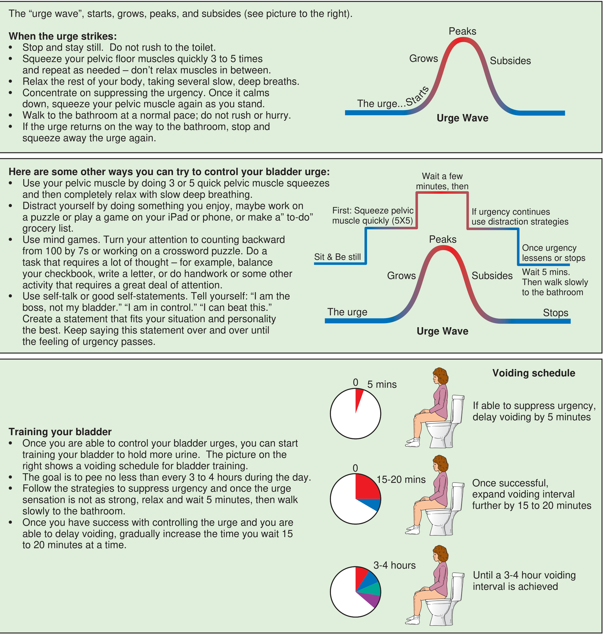
- Stop and stay still - do NOT rush to the toilet. Running actually worsens the urge.
- Squeeze your pelvic floor muscles quickly 3-5 times - rapid squeezes, not one long hold. This reflexively relaxes the detrusor (the bladder muscle).
- Relax the rest of your body and take slow, deep breaths - consciously slow your breathing.
- Distract your mind - count backwards from 100 by 7s, make a mental to-do list, think of something absorbing. This works because urgency has a strong mental component.
- Tell yourself: "I am in control. The urge will pass." - self-talk actually helps, this is evidence-based.
- Once the urge subsides, walk calmly and slowly to the bathroom - do not rush.
PILLAR 2: Pelvic Floor Muscle Training (Kegel Exercises)
- Find the right muscle - imagine you are stopping the flow of urine mid-stream. That squeezing is the pelvic floor.
- Do NOT squeeze your buttocks or stomach - only the pelvic floor.
| Type | How | Reps |
|---|---|---|
| Quick flicks | Squeeze fast and release immediately | 10-15 reps |
| Slow holds | Squeeze and hold for 5-10 seconds, then fully relax | 10 reps |
PILLAR 3: Bladder Retraining (Stretches Your Bladder's Capacity)
- Week 1-2: When you feel an urge, suppress it and delay voiding by just 5 minutes before going.
- Week 3-4: Extend the delay to 10-15 minutes.
- Week 5+: Keep extending by 15-20 minutes each week.
- End goal: Voiding every 3-4 hours during the day (not every 30-60 minutes).
PILLAR 4: Lifestyle Changes That Make a Big Difference
| Change | Why It Helps |
|---|---|
| Cut caffeine (tea, coffee, cola) | Caffeine directly irritates the bladder lining and worsens urgency |
| Cut alcohol | Diuretic + irritant effect |
| Reduce fizzy drinks | Carbonation irritates the bladder |
| Drink the right amount - about 1.5-2L water/day | Too little concentrates urine (more irritating); too much fills the bladder faster |
| Avoid drinking large amounts at once | Spread fluid intake evenly through the day |
| Stop drinking 2 hours before bed | Reduces nocturia (night-time urgency) |
| Lose weight if overweight | Excess abdominal weight increases bladder pressure - directly relevant to your breathing trigger |
| Treat constipation | A full bowel presses on the bladder |
For Your Specific Trigger (Breathing Out)
- Practice diaphragmatic breathing - train yourself to breathe from your belly rather than holding tension in your core. This reduces the sharp pressure changes that trigger your bladder.
- At the moment of exhale-triggered urge: immediately do 3-5 rapid pelvic floor squeezes. This is your personal "rescue move."
How Long Does This Take?
- Some improvement in 2-4 weeks
- Significant improvement in 6-12 weeks
- Full benefit in 3-6 months
How to find right muscle
pelvic floor muscle anatomy diagram male
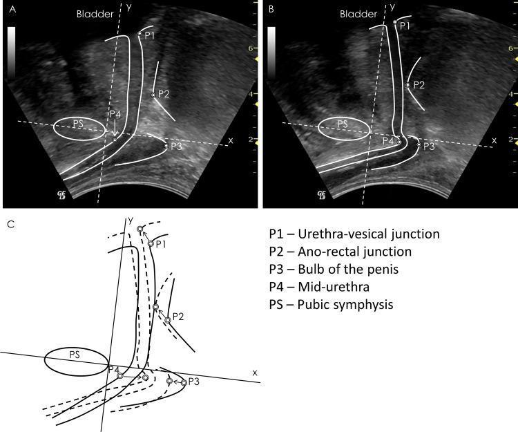
This composite educational graphic features diagnostic transperineal ultrasound images and a schematic diagram illustrating male pelvic floor dynamics. Panels A and B present sagittal ultrasound views in relaxed and contracted states, respectively, with superimposed anatomical markers. Key landmarks identified include the bladder, pubic symphysis (PS), urethra-vesical junction (P1), ano-rectal junction (P2), bulb of the penis (P3), and mid-urethra (P4). Panel C is a comparative line diagram that overlays the relaxed (dashed lines) and contracted (solid lines) positions of these structures relative to the pubic symphysis. Displacement vectors (arrows) demonstrate the cranial and ventral movement of the pelvic floor landmarks during voluntary contraction. The content is designed to teach pelvic floor functional anatomy and the calculation of landmark displacement, which is clinically relevant for assessing pelvic floor muscle function and treating conditions like urinary incontinence or pelvic floor dysfunction in males.
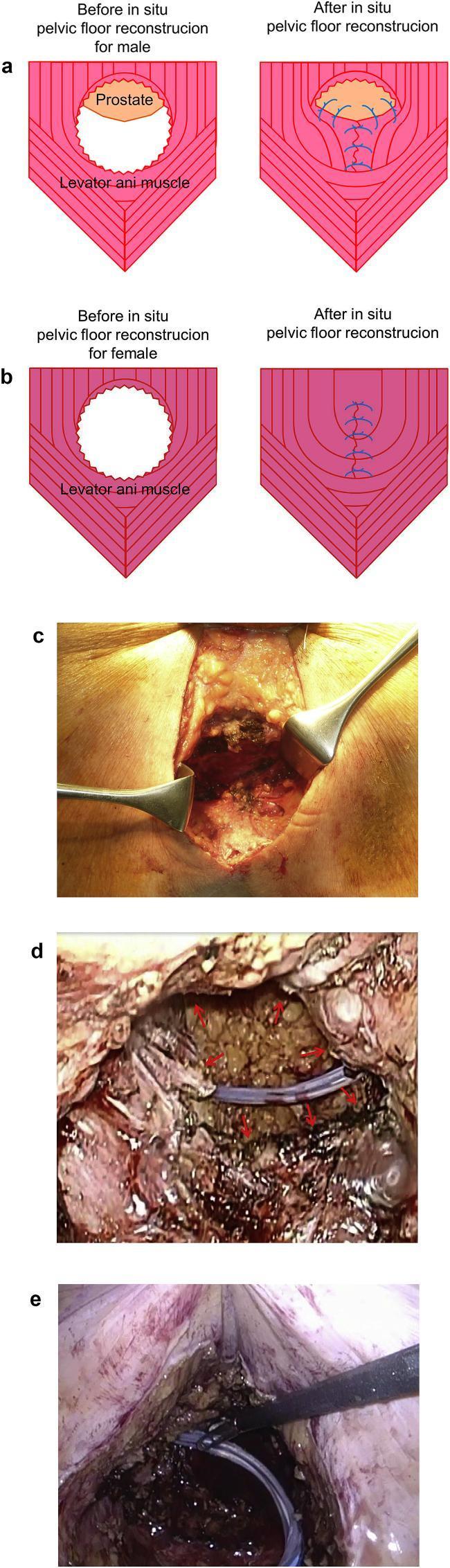
This multi-panel figure illustrates surgical techniques and intraoperative findings for in situ pelvic floor reconstruction following perineal surgery. Panels (a) and (b) are anatomical diagrams showing the schema for levator ani muscle closure in male and female patients, respectively. The male diagram highlights the relationship between the levator ani and the prostate, showing the placement of sutures to bridge the pelvic defect. Panel (c) is a clinical photograph of the perineal surgical site, with metal retractors exposing a deep pelvic wound where sutures are applied to the levator ani muscle. Panels (d) and (e) provide laparoscopic views of the pelvic cavity. Panel (d) shows the pelvic floor prior to closure, with arrows identifying the unsutured levator ani muscle and a visible intra-abdominal drainage tube. Panel (e) depicts the same surgical field after levator ani closure, showing the reconstructed pelvic floor, the drainage tube in situ, and surgical instruments used for manipulation. The content is designed for surgical education regarding pelvic floor integrity and hernia prevention post-resection.
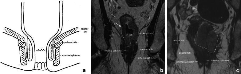
This composite educational image illustrates the pelvic floor and anal canal anatomy and its clinical relevance in rectal cancer staging. Panel (a) is an anatomical diagram depicting the coronal view of the anal canal, highlighting the levator ani, puborectalis muscle, external sphincter, and internal sphincter, with a dashed line marking the intersphincteric plane. Panel (b) is a coronal T2-weighted MRI showing a cT3a rectal tumor in a male patient. An arrow indicates irregular wall thickening with minimal (±1 mm) perirectal extension invading the right internal anal sphincter. Panel (c) is a coronal T2-weighted MRI of a female patient with a cT4b rectal tumor. The tumor (outlined by a dashed line) shows extensive infiltration, invading both internal sphincters and extending into the external sphincter, levator ani, and puborectalis muscles on the left side. Muscles and sphincters appear characteristically hypointense on T2 imaging, while the tumor presents as an intermediate-signal mass disrupting normal anatomical boundaries, essential for determining surgical management such as abdominoperineal resection.
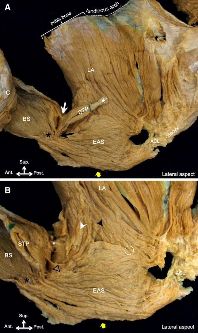
This clinical anatomical photograph displays a lateral cadaveric dissection of the human male pelvic floor muscles, divided into two panels (A and B). The imaging demonstrates the complex spatial relationships and intermuscular connections between the superficial and deep perineal layers. Panel A shows the superficial transverse perineal (STP) muscle originating near the lateral tendon (white star) and extending inferomedially to connect with the external anal sphincter (EAS, black arrow) and anteriorly with the bulbospongiosus (BS, white arrow). The BS and EAS are also linked (black asterisk). The levator ani (LA) is shown originating from the pubis bone and tendinous arch. Panel B, following lateral reflection of the STP, reveals the LA's bifurcation into anterior (white arrowhead) and posterior (black arrowhead) muscle bundles relative to the EAS. Key educational markers indicate the connection between the LA and BS (white asterisk), the blending point of the LA and EAS (white circle), and the connection between the STP and LA (black triangle). The anococcygeal ligament (ACL) is visible posteriorly, anchoring the EAS to the coccyx. This visual resource is intended for advanced medical education in surgical anatomy and pelvic floor functional pathology.
Method 1: The "Stop the Stream" Test (Easiest)
- Start peeing normally
- Mid-stream, try to stop the flow completely
- The muscle you just squeezed to stop it - that is your pelvic floor
Important: Only do this to identify the muscle, not as a regular exercise. Doing it repeatedly while urinating can interfere with normal bladder function.
Method 2: The "Lift and Squeeze" Feel
- Sit comfortably or lie on your back with knees bent
- Imagine you are trying to stop yourself passing gas (wind)
- You will feel a lifting, squeezing sensation deep inside your lower pelvis - between your sit bones
- For men: the base of your penis and scrotum will lift slightly upward - you may be able to feel this with your hand on the inner thigh area
Method 3: The Mirror/Hand Test (Confirms You Have It Right)
- Sit or stand and look at the base of your penis
- When you squeeze the pelvic floor correctly, the penis will pull slightly inward and upward (like a small lift)
- Your buttocks, thighs, and stomach should not move
How to Know You Have the WRONG Muscle
| Wrong muscle being used | Sign |
|---|---|
| Buttocks | Your bottom lifts off the seat |
| Thighs | Thighs press together or rotate inward |
| Abdomen | Your belly pushes outward or tightens |
| Holding your breath | You stop breathing while squeezing |
The Ultrasound View (What Happens Inside)
Once You Find It - How to Practice
| Exercise | What to do | Goal |
|---|---|---|
| Quick flick | Squeeze fast, release immediately | 10-15 times |
| Slow hold | Squeeze and hold for 5 seconds, then fully relax for 5 seconds | 10 times |
| Full relax | After each set, consciously let the muscle go completely limp | Always |
And when i sqeese suprapubic area urine sensation occurs
When i press suprapubic area urine sensation comes without full bladder