
Benign prostatic hypertrophy pathogenisis
"benign prostatic hyperplasia"[MeSH Terms] AND pathogenesis
benign prostatic hyperplasia pathogenesis DHT stromal epithelial diagram
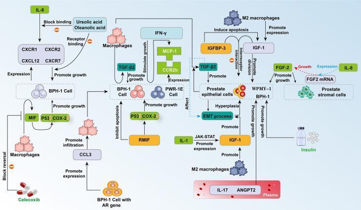
This medical pathophysiology diagram illustrates the complex cytokine signaling network and cellular interactions involved in Benign Prostatic Hyperplasia (BPH). It highlights the interplay between prostate epithelial cells (BPH-1, PWR-1E), stromal cells (WPMY-1), and infiltrating M2 macrophages. Key signaling pathways depicted include the IL-8 axis (involving receptors CXCR1/2 and CXCR7), which promotes BPH-1 growth and is inhibited by Ursolic and Oleanolic acids. The diagram shows M2 macrophages secreting TGF-β2 and IGF-1 to drive epithelial-mesenchymal transition (EMT), hyperplasia, and cell division. Pro-inflammatory signaling through IL-1 activates the JAK-STAT pathway to stimulate IGF-1, while recombinant MIF (rMIF) promotes growth by modulating P53 and COX-2 levels. Additionally, the role of metabolic factors like insulin and plasma cytokines (IL-17, ANGPT2) in stimulating BPH-1 and WPMY-1 cell proliferation is shown. Therapeutic interventions are represented by Celecoxib, which blocks macrophage-induced proliferation via CCL3 reversal. This schematic serves as an educational tool for understanding the immunoinflammatory and endocrine mechanisms driving prostate enlargement.
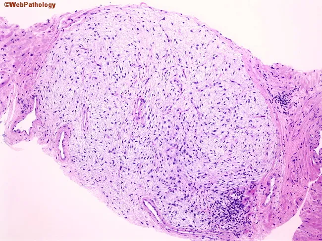
Light microscopy image of prostatic tissue stained with Hematoxylin and Eosin (H&E). The slide displays a well-circumscribed nodule composed of loose, fibromuscular stroma with sparse cellularity, representing an early stromal component of benign prostatic hyperplasia (BPH). Epithelial elements are scant within the nodule, with little glandular proliferation, and the surrounding prostate parenchyma shows a conventional fibromuscular stroma adjacent to residual glands. The stromal cells are spindle-shaped with elongated nuclei and pale cytoplasm embedded in a myxoid to fibrous extracellular matrix; mitotic activity is absent or minimal. There is no cytologic atypia, necrosis, or significant inflammatory infiltrate. This pure stromal nodularity represents one of the earliest histologic changes in BPH and can be encountered incidentally on prostate needle biopsies. Importantly, the diagnosis of prostatic hyperplasia (or nodular hyperplasia) should not be rendered on needle core biopsy; rather, specimens should be signed out as benign prostatic tissue with descriptive comment on the nodular stroma. Clinically, such findings may correlate with lower urinary tract symptoms in the patient, but histology alone cannot confirm severity. The differential considerations include stromal-rich lesions or scar tissue; malignant prostatic neoplasia is not suggested by these features. This pattern emphasizes strict sign-out criteria.
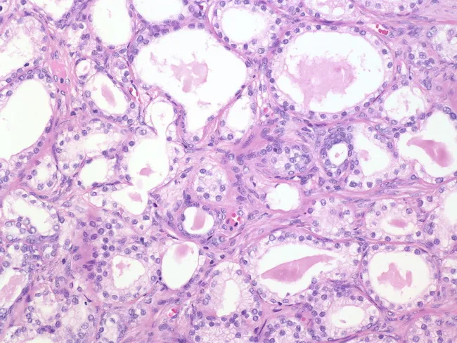
High magnification hematoxylin and eosin stained sections from a transurethral resection specimen show benign prostatic tissue with atypical adenomatous hyperplasia (adenosis). Glandular units vary in size and display round, oval, or irregular lumina. The epithelial cytoplasm is pale, contrasting with the amphophilic cytoplasm commonly seen in prostatic adenocarcinoma. Nuclei are not markedly enlarged and prominent nucleoli are generally absent; however, in up to about 30% of cases nucleoli may be conspicuous. The architecture consists of densely packed acini forming small, tight clusters without destructive invasion or stromal desmoplasia. Basal cell layering, when preserved, supports a benign process, and cytologic atypia is mild relative to carcinoma. The lesion overlaps with features that can mimic cancer, so careful assessment of invasion, nucleolar prominence, mitotic activity, and glandular polarity is essential. This image highlights a benign proliferative process, benign mimics of neoplasia, and the importance of correlating histology with clinical data. Clinically, distinguishing adenosis from prostatic adenocarcinoma influences management, PSA interpretation, and surveillance strategy. The findings are relevant for surgical pathology education, differential diagnosis discussions, and educational case repositories aiming to illustrate pitfalls in prostate lesion interpretation, particularly in TURP specimens. Educational value. Educational context for pathology residents and practicing clinicians worldwide today.
Benign Prostatic Hyperplasia (BPH) - Pathogenesis
Definition and Epidemiology
- Robbins & Kumar Basic Pathology, p. 659
- Histology: A Text and Atlas, p. 2145
Zone of Origin
Core Pathogenic Mechanisms
1. Dihydrotestosterone (DHT) - The Central Mediator
- Circulating testosterone enters prostatic stromal cells and is converted to DHT by the enzyme 5α-reductase type 2
- DHT is 10x more potent than testosterone and binds nuclear androgen receptors (ARs) in both stromal and epithelial cells
- DHT acts as:
- An autocrine agent on stromal cells (stimulates their own proliferation)
- A paracrine hormone on glandular epithelial cells (drives epithelial proliferation)
- DHT-induced growth factors increase proliferation of stromal cells and decrease apoptosis (cell death) of epithelial cells - a dual mechanism of net tissue accumulation
- Robbins & Kumar Basic Pathology, p. 659
- Histology: A Text and Atlas, p. 2145
2. Age-Related Hormonal Imbalance (Estrogen-Androgen Shift)
-
Testosterone levels decline
-
Estrogen levels remain unchanged or increase due to peripheral conversion of androgens (via aromatase in adipose tissue)
-
The resulting elevated estrogen:androgen ratio acts synergistically with DHT
-
Both stromal and epithelial cells express estrogen receptors, so estrogen drives additional proliferation
-
This hormonal shift explains why BPH is a disease of aging even as testosterone falls
-
Robbins & Kumar Basic Pathology, p. 659
3. Stromal-Epithelial Interactions
- Stromal cells (fibroblasts, smooth muscle cells) produce paracrine growth factors (e.g., FGF-7/KGF, IGF-1, TGF-β2) that stimulate epithelial proliferation
- Epithelial cells in turn signal back to stroma - this bidirectional crosstalk amplifies the hyperplastic process
- BPH nodules are composed of variable proportions of fibromuscular stroma and proliferating glandular elements, reflecting both components
4. Inflammatory / Immune Mechanisms (Emerging Evidence)
- M2 macrophages secreting TGF-β2 and IGF-1, driving epithelial-mesenchymal transition (EMT) and hyperplasia
- IL-8 axis (via CXCR1/2 receptors) promoting epithelial cell growth
- IL-1 → JAK-STAT → IGF-1 pathway stimulating proliferation
- COX-2 and pro-inflammatory cytokines (IL-17, ANGPT2) contributing to a pro-proliferative microenvironment
5. Cellular Senescence
- Aging prostatic epithelial cells undergo senescence and produce interleukin-19 (IL-19), which promotes BPH development
- The senescence-associated secretory phenotype (SASP) creates a pro-inflammatory microenvironment that drives further stromal and epithelial proliferation
6. Genetic and Familial Factors
-
A hereditary/familial form of BPH exists
-
Twin studies and family studies show genetic susceptibility
-
Men with a first-degree relative with symptomatic BPH have a significantly increased risk, particularly for early-onset disease (before age 60)
-
Campbell-Walsh-Wein Urology, block 79
Summary Table
| Factor | Mechanism | Effect |
|---|---|---|
| DHT (via 5α-reductase type 2) | Binds nuclear ARs in stroma + epithelium | Increased proliferation, decreased apoptosis |
| Estrogen (with aging) | Synergizes with DHT via estrogen receptors | Additional epithelial + stromal growth |
| Stromal growth factors (FGF-7, IGF-1) | Paracrine signaling | Epithelial proliferation |
| M2 macrophages, IL-8, IL-1 | Cytokine-mediated signaling | Inflammatory-driven hyperplasia |
| Cellular senescence (IL-19) | SASP creates pro-growth environment | Stromal and epithelial expansion |
| Genetic susceptibility | Inherited AR sensitivity or growth factor variants | Familial early-onset BPH |
Morphological Consequences
-
Prostate weight increases 3-5 fold (60-100 g or more; normal ~20 g)
-
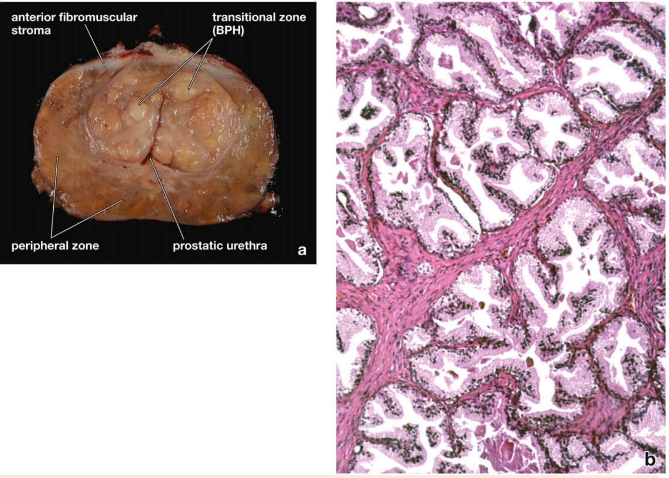
Hyperplastic nodules appear in the transition zone with variable solid/cystic architecture
-
Microscopically: nodules contain proliferating glands lined by tall columnar epithelial cells + outer flattened basal cells (bi-layered pattern - distinguishes BPH from carcinoma)
-
Glandular lumina may contain corpora amylacea (laminated proteinaceous material)
-
Smooth muscle hypertrophy in stroma adds a dynamic component to obstruction (targeted by α1-blockers)
-
Robbins & Kumar Basic Pathology, p. 659
Therapeutic Implications of Pathogenesis
| Pathogenic Target | Drug Class | Mechanism |
|---|---|---|
| DHT synthesis (5α-reductase) | 5α-reductase inhibitors (finasteride, dutasteride) | Block testosterone → DHT conversion; reduce gland volume by ~25% |
| α1-adrenergic receptors on smooth muscle | α1-blockers (tamsulosin, alfuzosin) | Relax prostatic smooth muscle; reduce dynamic obstruction |
| Surgical | TURP, HoLEP, laser PVP | Physically remove obstructing tissue |