
Lucid interval
extradural epidural haematoma lucid interval CT scan
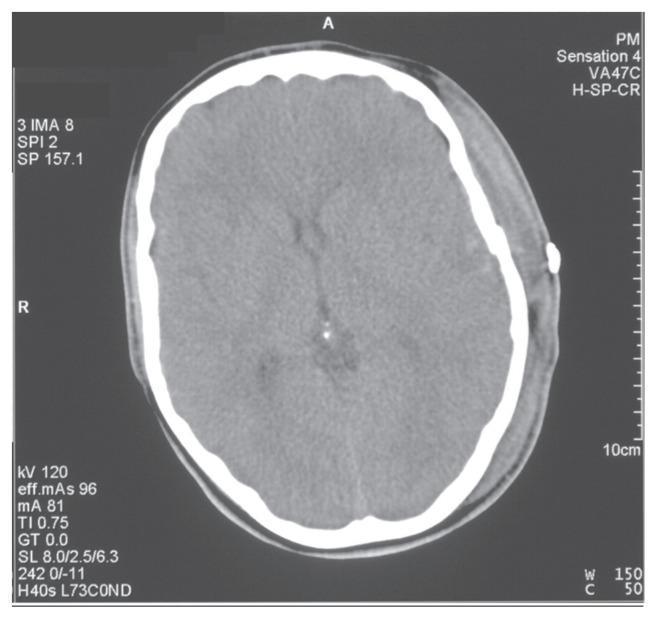
This diagnostic image is a non-contrast axial computed tomography (CT) scan of the head, representing a post-operative follow-up after the evacuation of a left temporoparietal extradural haematoma. The skull appears as a hyperdense peripheral ring, with a small, high-density metallic object visible on the left external scalp surface, likely a surgical staple or marker. The intracranial findings show that while the primary haematoma has been evacuated, residual mass effect is evident by a rightward midline shift and mild effacement of the left lateral ventricle. The brain parenchyma exhibits relatively uniform attenuation, though soft tissue swelling is noted in the left extracranial scalp region, corresponding to the surgical site. This image serves as an educational example of post-neurosurgical radiological assessment, illustrating the transition from an acute compressive lesion (extradural haematoma) to a post-evacuation state with resolving mass effect.
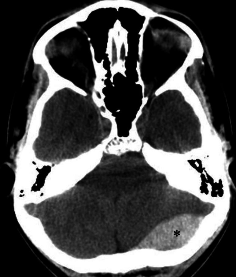
An axial non-contrast computerized tomography (CT) scan of the brain demonstrating an acute epidural (extradural) hematoma. The primary finding is a well-defined, biconvex (lens-shaped), hyperdense fluid collection located in the left occipital region, marked with an asterisk. This hyperdensity is characteristic of an acute hemorrhage within the epidural space, which typically does not cross cranial sutures. Secondary mass effect is visible, characterized by a mild left-to-right midline shift and compression of adjacent brain parenchyma. Anatomical landmarks include the skull base, posterior fossa structures, and the cerebellum. The image illustrates a classic neurosurgical emergency often associated with traumatic skull fractures and arterial bleeding, typically from the middle meningeal artery. This diagnostic image is intended for educational use in emergency medicine, radiology, and neurology to distinguish epidural hematomas from crescent-shaped subdural hematomas.
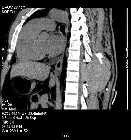
This diagnostic image is a sagittal CT scan of the dorsal spine, demonstrating a large spinal epidural hematoma. The imaging shows a localized, hyperdense collection (increased density) within the posterior extradural space at the thoracic level. This mass effect causes significant anterior displacement and compression of the spinal cord against the posterior aspect of the vertebral bodies. Anatomical landmarks include the thoracic vertebral column and the adjacent paraspinal tissues. Notably, the hematoma has displaced the overlying spinous process, pushing it posteriorly/superiorly from its normal alignment. This clinical finding is characteristic of an acute postoperative or traumatic complication, specifically a spinal epidural hematoma (SEH) resulting in spinal cord compression. The scan provides critical evidence for emergency surgical intervention, such as re-exploration and evacuation, to prevent permanent neurological deficit.
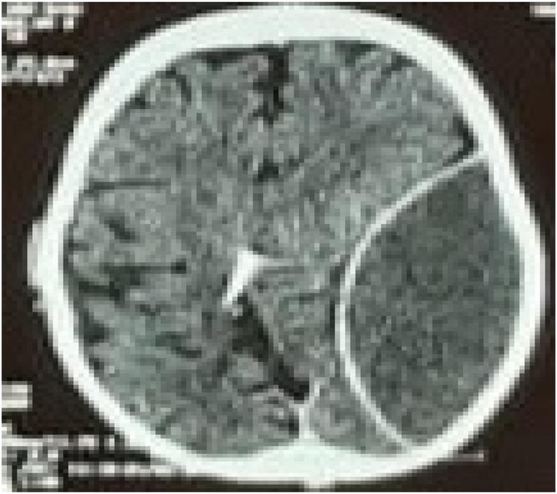
An axial non-contrast CT scan of the brain demonstrating a large, acute epidural (extradural) hematoma in the left hemisphere. The hematoma presents as a well-defined, biconvex (lenticular), hyperdense collection located between the skull and the dura mater. There is a significant mass effect evidenced by the compression of the left cerebral parenchyma, effacement of the adjacent cortical sulci, and midline shift toward the right. In the contralateral hemisphere, a radiopaque ventricular catheter is visible, extending into the posterior aspect of the right lateral ventricle near the occipital horn. This diagnostic image is representative of neurosurgical trauma emergencies, illustrating the classic appearance of arterial bleeding (typically from the middle meningeal artery) and secondary intracranial hypertension markers. The image serves as an educational resource for identifying acute traumatic intracranial hemorrhage and the presence of neurosurgical shunting devices.
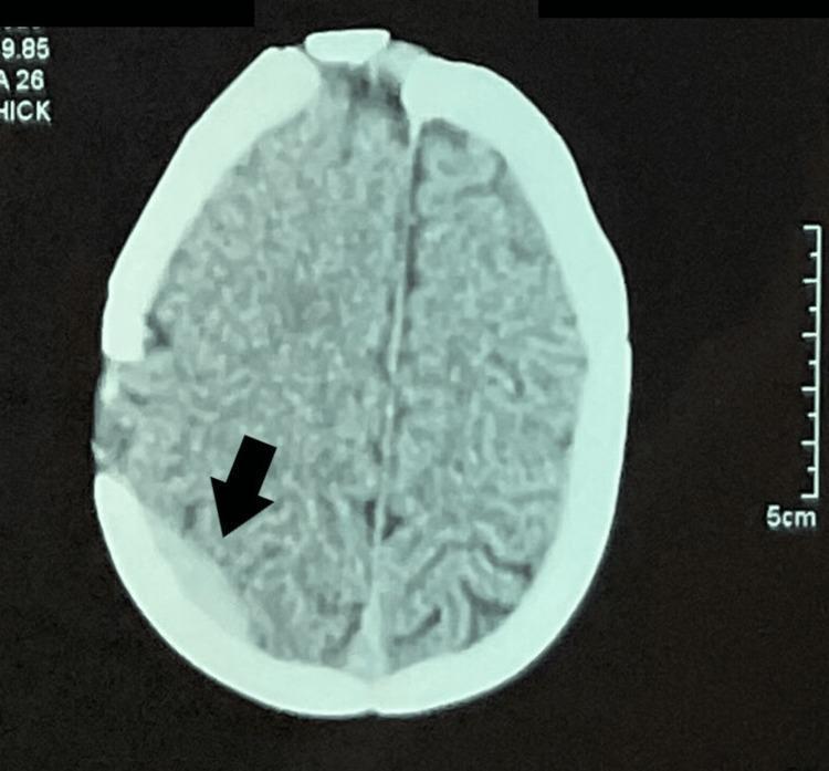
An axial non-contrast Computed Tomography (CT) scan of the head at the level of the upper cerebral convexities. The primary finding is a right-sided parietal extradural (epidural) hematoma, indicated by a black arrow. The hematoma appears as a focal, hyperdense, biconvex/lenticular-shaped collection adjacent to the inner table of the skull. A focal disruption or surgical alteration of the skull (bone flap/cranioplasty) is visible near the site of the hematoma. The surrounding brain parenchyma shows preserved gray-white matter differentiation, and there is no significant evidence of midline shift or obstructive hydrocephalus at this level. The falx cerebri remains in a midline position. This diagnostic image illustrates a postoperative complication following cranial surgery, specifically an acute extradural hemorrhage. The content is suitable for neurological, radiological, and surgical education regarding intracranial hemorrhages and postoperative monitoring.
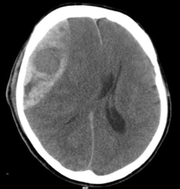
This diagnostic image is an axial non-contrast Computed Tomography (CT) scan of the brain, demonstrating a large, acute epidural (extradural) hematoma. The lesion is located on the right side of the skull and exhibits a classic biconvex or lens-shaped (lentiform) morphology, characterized by a hyperdense appearance relative to the brain parenchyma. The hematoma is clearly demarcated and constrained by the cranial sutures, positioned between the inner table of the skull and the dura mater. The image illustrates a significant mass effect, resulting in the effacement of adjacent sulci, partial compression of the right lateral ventricle, and a notable leftward midline shift of the falx cerebri. These findings are highly characteristic of an acute arterial bleed, typically involving the middle meningeal artery. This scan serves as a critical educational tool for identifying neurosurgical emergencies, understanding intracranial pressure dynamics, and recognizing the radiological hallmarks that differentiate epidural hematomas from crescent-shaped subdural hematomas.
Lucid Interval
Definition
Unconsciousness (from concussion) → Lucid interval (patient wakes, appears normal) → Second unconsciousness (from rising intracranial pressure)
- S Das Manual on Clinical Surgery, 13th Ed.: "Lucid interval is the short period of consciousness between initial unconsciousness which occurs immediately after the accident and unconsciousness at a later stage after the lucid interval."
Pathophysiology (Extradural / Epidural Haematoma)
- Phase 1 - Initial concussion: brief loss of consciousness from the primary impact.
- Lucid interval: The dura is slowly stripped off the skull by accumulating arterial blood. However, intracranial pressure (ICP) remains normal during this phase because CSF is displaced into the spinal canal - this compensatory mechanism temporarily buffers the pressure rise.
- Phase 3 - Deterioration: When compensation fails, ICP rises sharply. The medial temporal lobe herniates through the tentorial hiatus, compressing the midbrain's reticular activating system. The patient re-enters coma.
Key Clinical Features of Extradural Haematoma
| Feature | Detail |
|---|---|
| Classic sequence | Concussion → lucid interval → coma |
| Duration of lucid interval | Few hours (2-4) up to a week |
| Frequency | Present in only 30-40% of extradural haematoma cases (Essentials of Forensic Medicine 2026) |
| Pupil | Ipsilateral (same side as bleed) fixed and dilated pupil; later bilateral |
| Contralateral hemiplegia | Motor cortex compression; initially face → arm → trunk → leg |
| Late ipsilateral hemiplegia | Kernohan's notch - contralateral peduncle pressed against tentorial edge |
| Deterioration signs | Confusion (appears "drunk") → drowsiness → coma → decerebrate rigidity → death |
| Cause of death | Respiratory failure from brainstem compression |
| CT finding | Biconvex (lens-shaped / lentiform) hyperdense collection; does not cross suture lines |
| Mortality | 20-50% of cases are fatal |
Why Lucid Interval May Be Absent
Lucid Interval vs. Subdural Haematoma
| Feature | Extradural | Subdural |
|---|---|---|
| Lucid interval | Classic; well-defined | Short or absent |
| Onset of compression | Later (slower arterial bleed) | Earlier (blood accumulates faster) |
| Compensation | CSF displacement buys time | Less time for compensation |
| Incidence | Less common | ~6x more common |
"Talk and Die" Phenomenon
- Expanding subdural haematoma
- Expanding epidural haematoma
- Worsening cerebral oedema around a contusion
- Delayed appearance of an intracerebral clot
Lucid Interval in Psychiatry (Forensic Context)
Difference: Head Injury vs. Insanity
| Feature | Head Injury | Insanity |
|---|---|---|
| History | Head trauma | Mental illness |
| Preceding phase | Cerebral concussion | Symptoms of insanity |
| Following phase | Cerebral irritation/compression | Recurrence of insanity |
| Frequency | Only once | More than once |
Classic CT Appearance