
Write short note on the topic anophthalmic socket for pg ophthalmology exam
Reading File
Now let me search the medical library for anophthalmic socket content in parallel:
Finding Sources
Finding Sources
Found it in Kanski's. Let me read the dedicated section:
Reading File
Finding Sources
I can see the section starts at line 6884. The output was truncated - let me read more of this section:
Reading File
Reading File
Reading File
I now have comprehensive content from Kanski's. Let me also fetch a couple of the key clinical images to include:
Loading Image
Loading Image
I now have all the content needed. Here is the comprehensive short note:
The Anophthalmic Socket
(Source: Kanski's Clinical Ophthalmology: A Systematic Approach, 10th Ed.)
Definition
The anophthalmic socket is the orbital cavity that remains after removal of an eye (enucleation or evisceration) or the entire orbital contents (exenteration). Management focuses on restoring orbital volume, maintaining socket anatomy, and achieving a cosmetically acceptable prosthesis.
Surgical Procedures for Eye Removal
Three main procedures create an anophthalmic socket:
1. Enucleation
Removal of the entire globe with the extraocular muscles detached.
Indications:
- Primary intraocular malignancy (e.g., retinoblastoma, uveal melanoma) where tissue must remain intact for histopathology
- Severe trauma with risk of sympathetic ophthalmia outweighing visual recovery
- Blind, painful, or unsightly eye (though evisceration is now generally preferred for this indication)
2. Evisceration
Removal of the intraocular contents while leaving the sclera and extraocular muscle insertions intact. The cornea is removed to gain access.
Advantages over enucleation:
- Better prosthesis motility (extraocular muscles remain undisturbed)
- Better cosmetic outcome
Contraindication: Suspected intraocular malignancy (incomplete tissue for histology)
3. Exenteration
Removal of the globe and all orbital soft tissues.
Indications:
- Orbital malignancy (primary, or invasion from eyelid/conjunctiva/adnexa)
- Rarely: non-malignant disease such as orbital mucormycosis
Rehabilitation after exenteration: Prostheses can be attached to surrounding skin with adhesive, mounted on glasses, or secured with osseointegrated magnets on the orbital rim.
Orbital Implants
After enucleation or evisceration, an orbital implant is placed to restore volume and improve motility.
| Feature | Non-integrated (Solid) | Integrated (Porous) |
|---|---|---|
| Material | Silicone, acrylic | Hydroxyapatite, porous polyethylene |
| Fibrovascular ingrowth | No | Yes |
| Motility transfer | Limited | Better |
| Peg insertion possible | No | Yes (improves motility further) |
Key point: Careful placement (sufficiently deep, well-covered by vascularized tissue) is more important than the choice of implant material to prevent extrusion.
A peg can be inserted into porous implants later to improve motility; the peg must be covered by socket tissue and cannot attach directly to the overlying prosthesis.
Post-Enucleation Socket Syndrome (PESS)
A well-known complication resulting from orbital volume deficit, especially when no implant is used.
Pathophysiology: Volume loss causes loss of upper lid support → upper lid droops → prosthesis tilts backward and rotates upward → forward pressure on lower lid → inferior fornix shallowing → lower lid sag.
Classic features (PESS tetrad):
- Enophthalmos of the prosthesis
- Upper lid ptosis
- Superior sulcus hollowing / deepening
- Sagging lower lid (ectropion tendency)
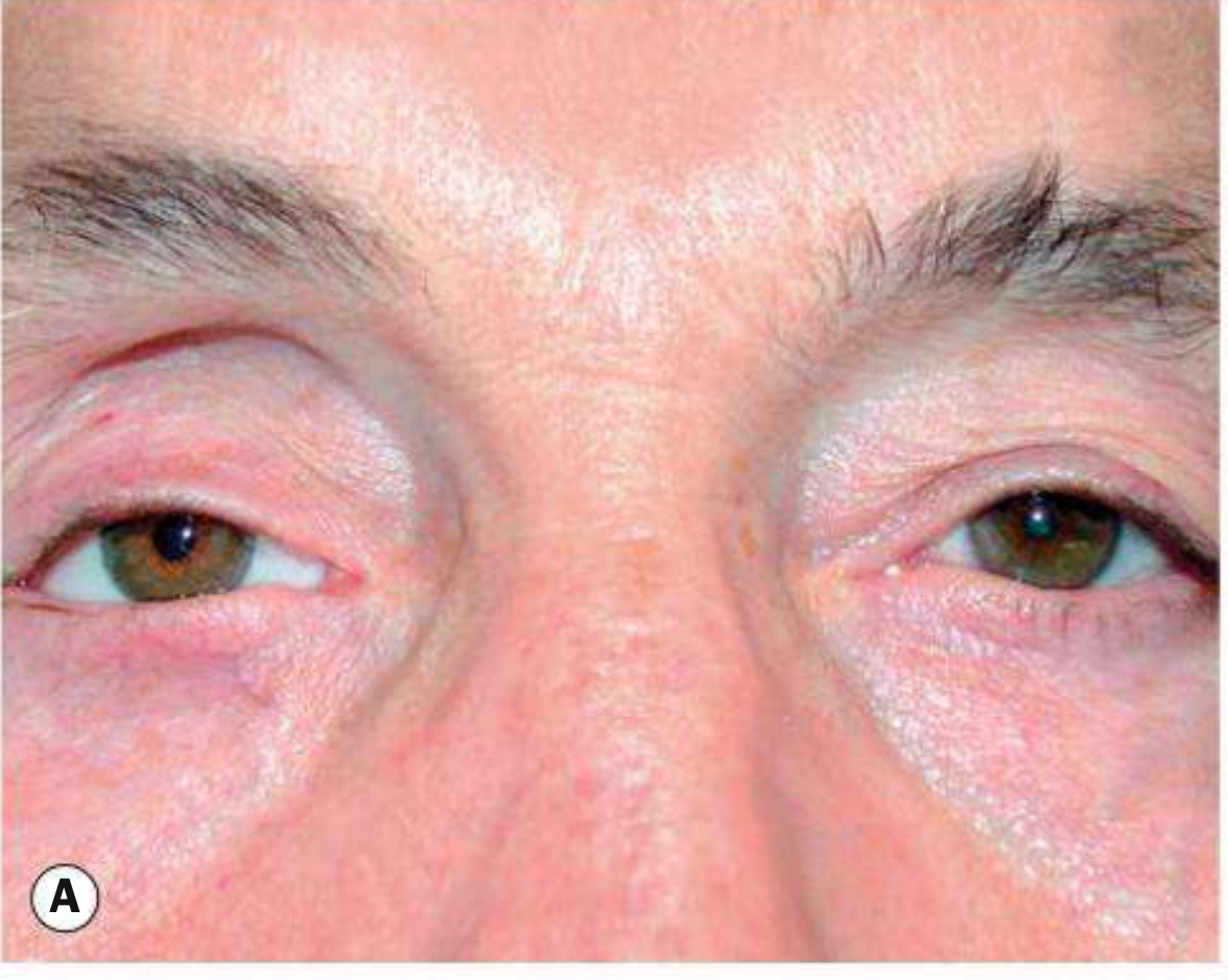
Fig. 4.52A: Right post-enucleation socket syndrome (PESS) - note upper lid ptosis and superior sulcus hollowing
Management of PESS:
- No implant present: insert a secondary orbital implant
- Implant present but volume insufficient: insert a silicone block into the inferior orbit
- Adjunct: upper lid ptosis surgery as needed
Implant Extrusion
A significant complication of all implants.
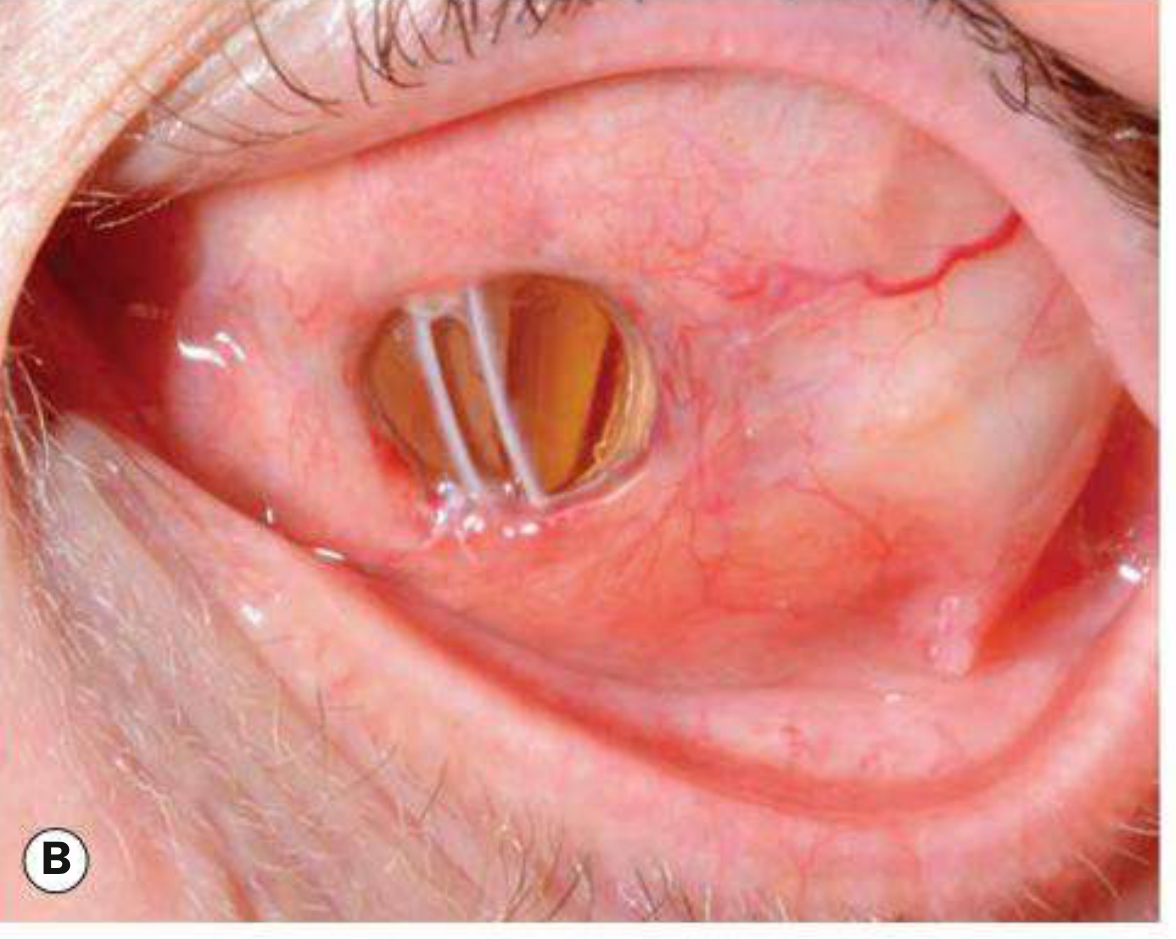
Fig. 4.52B: Extruding ball implant
Prevention: Ensure implant is placed sufficiently deep and well-covered with vascularized tissue (use donor sclera or mesh wrap to secure extraocular muscles over the implant surface).
Ocular Prosthesis (Artificial Eye) Fitting
- Immediately post-surgery: a conformer (silicone or acrylic) is placed to support the conjunctival fornices
- At ~6-8 weeks: socket impression moulds are taken
- A temporary artificial eye is fitted while waiting for the custom prosthesis
- Final prosthesis: custom-shaped to the individual socket, matched to the fellow eye in color and appearance
Cosmetic Shell
Used to cover a phthisical or unsightly eye (not an anophthalmic socket per se, but a related prosthetic rehabilitation). The shell restores volume and provides good cosmesis with transmitted motility from the underlying globe.
Summary Table
| Procedure | What is removed | Globe intact? | Muscles attached? | Use in malignancy |
|---|---|---|---|---|
| Enucleation | Entire globe | No | No (detached) | Yes |
| Evisceration | Intraocular contents | Sclera left | Yes (intact) | No |
| Exenteration | Globe + orbital contents | No | No | Yes (orbital) |
High-yield points for PG exam:
- Evisceration preferred for blind painful eye; enucleation for intraocular malignancy
- PESS = enophthalmos + ptosis + superior sulcus hollowing + lower lid sag - classically seen without an implant
- Integrated (porous) implants (hydroxyapatite/porous polyethylene) allow fibrovascular ingrowth and better motility
- Implant extrusion is prevented by depth of placement and vascular tissue coverage, not by implant material choice
- Conformer placed immediately; custom prosthesis fitted at 6-8 weeks
This is a shared conversation. Sign in to Orris to start your own chat.