
Brachial plexus
brachial plexus anatomy diagram roots trunks divisions cords branches
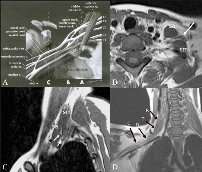
This composite educational graphic illustrates the normal anatomy of the brachial plexus through anatomical diagrams and Magnetic Resonance Imaging (MRI). Panel A is an anatomical diagram depicting the plexus architecture from roots (C5-T1) to terminal branches, labeled with musculoskeletal landmarks like the scalene muscles and coracoid process. Panel B (axial) and Panel C (sagittal) are T1-weighted MRI scans demonstrating the interscalene triangle, defined by the anterior scalene (sa) and middle scalene (sm) muscles. These muscles serve as primary radiological landmarks for identifying the neural roots and trunks (indicated by arrows). Panel D provides an oblique coronal T1-weighted MRI view, tracing the sequential segments of the right brachial plexus: roots (R), trunks (T), divisions (D), and cords (C). This resource is designed for medical education in radiology and neurology to assist in the localization of neural structures and the diagnostic evaluation of conditions like brachial plexopathy, neurogenic tumors, or Pancoast tumors.
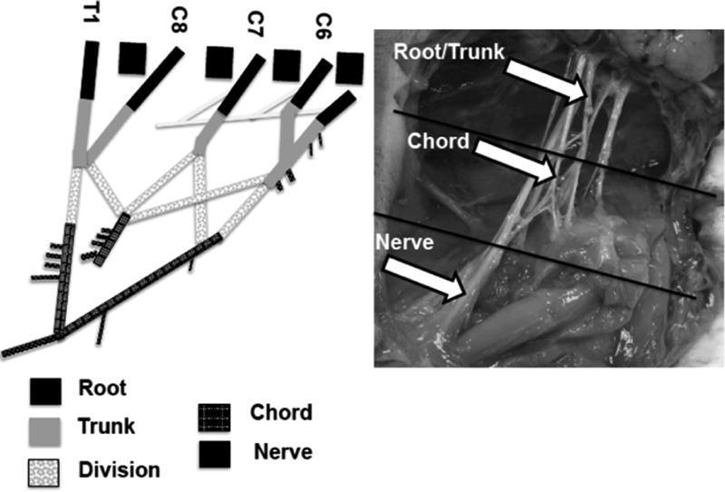
This composite educational material consists of a schematic diagram and a corresponding intraoperative clinical photograph illustrating the anatomical segments of the brachial plexus. The schematic on the left delineates the progression from spinal nerve roots (C6, C7, C8, and T1) through trunks, divisions, and chords to terminal nerves, using distinct grayscale patterns for each segment. It highlights the convergence of roots into upper, middle, and lower trunks and the subsequent formation of the medial and lateral cords through ventral divisions. The intraoperative photograph on the right provides clinical correlation in a neonatal model, with white arrows and horizontal lines demarcating the supraclavicular root/trunk region, the infraclavicular chord region, and the distal nerve branches. This comparison is used to facilitate identification of nerve bifurcations—such as the 'M' shape of the terminal branches—and to define anatomical boundaries for biomechanical or surgical study. The content is designed for intermediate to advanced medical education in peripheral nerve anatomy and neurosurgery.
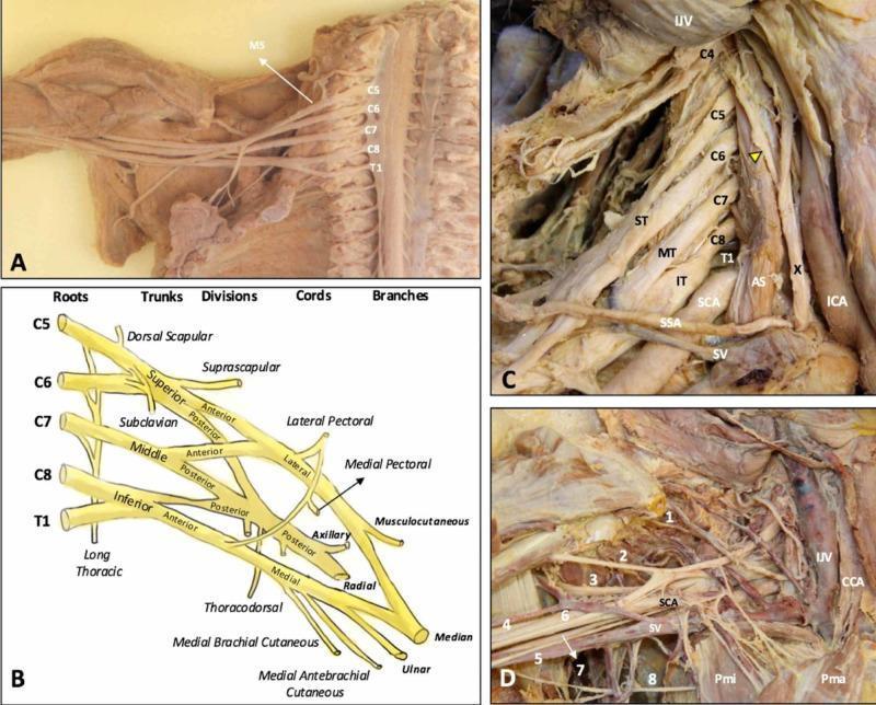
This composite educational graphic details the anatomy of the human brachial plexus (BP) through cadaveric dissections and a schematic diagram. (A) Cadaveric dissection of the cervical spine and axilla with the anterior scalene muscle removed, exposing the C5-T1 nerve roots emerging laterally to the middle scalene muscle (MS). (B) A structured schematic diagram illustrating the hierarchical organization of the BP from roots (C5-T1), trunks (Superior, Middle, Inferior), divisions (Anterior, Posterior), and cords (Lateral, Posterior, Medial) to terminal branches. (C) Dissection showing the roots in situ within the scalene triangle, formed between the anterior (AS) and middle scalene muscles, alongside major vessels including the subclavian artery (SCA) and internal jugular vein (IJV). The phrenic nerve (yellow arrowhead) is seen traversing the anterior scalene. (D) Detailed view of the infraclavicular and axillary regions showing terminal branches (1-8: suprascapular, musculocutaneous, axillary, radial, medial brachial cutaneous, median, ulnar, and intercostobrachial nerves) in relationship to the pectoralis minor (Pmi), pectoralis major (Pma), and subclavian vessels. This content provides essential topographical and clinical anatomy for understanding thoracic outlet syndrome and regional anesthesia.
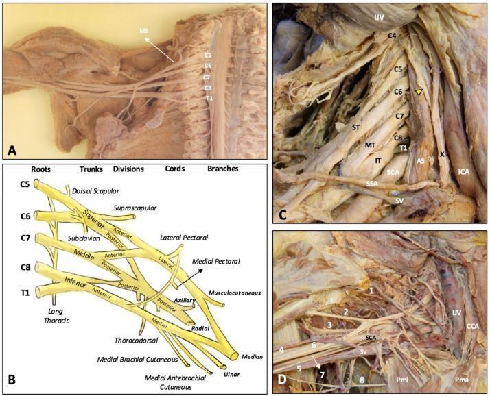
Educational anatomical resource illustrating the brachial plexus through cadaveric dissections (Panels A, C, D) and a schematic diagram (Panel B). Panel A shows the overview of the plexus and middle scalene muscle (MS) relative to the spinal cord. Panel B provides a detailed schematic of the classical brachial plexus organization: roots (C5-T1), superior (ST), middle (MT), and inferior (IT) trunks, anterior and posterior divisions, cords (lateral, medial, posterior), and terminal branches. Panel C displays a deep neck dissection highlighting the relationship between roots C4-T1 and the anterior scalene (AS), subclavian artery (SCA), internal jugular vein (IJV), and vagus nerve (X). A yellow arrowhead identifies the phrenic nerve. Panel D focuses on the infraclavicular and axillary regions, showing terminal branches: 1 (suprascapular), 2 (musculocutaneous), 3 (axillary), 4 (radial), 6 (median), and 7 (ulnar) nerves. Vascular landmarks include the common carotid artery (CCA) and subclavian vein (SV), alongside the pectoralis major (Pma) and minor (Pmi) muscles, providing clinical context for regional anesthesia and surgical approaches.
Brachial Plexus
Overview
Structure: Roots → Trunks → Divisions → Cords → Branches

1. Roots (anterior rami C5–T1)
- Lie between the anterior scalene and middle scalene muscles
- Receive gray rami communicantes from the sympathetic trunk (postganglionic sympathetic fibers)
- Branches directly from the roots:
- Dorsal scapular nerve (C5) - rhomboid major & minor
- Long thoracic nerve (C5-C7) - serratus anterior
- Small contribution of C5 to the phrenic nerve
2. Trunks (in the posterior triangle & over rib I)
| Trunk | Root Contributions |
|---|---|
| Superior (upper) | C5 + C6 |
| Middle | C7 alone |
| Inferior (lower) | C8 + T1 |
- Suprascapular nerve (C5, C6) - supraspinatus & infraspinatus
- Nerve to subclavius (C5, C6) - subclavius muscle
3. Divisions (behind the clavicle)
- Anterior divisions → supply anterior (flexor) compartments
- Posterior divisions → supply posterior (extensor) compartments
4. Cords (in the axilla, named relative to the 2nd part of axillary artery)
| Cord | Formation | Spinal levels |
|---|---|---|
| Lateral | Anterior div. of upper + middle trunks | C5-C7 |
| Medial | Anterior div. of inferior trunk | C8, T1 |
| Posterior | All three posterior divisions | C5-T1 |
5. Terminal Branches

- Lateral pectoral nerve (C5-C7) - pectoralis major
- Musculocutaneous nerve (C5-C7) - coracobrachialis, biceps brachii, brachialis; sensation to lateral forearm (as lateral cutaneous nerve of forearm)
- Lateral root of the median nerve
- Medial pectoral nerve (C8-T1) - pectoralis major & minor
- Medial cutaneous nerve of arm (C8, T1)
- Medial cutaneous nerve of forearm (C8, T1)
- Ulnar nerve (C7-T1) - intrinsic hand muscles, flexor carpi ulnaris, medial FDP
- Medial root of the median nerve
- Superior subscapular nerve - subscapularis
- Thoracodorsal nerve (C6-C8) - latissimus dorsi
- Axillary nerve (C5, C6) - deltoid, teres minor; sensation over "regimental badge" area
- Radial nerve (C5-T1) - all posterior compartment muscles of arm and forearm
- Inferior subscapular nerve - subscapularis, teres major
Key Branches Summary Table
| Branch | Origin | Spinal Levels | Main Function |
|---|---|---|---|
| Dorsal scapular | C5 root | C5 | Rhomboids |
| Long thoracic | C5-C7 roots | C5-C7 | Serratus anterior |
| Suprascapular | Superior trunk | C5, C6 | Supra/infraspinatus |
| Musculocutaneous | Lateral cord | C5-C7 | Arm flexors |
| Axillary | Posterior cord | C5, C6 | Deltoid, teres minor |
| Radial | Posterior cord | C5-T1 | All posterior extensors |
| Median | Lateral + medial cords | C5-T1 | Anterior forearm/thenar |
| Ulnar | Medial cord | C7-T1 | Intrinsic hand/hypothenar |
Clinically Important Lesions
Erb-Duchenne Palsy (Upper plexus - C5, C6)
- Causes: Birth trauma (traction on shoulder during delivery), motorcycle accidents, downward traction of shoulder
- Deformity: "Waiter's tip" - arm hangs adducted, medially rotated, forearm pronated
- Deficits: Weakness of shoulder abduction, lateral rotation, elbow flexion, forearm supination
- Best prognosis of brachial plexus injuries; most common type
Klumpke's Palsy (Lower plexus - C8, T1)
- Causes: Upward traction (grabbing a branch during a fall), Pancoast tumor, thoracic outlet syndrome
- Deficits: Weakness of intrinsic hand muscles (thenar + hypothenar atrophy), weakness of finger/wrist flexors; sensory loss on ulnar aspect of hand/forearm
- Horner's syndrome (ptosis, miosis, anhidrosis) may accompany if T1 root is damaged proximal to the sympathetic trunk
- Poor prognosis
Thoracic Outlet Syndrome
- Compression of the lower brachial plexus (and subclavian vessels) between the clavicle and rib I
- Symptoms worsen with arm elevation and external rotation
- Investigation: EMG, X-ray (cervical rib?); management: shoulder strengthening, surgical decompression for refractory cases
Pancoast Syndrome
- Apical lung tumor (usually non-small cell) invades the lower brachial plexus
- Lower plexus signs + possible Horner's syndrome (T1 root); occasional hoarseness (recurrent laryngeal nerve)
- May ultimately involve the entire plexus, causing a flail, insensate arm
Parsonage-Turner Syndrome (Brachial Neuritis/Brachial Plexitis)
- Inflammatory/idiopathic cause; onset in adulthood with severe burning shoulder/lateral neck pain followed by weakness
- Most patients recover fully within 6-12 weeks; some have residual atrophy
Long Thoracic Nerve - Special Note
Regional Anesthesia Approaches
- Interscalene block - targets the roots/trunks at the scalene triangle; good for shoulder/proximal arm surgery; risks phrenic nerve palsy (common), Horner's syndrome, pneumothorax
- Supraclavicular block - targets the trunks/divisions at their most compact point ("choke point"); efficient for entire upper limb surgery
- Infraclavicular block - targets the cords around the axillary artery
- Axillary block - targets terminal branches in the axilla; safest approach but misses musculocutaneous and axillary nerves unless specifically sought
Additional Diagram
