
PTA
Asking for Preferences
What does PTA mean in your context?
PDA
Patent Ductus Arteriosus (PDA) is a congenital heart defect where the ductus arteriosus - a fetal blood vessel connecting the pulmonary artery to the aorta - fails to close after birth. In fetuses, it bypasses the lungs (since oxygenation occurs via placenta). After birth it normally closes within 24-72 hours due to rising oxygen levels and falling prostaglandins. If it stays open, blood shunts left-to-right (aorta -> pulmonary artery), causing pulmonary overcirculation.
Searching sources...
Reading File
Finding Sources
Searching Images
patent ductus arteriosus diagram shunt
Searching PubMed
"patent ductus arteriosus"[MeSH Terms] AND treatment
Reading File
Reading File
Reading File
Reading File
Searching Images
PDA patent ductus arteriosus echocardiogram chest xray
Loading Image
Here is a comprehensive overview of Patent Ductus Arteriosus (PDA):
Patent Ductus Arteriosus (PDA)
Anatomy
The ductus arteriosus connects the descending aorta (just distal to the left subclavian artery) to the main pulmonary trunk near the origin of the left pulmonary artery. The arrow in the diagram below shows the direction of left-to-right shunting through the PDA:
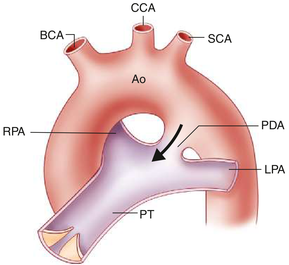
Ao = aorta; BCA = brachiocephalic artery; CCA = common carotid; LPA = left pulmonary artery; PDA = patent ductus arteriosus; PT = pulmonary trunk; RPA = right pulmonary artery; SCA = subclavian artery.
Fetal Role and Normal Closure
During intrauterine life, the ductus allows blood to bypass the unoxygenated lungs, flowing from the pulmonary artery to the aorta. After birth, three triggers drive closure within 24-72 hours in term infants:
- Rising arterial oxygen tension
- Falling pulmonary vascular resistance
- Declining prostaglandin E2 levels
Normal complete obliteration occurs within the first few months, leaving the ligamentum arteriosum. Failure of TGF-β induction after birth may play a role in persistent patency. - The Developing Human, p. 881
Epidemiology & Risk Factors
- Accounts for ~7% of all congenital heart defects; 90% are isolated
- 2-3x more common in females than males
- Strong association with maternal rubella in early pregnancy
- Present in virtually all preterm neonates ≤28 weeks with birth weight <1750 g (due to hypoxia and surfactant deficiency)
- High-altitude living (hypoxia delays closure)
- Associated with chromosomal anomalies, coarctation of the aorta, transposition of the great arteries, pulmonary stenosis/atresia
Sources: Robbins Basic Pathology, p. 262; The Developing Human, p. 882
Pathophysiology
Blood shunts left-to-right (aorta → pulmonary artery) because systolic and diastolic pressure in the aorta normally exceeds that in the pulmonary artery. This causes:
- Pulmonary overcirculation
- Left atrial and left ventricular dilation (volume overload)
- Progressive pulmonary hypertension
Severity depends on the size of the PDA and the pulmonary-to-systemic vascular resistance ratio:
| PDA Size | Hemodynamic Effect |
|---|---|
| Small | Continuous flow, no chamber dilation, no pulmonary HTN, no symptoms - but ~0.45%/year risk of endarteritis |
| Moderate/Large | Variable L-to-R shunt, LA/LV dilation, pulmonary HTN, symptoms by 2nd-3rd decade |
| Eisenmenger | Shunt reversal (R-to-L), cyanosis of lower extremities > upper, contraindication to closure |
Goldman-Cecil Medicine, p. 611
Clinical Features
- Murmur: Classic continuous "machinery" murmur at the 1st-2nd left intercostal space below the left clavicle (harsh, "machinery-like")
- Diastolic component shortens as pulmonary pressure rises
- Murmur disappears entirely with Eisenmenger physiology
- Bounding/wide pulse pressure: Diastolic flow into pulmonary artery lowers systemic diastolic BP
- Differential cyanosis and clubbing: Toes > fingers (in Eisenmenger - lower extremities have deoxygenated blood from pulmonary artery via PDA)
- Infective endocarditis/endarteritis risk (small PDAs: ~0.45%/year after 2nd decade)
- Without treatment, mortality rises to 3-4%/year by the 4th decade; two-thirds die by age 60
Diagnosis
- Echocardiography: Gold standard - Doppler signal identifies the shunt even when direct visualization is difficult; shows LA/LV dilation
- Chest X-ray: Dilated ascending aorta and pulmonary artery, enlarged left-sided chambers; calcification at the PDA site in older patients
- Cardiac catheterization: Indicated to assess pulmonary vascular resistance before closure when pulmonary HTN is suspected
Goldman-Cecil Medicine, p. 612
Treatment
Premature Neonates
- Indomethacin or Ibuprofen (COX inhibitors) - reduce prostaglandin synthesis to promote ductal closure
- A 2025 Bayesian meta-analysis (PMID 39815089) found pharmacological treatment may reduce PDA but has heterogeneous effects and may not always reduce need for interventional procedures
- A 2025 meta-analysis (PMID 40240526) confirmed heterogeneity in treatment response to indomethacin, ibuprofen, and acetaminophen
- Acetaminophen is an emerging alternative with fewer side effects
Procedural/Surgical Closure (all ages)
- Indications: Left-sided heart enlargement, prior endarteritis, or symptomatic PDA
- Contraindication: Eisenmenger complex (right-to-left shunt - closure would be fatal)
- Options:
- Transcatheter device or coil occlusion - accepted procedure in adults; residual shunt rates 0.5-8%
- Surgical ligation - operative mortality <1% to 8% depending on calcification and pulmonary HTN severity
- A 2024 meta-analysis (PMID 38329109) comparing percutaneous closure vs. surgery in low-birth-weight preterms found both are viable options
After ligation in infancy/early childhood, cardiac function is typically normal with no special follow-up needed.
Goldman-Cecil Medicine, p. 612; Robbins Basic Pathology, p. 263
Key Summary
| Feature | Detail |
|---|---|
| Shunt direction | L → R (aorta → pulmonary artery) |
| Key murmur | Continuous "machinery" murmur, left 2nd ICS |
| Key sign | Bounding pulses, wide pulse pressure |
| Complication | Eisenmenger syndrome, endarteritis, heart failure |
| Medical Rx | Indomethacin/Ibuprofen (neonates) |
| Definitive Rx | Transcatheter coil/device or surgical ligation |
| Contraindication | Closure in Eisenmenger physiology |
This is a shared conversation. Sign in to Orris to start your own chat.