
Tell me everything about phenytoin drug as much as you can from all apsects
phenytoin epilepsy pharmacology
phenytoin toxicity nystagmus ataxia gingival hyperplasia
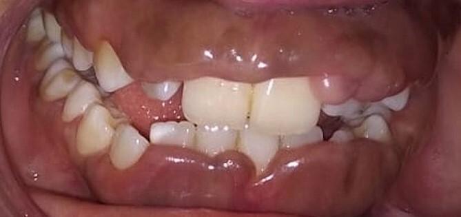
This clinical intraoral photograph demonstrates severe, generalized gingival enlargement, consistent with medication-induced gingival overgrowth (specifically phenytoin-induced). The gingiva exhibits significant hypertrophy and puffiness, primarily localized to the maxillary and mandibular anterior regions. The tissue is characterized by a distinctive bluish-red discoloration and a smooth, shiny surface texture indicating underlying edema. The hyperplastic gingiva significantly obscures the dental crowns, covering approximately two-thirds of the tooth surfaces. This presentation illustrates the classic clinical manifestation of drug-induced gingival hyperplasia, which often results in altered aesthetics, increased risk of periodontal disease due to difficult oral hygiene, and tissue friability during mastication. This image serves as a high-quality educational resource for dental and medical professionals studying oral pathology and the systemic side effects of anticonvulsant therapy.
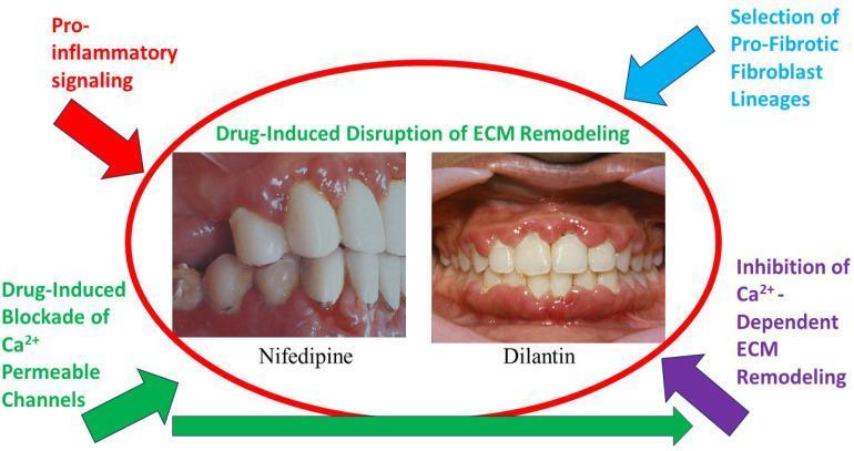
This medical visual combines clinical intraoral photographs with a conceptual diagram to illustrate drug-induced gingival overgrowth (DIGO). The central photographs compare the gingival manifestations of two specific pharmacological agents: Nifedipine (a calcium channel blocker) and Dilantin (Phenytoin, an anticonvulsant). The Nifedipine clinical image displays erythematous, swollen gingival margins with moderate hyperplasia. The Dilantin image demonstrates more severe, generalized gingival overgrowth characterized by bulbous interdental papillae and extensive coverage of the clinical crowns by thickened, lobulated gingival tissue. Surrounding these images is a pathophysiological flow diagram explaining the mechanism of extracellular matrix (ECM) disruption. Key educational concepts mapped include the drug-induced blockade of calcium-permeable channels (such as TRPV4), the inhibition of calcium-dependent ECM remodeling, and the selection of pro-fibrotic fibroblast lineages. The diagram highlights how pro-inflammatory signaling synergizes with these drug effects to disturb the homeostatic balance of collagen synthesis and degradation in periodontal tissues, leading to the observed pathological tissue enlargement.
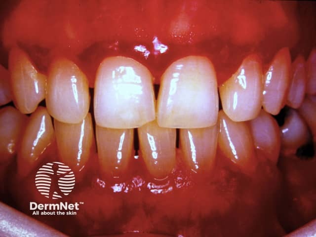
Imaging modality: Intraoral clinical photograph (frontal view) of the maxillary and mandibular anterior segments. Anatomical context: oral cavity, specifically the gingival mucosa around the incisors and canines in both arches. Visual features: generalized gingival hyperplasia with bulky, lobulated, fibrous-appearing tissue overlying the crowns of the central and lateral incisors; margins are relatively rounded with mild generalized erythema and slight edema; interdental papillae appear swollen; margin-to-tooth relationship is largely preserved despite enlargement; no radiographic bone detail. The photo shows a reddish-pink gingival color, indicative of inflammation; surface is smooth, with some subtle gloss from lighting. Clinical interpretation: this appearance is characteristic of gingival hyperplasia, which can be plaque-induced, drug-induced (phenytoin, cyclosporine, calcium channel blockers), or hereditary gingival fibromatosis. Not a routine normal variation. Diagnostic significance: highlights need for periodontal evaluation, plaque control, and medical history review; may require gingivoplasty/gingivectomy for functional and aesthetic improvement if fibrous component dominates. Differential considerations include inflammatory gingivitis, drug-related overgrowth, hereditary fibromatosis. Clinical correlation: assess oral hygiene, medication exposure, and systemic conditions; management includes dental cleaning, antimicrobial rinses, periodontal therapy, and potential collaboration with the prescribing clinician to modify contributing medications. This image supports dental education, periodontal training, and case-based learning in gingival enlargement.
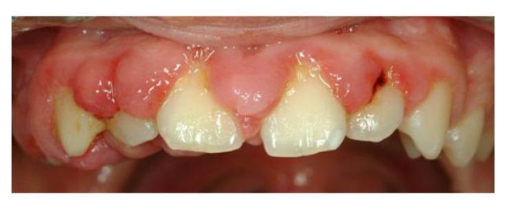
This clinical intraoral photograph demonstrates severe drug-induced gingival enlargement (overgrowth) affecting the maxillary arch. The gingiva appears significantly inflamed, characterized by a bright red hue, bulbous interdental papillae, and an uneven, lobulated texture. The overgrowth extends coronally, covering approximately one-third to one-half of the clinical crowns of the maxillary incisors and canines, resulting in a shortened appearance of the teeth and loss of the normal scalloped gingival contour. Areas of yellow-white plaque accumulation and material alba are visible along the gingival margins, particularly at the junction of the hypertrophied tissue and the tooth surface. The presentation is classic for gingival hyperplasia secondary to medications such as anticonvulsants (phenytoin), calcium channel blockers (nifedipine), or immunosuppressants (cyclosporine). This image serves as an educational example of medication-related adverse effects in the oral cavity and the importance of oral hygiene in managing periodontal inflammation during systemic therapy.
1. Overview & History
- Katzung's Basic and Clinical Pharmacology, 16th Ed.
- Goodman & Gilman's Pharmacological Basis of Therapeutics
2. Chemical Structure & Formulations
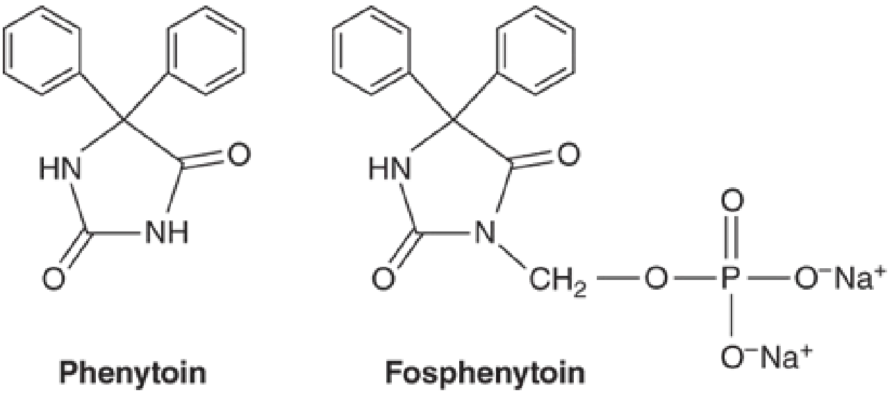
- Phenytoin is the 5,5-diphenyl-substituted analog of hydantoin - a five-membered ring similar to barbiturates (six-membered ring)
- pKa = 8.06-8.33; the free base is poorly water-soluble
- Phenytoin sodium dissolves in water at ~17 mg/mL
- Available as:
- Extended-release capsules (peak at 4-12 hours; can be dosed once daily)
- Immediate-release capsules (peak at 1.5-3 hours)
- Oral suspension and chewable tablets (free base; TID dosing)
- IV solution (contains propylene glycol and alcohol, pH 12)
- Fosphenytoin (water-soluble prodrug; preferred IV/IM route)
3. Mechanism of Action
- Primary mechanism: Slows the rate of recovery of voltage-activated Na⁺ channels from inactivation. This reduces the ability of neurons to fire repetitively at high frequency - the hallmark of seizure discharges.
- The effect is greater when the membrane is depolarized (voltage-dependent), targeting neurons that are pathologically active.
- At therapeutic concentrations: selective Na⁺ channel effect only - no change in spontaneous activity or GABA/glutamate responses.
- At supratherapeutic concentrations (5-10× higher): reduction of spontaneous activity + enhancement of GABA responses - this contributes to toxicity.
- Also blocks secondary calcium influx into partially depolarized neurons, reducing their excitability and prolonging their refractory period.
- In cardiac tissue: acts as a Class IB antiarrhythmic - slows conduction in the His-Purkinje system.
- Goodman & Gilman's, p. 411-412
- Henry's Clinical Diagnosis, p. 424
- Tietz Textbook of Laboratory Medicine, 7th Ed.
4. Pharmacokinetics
Absorption
- Oral bioavailability is highly formulation-dependent (ranges from 30-95%)
- Particle size and pharmaceutical additives significantly affect absorption rate and extent
- Time to peak: 3-12 hours (oral), 1-2 hours (IV/fosphenytoin)
- IM injection of phenytoin: NOT recommended - drug precipitates at injection site with slow, unpredictable absorption
Distribution
- ~90-95% protein-bound (primarily to albumin)
- Only free (unbound) drug is pharmacologically active and crosses membranes
- Protein binding is reduced by: hypoalbuminemia, uremia, hyperbilirubinemia, elderly state, competing drugs (valproate, salicylates, sulfonylureas)
Metabolism - THE KEY FEATURE
- Metabolized by hepatic microsomal enzymes: primarily CYP2C9/2C10, secondarily CYP2C19
- Principal metabolite: 5-(p-hydroxyphenyl)-5-phenylhydantoin - inactive, excreted as glucuronide
- SATURABLE (zero-order/Michaelis-Menten) kinetics within the therapeutic range:
- At LOW concentrations (<5 μg/mL): first-order kinetics apply
- As levels rise, hepatic metabolism becomes saturated → small dose increases cause disproportionately large plasma level increases
- This explains why 300 mg/day and 400 mg/day can produce vastly different blood levels
- Plasma t½ ranges from 6-24 hours at concentrations below 10 μg/mL
Elimination
- Only ~5% excreted unchanged in urine
- Half-life is dose-dependent: t½ = 24 ± 12 hours (standard dosing)
- Steady state: ~7-10 days of continuous dosing
- Not effectively removed by dialysis (high protein binding)
Fosphenytoin (Prodrug)
-
Water-soluble phosphate ester; converted to phenytoin by plasma and RBC phosphatases
-
Conversion t½: 8-15 minutes
-
Protein binding: 95-99% (even higher than phenytoin)
-
Can be given IM or IV; preferred over phenytoin IV due to better tolerability
-
Dosed in "phenytoin equivalents" (PE)
-
Katzung's Basic and Clinical Pharmacology, 16th Ed.
-
Goodman & Gilman's, p. 412
-
Henry's Clinical Diagnosis, p. 421
5. Therapeutic Uses & Indications
| Indication | Notes |
|---|---|
| Generalized tonic-clonic seizures | Primary chronic use |
| Focal (partial) seizures | Simple and complex partial |
| Status epilepticus | IV/fosphenytoin; 2nd/3rd-line agent |
| Post-neurosurgical seizure prophylaxis | Short-term use |
| Post-traumatic epilepsy prevention | Prophylactic; early post-injury period |
| Cardiac arrhythmias | Esp. digitalis-induced; Class IB antiarrhythmic |
| Trigeminal neuralgia | Historically; largely replaced by carbamazepine |
| Neuropathic pain | Off-label |
-
Absence seizures (petite mal)
-
May worsen absence epilepsy, juvenile myoclonic epilepsy (JME), and Dravet syndrome
-
Katzung, p. 855
-
Tietz Textbook of Laboratory Medicine, 7th Ed.
-
Forensic Medicine and Toxicology, 36th Ed.
6. Therapeutic Drug Monitoring (TDM)
| Parameter | Value |
|---|---|
| Therapeutic range (total) | 10-20 μg/mL (40-79 μmol/L) |
| Therapeutic range (free) | 1-2 μg/mL (1-8 μmol/L) |
| Toxic level | >20-30 μg/mL |
| Paradoxical seizure induction | >35 μg/mL |
- At 10 μg/mL: ~50% seizure response rate
- At 15 μg/mL: ~86% seizure suppression
- Correction for hypoalbuminemia is mandatory: use the Winter-Tozer equation
- Corrected phenytoin = measured total / [(0.2 × albumin) + 0.1]
- Monitor free phenytoin in: hypoalbuminemia, renal insufficiency, hepatic disease, pregnancy, elderly
- Sampling: trough level (within 30 min before next dose) for routine monitoring; 1 hr post-IV for peak
7. Adverse Effects
Concentration-Dependent (Dose-Related)
| Level | Signs/Symptoms |
|---|---|
| 20-30 μg/mL | Nystagmus (horizontal gaze nystagmus - earliest sign) |
| 30-40 μg/mL | Ataxia, dysarthria, slurred speech, coarse tremors, diplopia, vertigo |
| >40 μg/mL | Confusion, disorientation, oscillopsia, progressive CNS depression |
| Very high | Coma, paradoxical increase in seizure frequency, encephalopathy with opisthotonic posturing |
Chronic/Long-Term Adverse Effects
- Peripheral neuropathy (long-term)
- Cerebellar atrophy (with chronic high doses)
- Cognitive impairment, behavioral changes
- Gingival hyperplasia - occurs in ~20% of patients on chronic therapy; due to altered collagen turnover; NOT plasma-level dependent; worse in children/adolescents; managed by good oral hygiene; may require gingivoplasty
- Hirsutism - particularly problematic in females
- Coarsening of facial features
- Osteomalacia (hypocalcemia + elevated alkaline phosphatase): due to accelerated vitamin D metabolism AND impaired intestinal calcium absorption AND increased metabolism of vitamin K-dependent Ca²⁺ proteins in bone
- Hyperglycemia / glycosuria: inhibition of insulin secretion
- Hyponatremia: inhibition of ADH release
- Macrocytic/megaloblastic anemia (folate antagonism)
- Autoimmune thyroiditis
- Neutropenia, leukopenia
- Thrombocytopenia
- Red cell aplasia (rare)
- Agranulocytosis (rare)
- Transient elevation of hepatic transaminases
- Hepatic necrosis (rare, hypersensitivity-related)
Idiosyncratic/Hypersensitivity Reactions
- Morbilliform rash: 2-5% of patients
- Stevens-Johnson syndrome (SJS) - rare but life-threatening
- Toxic epidermal necrolysis (TEN) - rare
- Drug reaction with eosinophilia and systemic symptoms (DRESS / Phenytoin Hypersensitivity Syndrome): fever + rash + lymphadenopathy + hepatosplenomegaly; appears 3 weeks to 3 months after initiation
- Purple Glove Syndrome: purplish-black discoloration + edema + pain distal to injection site; may progress to skin necrosis; related to propylene glycol vehicle; less common with fosphenytoin
- Cardiovascular collapse with rapid IV infusion: bradycardia, heart block, ventricular fibrillation, hypotension - particularly in elderly and those with cardiac disease
8. Drug Interactions
Drugs that INCREASE Phenytoin Levels (inhibit CYP2C9/2C19)
- Isoniazid (especially in slow acetylators)
- Fluoxetine, fluvoxamine
- Metronidazole, miconazole
- Fluorouracil
- Chloramphenicol, cimetidine, disulfiram
- Dicumarol (warfarin)
- Valproate (dual mechanism: displaces from protein binding AND inhibits metabolism - sustained increase in free phenytoin)
Drugs that DECREASE Phenytoin Levels (induce CYP)
- Alcohol (chronic), barbiturates, carbamazepine: enzyme induction → increased metabolism
- Rifampin: potent inducer
Drugs that Displace Phenytoin from Protein Binding (increase free fraction temporarily)
- Salicylates, valproate, phenylbutazone, sulfonamides, sulfonylureas
- Note: total phenytoin level decreases but free fraction stays similar (until re-equilibration)
Drugs whose Levels are DECREASED by Phenytoin (phenytoin induces CYP3A4, 2C9, and others)
-
Oral contraceptives (risk of unplanned pregnancy - clinically significant given teratogenicity)
-
Warfarin (risk of bleeding disorder - bidirectional interaction)
-
Corticosteroids
-
Carbamazepine, cyclosporine, itraconazole
-
Methadone, methotrexate, doxycycline
-
Thyroid hormones (decreases T3 and T4 without causing clinical hypothyroidism usually)
-
Goodman & Gilman's, p. 423
-
Tietz Textbook, p. 1518-1519
-
Katzung, p. 880-881
9. Toxicology & Overdose Management
Toxicological Thresholds
- Therapeutic: up to 20 μg/mL
- Toxic effects: typically >30 μg/mL
- Paradoxical seizures: >35 μg/mL
- Lethal dose: not clearly defined; death usually results from ventricular fibrillation or cardiac arrest (especially with rapid IV administration)
Features of Overdose
- Early: horizontal gaze nystagmus, gait ataxia, drowsiness
- Progressive: vertical nystagmus, oscillopsia, slurred speech, coarse tremors, confusion
- Severe: coma (rare), paradoxical increase in seizures, encephalopathy
- Cardiac: hypotension (dose/concentration-dependent), bradycardia, heart block, VF/cardiac arrest (especially IV route)
Management
- Decontamination: gastric lavage (within a few hours), activated charcoal
- Supportive care + continuous cardiovascular monitoring
- Hypotension: Trendelenburg positioning, IV fluids, vasopressors (dopamine, norepinephrine)
- Heart block: atropine or temporary cardiac pacing
- Paradoxical seizures: diazepam first-line, phenobarbitone second-line
- Hypersensitivity reactions: systemic corticosteroids
- Forensic Medicine and Toxicology, 36th Ed., p. 6774-6788
- Washington Manual of Medical Therapeutics
10. Use in Special Populations
Pregnancy
- Category D teratogen
- Causes Fetal Hydantoin Syndrome: neural tube defects, orofacial clefts, cardiovascular malformations, urogenital anomalies, limb deformities, digit/nail hypoplasia
- Risk of neonatal hemorrhage (inhibits vitamin K-dependent clotting factors) - give maternal vitamin K before delivery
- Recent 2023 Cochrane systematic review (PMID 37647086) confirmed elevated congenital malformation risk with phenytoin monotherapy in pregnancy
- A 2024 systematic review in Neurology (PMID 38531021) found adverse neurodevelopmental outcomes in children exposed in utero
- Use only when benefit outweighs risk; newer agents preferred
- Oral contraceptive failure: phenytoin induces CYP3A4, reducing OC efficacy
Renal Disease
- No significant renal clearance (only 5% excreted unchanged)
- But: uremia reduces albumin binding → higher free fraction → toxicity at seemingly therapeutic total levels
- Monitor free phenytoin in renal patients
Hepatic Disease
- Reduced CYP-mediated metabolism → drug accumulation
- Hypoalbuminemia (cirrhosis) → higher free fraction
Elderly
- Reduced albumin → increased free fraction
- Decreased CYP activity → slower metabolism
- Increased CNS sensitivity
- Higher risk of osteomalacia
Pediatric Dosing (Harriet Lane)
- Oral: extended release capsules (BID); chewable tablets and suspension (TID)
- IV push rate: ≤0.5 mg/kg/min in neonates; ≤1 mg/kg/min in infants/children/adults (max 50 mg/min)
- Ideal body weight for dosing calculations
- Therapeutic levels: 10-20 mg/L (total) or 1-2 mg/L (free)
- Oral absorption reduced in neonates; t½ is variable (7-42 hours)
Pharmacogenomics
- CYP2C9 intermediate metabolizer: 25% dose reduction + TDM
- CYP2C9 poor metabolizer: 50% dose reduction + TDM
- HLA-B*1502 carriers: associated with increased risk of SJS/TEN (especially in Asian populations); some guidelines recommend avoiding phenytoin in these patients
- HLA-B1502 or CYP2C93 carriers: consider avoiding phenytoin
11. Cardiac Uses (Antiarrhythmic)
- Stabilizes cardiac sodium channels in the His-Purkinje system
- Most useful for digitalis-induced arrhythmias (including digoxin toxicity causing atrial/ventricular arrhythmias)
- Therapeutic concentration range for antiarrhythmic effect mirrors the antiseizure range: 10-20 μg/mL
- IV use is associated with significant risk (bradycardia, hypotension, VF); requires ECG monitoring
- Rarely used today for this purpose given safer alternatives
12. Seizure Prophylaxis in Brain Injury (Current Guidelines)
13. Summary Reference Table
| Feature | Detail |
|---|---|
| Drug class | Hydantoin / sodium channel blocker |
| Mechanism | Prolongs Na⁺ channel inactivation; use- and voltage-dependent |
| Indications | Focal seizures, tonic-clonic seizures, status epilepticus, post-TBI prophylaxis, digitalis arrhythmias |
| NOT used for | Absence seizures, JME, Dravet syndrome |
| Oral bioavailability | 30-95% (formulation-dependent) |
| Protein binding | 90-95% (albumin) |
| Metabolism | Hepatic CYP2C9/CYP2C19; saturable (zero-order) kinetics |
| Half-life | 6-24 h (dose-dependent) |
| Therapeutic range | 10-20 μg/mL (total); 1-2 μg/mL (free) |
| Major side effects | Nystagmus, ataxia, gingival hyperplasia, hirsutism, osteomalacia, SJS/TEN, teratogenicity |
| Pregnancy risk | Category D - Fetal Hydantoin Syndrome |
| Dialyzable | No (high protein binding) |
| Key drug interactions | Valproate (↑ free), OCP (↓ OCP), warfarin (bidirectional), INH (↑ phenytoin) |
| Prodrug | Fosphenytoin (water-soluble, preferred IV/IM form) |
| Pharmacogenomics | CYP2C9 variants; HLA-B*1502 (SJS/TEN risk) |