
SAQs – Describe the Etiopathogenesis, Morphology (Gross & Microscopy), Clinical Features & Complications of – 1. ***** Turner’s Syndrome 2. ***** Down’s Syndrome 3. ***** Klinefelter’s Syndrome 4. ***** Tay Sachs’ Disease 5. ***** Gaucher’s Disease 6. ***** Niemann Pick Disease Answer these questions according to the latest edition of the Harsh mohan pathology text book
SAQ Answers - Genetic & Metabolic Diseases
1. TURNER'S SYNDROME
Etiopathogenesis
| Type | Karyotype | Frequency |
|---|---|---|
| Classic monosomy | 45,X | ~57% |
| Structural abnormalities | 46,X,i(Xq); 46,X,r(X); 46,X,del(Xp/Xq) | ~14% |
| Mosaics | 45,X/46,XX; 45,X/46,XY; 45,X/47,XXX | ~29% |
- In 57% of cases, an entire X chromosome is missing - resulting in a 45,X karyotype
- Structural abnormalities include: (1) isochromosome of the long arm i(Xq) - loss of short arm; (2) ring chromosome r(X); (3) deletions del(Xp) or del(Xq)
- In approximately 80% of cases, the retained X chromosome is of maternal origin, suggesting abnormality in paternal gametogenesis
- 5-10% of Turner patients have Y chromosome sequences - these carry a higher risk of gonadoblastoma
- Both X chromosomes are required for normal oocyte development
- During normal fetal development, ovaries contain up to 7 million oocytes
- In Turner syndrome, fetal ovaries develop normally in the first 18 weeks of gestation
- After 18 weeks, absence of the second X chromosome leads to accelerated oocyte attrition - complete by age 2 years
- Result: "Menopause before menarche" - ovaries reduced to fibrotic, atrophic streaks devoid of ova and follicles
Morphology
- Streak gonads - ovaries replaced by fibrotic, pale streaks (streak ovaries), 2-3 cm long, lying in the broad ligament; devoid of follicles and ova
- Uterus is infantile (hypoplastic)
- Cystic hygroma in neonates - markedly distended lymphatic channels in the neck
- Coarctation of aorta - left-sided cardiovascular malformations
- Horseshoe kidney - renal abnormality (rotational defect)
- Webbed neck - residue of resolved cystic hygroma
- Short stature (rarely exceeds 150 cm)
- Widely spaced nipples, broad (shield) chest
- Cubitus valgus (increased carrying angle of elbow)
- Streak ovaries show dense fibrous stroma with complete absence of primordial follicles and ova
- Lymphedematous changes in skin and subcutaneous tissue in neonates
- Aorta may show cystic medial necrosis (in cases with aortic dissection risk)
Clinical Features
| Feature | Description |
|---|---|
| At birth/infancy | Edema of dorsum of hands and feet (lymphedema); cystic hygroma; low birth weight |
| Childhood | Short stature (most consistent feature); webbing of neck; low posterior hairline |
| Adolescence/Adults | Primary amenorrhea (Turner syndrome = single most important cause of primary amenorrhea, accounting for ~1/3 of all cases) |
| Failure of secondary sex characteristics - infantile genitalia, minimal breast development, sparse pubic hair | |
| Musculoskeletal | Short stature (height rarely exceeds 150 cm); cubitus valgus; short 4th metacarpal |
| Cardiovascular | Congenital heart disease in 25-50% - coarctation of aorta most common; bicuspid aortic valve; aortic root dilation in 30%; 100-fold higher risk of aortic dissection |
| Mental | Usually normal; subtle defects in nonverbal, visual-spatial processing |
| Endocrine | ~50% develop thyroid autoantibodies; up to half develop clinical hypothyroidism |
| Metabolic | Glucose intolerance, obesity, NAFLD, insulin resistance, metabolic syndrome in a subset |
Complications
- Infertility - complete sterility in 45,X patients; rare conception possible in mosaics
- Cardiovascular - aortic dissection (100-fold increased risk); coarctation of aorta; bicuspid aortic valve (most important cause of mortality in children)
- Gonadoblastoma - in patients with Y chromosome sequences (5-10%)
- Hypothyroidism - autoimmune thyroiditis
- Osteoporosis - due to estrogen deficiency
- Metabolic syndrome - insulin resistance, type 2 diabetes, NAFLD
- Recurrent otitis media - due to anatomical abnormalities
2. DOWN'S SYNDROME (Trisomy 21)
Etiopathogenesis
| Type | Mechanism | Frequency |
|---|---|---|
| Trisomy 21 | Meiotic nondisjunction | ~95% |
| Robertsonian translocation | Extra chr 21 material translocated to chr 14 or 22 | ~4% |
| Mosaic | Mitotic nondisjunction in early embryogenesis | ~1% |
- Nondisjunction during meiosis I or II - extra chromosome 21 fails to separate
- In 95% of cases, the extra chromosome 21 is of maternal origin
- Maternal age is the strongest risk factor: incidence is 1 in 1550 below age 20, rising to 1 in 25 above age 45
- The reason for increased susceptibility of the ovum to nondisjunction at advanced maternal age remains unknown
- Long arm of chromosome 21 translocated to another acrocentric chromosome (chromosome 14 most commonly)
- The fertilized ovum already has two normal copies + translocated material = triple gene dosage
- Often familial - carrier parent (usually mother) has karyotype 45,XX,del(14;21)(q10;q10)
- Maternal age is not a factor in translocation or mosaic Down syndrome
Morphology
- Flat facial profile with depressed nasal bridge
- Oblique (upward slanting) palpebral fissures
- Epicanthic folds (folds of skin at inner canthus of each eye)
- Brushfield spots - speckling of the iris
- Protruding tongue - macroglossia; mouth is usually open
- Single palmar crease (simian crease)
- Sandal gap - wide space between 1st and 2nd toes
- Hypotonia - generalized muscle hypotonia
- Congenital heart defects (40%): atrioventricular septal defect (43%), VSD (32%), ASD (19%), Tetralogy of Fallot (6%)
- Short stature, brachydactyly (short broad hands)
- Duodenal atresia and other GI malformations
- Senile plaques (amyloid beta deposits) - identical to Alzheimer disease
- Neurofibrillary tangles
- Neuronal loss and gliosis
- These changes appear by age 35-40 in virtually all trisomy 21 patients
Clinical Features
| System | Features |
|---|---|
| Facies | Flat facial profile; upward slanting palpebral fissures; epicanthic folds; Brushfield spots; small nose; open mouth with protruding tongue |
| CNS | Intellectual disability (IQ usually 20-50); hypotonia at birth; delayed developmental milestones |
| Cardiovascular | Congenital heart disease in 40%; AV septal defects most common |
| GI | Duodenal atresia, Hirschsprung disease, esophageal atresia |
| Musculoskeletal | Short stature; broad hands; clinodactyly (curved 5th finger); single palmar crease; sandal gap |
| Immune | Abnormal T-cell function; susceptibility to serious infections (especially lung) |
| Endocrine | Thyroid autoimmunity and hypothyroidism |
| Hematology | 20-fold increased risk of precursor B-cell ALL; 500-fold increased risk of AML |
| Reproductive | Males: sterile (though females can sometimes reproduce) |
Complications
- Intellectual disability - the most characteristic complication; IQ 20-50
- Congenital heart disease - most common cause of mortality in infancy (especially AV septal defect)
- Leukemia - 20-fold increased ALL; 500-fold increased AML
- Alzheimer disease - virtually all patients >40 years develop neuropathological changes of Alzheimer disease (due to triplication of APP gene on chr 21)
- Recurrent infections - due to abnormal immune responses (especially pulmonary infections)
- GI malformations - duodenal atresia, Hirschsprung disease
- Atlantoaxial instability - cervical spine instability (relevant before surgery/sport)
- Hypothyroidism - autoimmune
- Obstructive sleep apnea - due to macroglossia and tonsillar/adenoid hypertrophy
- Infertility in males; reduced fertility in females
3. KLINEFELTER'S SYNDROME
Etiopathogenesis
| Karyotype | Frequency |
|---|---|
| 47,XXY (classic) | ~90% |
| Mosaics: 46,XY/47,XXY | ~15% overall |
| 48,XXXY; 48,XXYY; 49,XXXXY | Rare (more severe) |
- Supernumerary X chromosome results from nondisjunction during meiosis
- Maternal and paternal nondisjunction contribute equally
- Unlike Down syndrome, maternal age plays a lesser role
- Mosaic patterns (46,XY/47,XXY) are usually associated with milder clinical condition because the 46,XY cell line allows some normal spermatogenesis
- Aneuploidy and increased gene dosage from the supernumerary X
- Hypogonadism - testicular dysfunction is the cardinal feature
Morphology
- Testes markedly reduced in size - sometimes as small as 2 cm (normal: 4-5 cm); firm
- Eunuchoid body habitus:
- Elongated body with increased distance between soles and pubic bone
- Abnormally long legs
- Small atrophic testes with small penis
- Sparse facial, body, and pubic hair
- Gynecomastia (breast enlargement)
- Reduced body hair; female distribution of pubic hair
- Hyalinization of seminiferous tubules - tubules appear as "ghostlike" pink hyaline structures (most characteristic histological finding)
- In some patients: all tubules are primitive and embryonic - consist of cords of cells, never developed a lumen
- Leydig cells appear prominent (due to: atrophy of germ cell compartment + elevation of gonadotropin concentrations + crowding from tubular loss)
- Azoospermia or severe oligospermia is typical
- Some patients show interspersed apparently normal tubules among atrophic ones (especially in mosaics)
Clinical Features
| Feature | Description |
|---|---|
| Body habitus | Elongated body; eunuchoid proportions; long legs; tall stature |
| Testes | Small atrophic testes (sometimes only 2 cm); firm in consistency |
| Secondary sex characteristics | Reduced facial, body, and axillary hair; small penis; female distribution of pubic hair |
| Gynecomastia | Present; related to elevated estrogen:testosterone ratio |
| Cognitive | Average to below-average; modest deficit in verbal skills and reading/language comprehension |
| Hormonal | Low serum testosterone; elevated FSH and LH; elevated plasma estradiol |
| Cardiovascular | ~50% of adults have mitral valve prolapse; increased prevalence of ASD and VSD |
| Metabolic | Increased incidence of type 2 diabetes and metabolic syndrome (insulin resistance) |
| Bone | Increased incidence of osteoporosis and fractures (due to sex hormone imbalance) |
Complications
- Infertility / Sterility - most important complication; azoospermia in most; the most common cause of male sterility due to a genetic disorder
- Gynecomastia - increased risk of breast cancer (same as female population risk)
- Extragonadal germ cell tumors - 20 to 30-fold higher risk; mostly mediastinal teratomas
- Breast cancer - incidence similar to females
- Autoimmune diseases - increased incidence of SLE and other autoimmune disorders
- Type 2 diabetes and metabolic syndrome
- Osteoporosis - fractures due to sex hormonal imbalance
- Congenital heart disease - particularly mitral valve prolapse (~50% of adults)
- Learning difficulties - verbal processing deficits
4. TAY-SACHS DISEASE (GM2 Gangliosidosis, Type B)
Etiopathogenesis
- Autosomal recessive inheritance
- Caused by mutations in the HEXA gene on chromosome 15 - encodes the alpha (α) subunit of hexosaminidase A
- There are two isoenzymes of beta-hexosaminidase:
- Hex A = α subunit + β subunit (heterodimer)
- Hex B = β subunit + β subunit (homodimer)
- Degradation of GM2 gangliosides requires 3 polypeptides encoded by 3 genes: HEXA (chr 15), HEXB (chr 5), GM2A (chr 5 - activator protein)
- Tay-Sachs results specifically from mutations in HEXA - causing severe deficiency of Hex A only (Hex B remains functional)
- More than 100 mutations have been described in HEXA; most affect protein folding
- Especially prevalent among Ashkenazic (Eastern European) Jews - carrier rate of 1 in 30
- Also seen in French Canadians and Cajun population
- Mutant HEXA protein is misfolded → induces the unfolded-protein response
- Misfolded enzyme undergoes proteasomal degradation
- Result: accumulation of GM2 ganglioside (a complex sphingolipid) within lysosomes of neurons
- Neurons in CNS, autonomic nervous system, and retina are predominantly affected (because these cells are richest in gangliosides and cannot transport them to other cells for breakdown)
Morphology
- Brain: appears normal early; later shows cerebral atrophy as neurons are destroyed
- Retina: cherry-red spot at the macula (visible ophthalmoscopically)
- Neurons are markedly ballooned with distended, vacuolated cytoplasm - each vacuole represents a markedly distended lysosome filled with GM2 ganglioside
- Progressive destruction of neurons with proliferation of microglia
- Complex lipids accumulate in phagocytes within brain substance
- Retinal ganglion cells at the periphery of the macula are similarly swollen with GM2 ganglioside
- The cherry-red spot: normal color of the macular choroid is accentuated because the surrounding swollen pale ganglion cells create contrast (the fovea has no ganglion cells, so shows normal red color surrounded by pale halo)
- Whorled configurations ("onion-skin" membranes) within lysosomes - the most characteristic ultrastructural finding
- Also "membrane-bound vacuoles" in neurons
Clinical Features
| Feature | Description |
|---|---|
| Onset | Infants appear normal at birth; symptoms begin at ~6 months |
| Motor | Progressive motor deterioration; flaccidity; loss of head control |
| Mental | Progressive mental deterioration; eventually reaches vegetative state |
| Vision | Cherry-red spot at macula (appears early in disease course; characteristic but not pathognomonic); later blindness |
| Startle reflex | Exaggerated startle response to sound (hyperacusis) - characteristic early finding |
| Neurological | Seizures; progressive neurological regression; muscular flaccidity |
| Progression | By 2-3 years: completely vegetative; death usually by age 2-3 years |
Complications
- Progressive neurological deterioration leading to complete vegetative state
- Blindness - due to retinal ganglion cell destruction
- Seizures - refractory epilepsy
- Recurrent pulmonary infections (aspiration pneumonia) - due to bulbar dysfunction
- Death - invariably fatal; usually by age 2-3 years
- No treatment available; enzyme replacement does not cross the blood-brain barrier (unlike Gaucher type I)
5. GAUCHER'S DISEASE
Etiopathogenesis
- Autosomal recessive inheritance
- Mutations in the GBA gene (glucocerebrosidase gene) on chromosome 1q21
- More than 150 mutations have been described
- Glucocerebrosidase normally cleaves glucose from the ceramide portion of glucocerebroside
- Deficiency leads to accumulation of glucocerebroside in lysosomes of macrophages throughout the body
| Type | Name | Features |
|---|---|---|
| Type I | Chronic non-neuropathic | Most common (99% of cases); no CNS involvement; spleen, liver, bone marrow |
| Type II | Acute neuropathic | Infantile; severe CNS involvement; rapidly fatal |
| Type III | Subacute neuropathic | Juvenile/adult; CNS + visceral involvement; slower progression |
- Type I is especially common in Ashkenazic Jews (carrier frequency 1 in 12)
- GBA mutation is the most common known genetic risk factor for Parkinson disease
- Gaucher disease patients have a 20-fold higher risk of developing Parkinson disease
- 5-10% of all Parkinson disease patients have mutations in GBA
- Mechanism: inverse relationship between glucocerebrosidase level and α-synuclein aggregation
Morphology
- Splenomegaly - massive, the most striking feature; spleen can weigh several kilograms
- Hepatomegaly - liver enlarged, pale-yellowish
- Bone marrow replacement - expansion into cortical bone; pathological fractures
- Lymphadenopathy
- Skin: yellowish-brown pigmentation (in some patients)
- Gaucher cells = massively enlarged macrophages (20-100 μm in diameter) with distinctive "crumpled tissue paper" or "wrinkled silk" cytoplasm
- This appearance is due to the fibrillar (non-foamy) nature of the accumulated glucocerebroside - the storage material is fibrillar, not lipid droplets (unlike foam cells)
- Nucleus is eccentric and not foamy
- Stains positively with PAS (Periodic Acid-Schiff) stain
- Gaucher cells are found in: liver (Kupffer cells), spleen (red pulp), bone marrow, lymph nodes, and occasionally lung
- In type II and III: Gaucher cells also found in brain (perivascular spaces)
- Bone marrow biopsy shows Gaucher cells with associated bone remodeling
Clinical Features
| Feature | Description |
|---|---|
| Splenomegaly | Massive; often the presenting sign; causes abdominal fullness |
| Hepatomegaly | Moderate to marked |
| Bone involvement | Bone pain; pathological fractures; Erlenmeyer flask deformity of distal femur (on X-ray); avascular necrosis of femoral head |
| Hematological | Pancytopenia / thrombocytopenia due to hypersplenism |
| No CNS | No neurological involvement |
| Onset | Symptoms first appear in adult life; compatible with long life |
- Infantile onset
- CNS dysfunction: convulsions, progressive mental deterioration
- Liver, spleen, lymph nodes also affected
- Rapidly fatal (usually before age 2)
- Juvenile/young adult onset
- Both CNS dysfunction and visceral involvement
- Slower progression than type II
Complications
- Pathological fractures - due to bone marrow infiltration and cortical thinning
- Avascular necrosis of femoral head
- Hypersplenism - pancytopenia leading to bleeding diathesis and recurrent infections
- Hepatic fibrosis / cirrhosis - in long-standing disease
- Pulmonary involvement - interstitial lung disease (in severe cases)
- Parkinson disease - 20-fold increased risk
- CNS deterioration (Types II and III) - seizures, dementia, death
- Hepatocellular carcinoma - increased risk with hepatic involvement
6. NIEMANN-PICK DISEASE
Etiopathogenesis
- Autosomal recessive disorder
- Caused by mutations in the SMPD1 gene encoding acid sphingomyelinase (ASM)
- Deficiency of sphingomyelinase → accumulation of sphingomyelin (a phospholipid) within lysosomes
- Sphingomyelin accumulates in cells of the mononuclear phagocyte system: liver, spleen, bone marrow, lymph nodes, and CNS neurons
- Type A: severe deficiency of ASM (<1% of normal); infantile; severe CNS involvement
- Type B: partial deficiency of ASM; visceral involvement only; less severe
- Caused by mutations in NPC1 (95% of cases) or NPC2 gene
- Results in defective intracellular cholesterol transport (NPC1 protein is required for export of cholesterol from lysosomes)
- Leads to accumulation of unesterified cholesterol and sphingomyelin in lysosomes
- Progressive neurodegeneration is the dominant feature in Type C
| Type | Enzyme/Defect | Accumulation | CNS | Severity |
|---|---|---|---|---|
| Type A | Acid sphingomyelinase | Sphingomyelin | Severe | Infantile; fatal by age 3 |
| Type B | Acid sphingomyelinase (partial) | Sphingomyelin | None | Milder; compatible with adult life |
| Type C | NPC1/NPC2 (cholesterol transport) | Cholesterol + sphingomyelin | Severe | Late infantile/juvenile |
Morphology
- Splenomegaly - massive; the dominant gross finding
- Hepatomegaly - liver enlarged, pale-yellowish
- Lymphadenopathy
- Brain (Type A and C): cerebral atrophy; gyral narrowing
- Skin: yellowish-brown discoloration in some patients
- Niemann-Pick cells = massively enlarged macrophages/histiocytes (20-90 μm in diameter)
- Cytoplasm is distended with numerous small lipid droplets giving a foamy, vacuolated appearance ("foam cells")
- The foamy appearance contrasts with the "crumpled tissue paper" appearance of Gaucher cells
- Stains: PAS-positive; lipid stains (Sudan black, oil red O) positive; sphingomyelin stains blue with Nile blue sulfate
- Liver: Kupffer cells and hepatocytes are foamy
- Spleen: red pulp macrophages are foamy; white pulp is effaced
- Bone marrow: foamy macrophages throughout
- Lungs: foamy macrophages in alveolar spaces
- Brain (Type A): neurons contain zebra bodies on electron microscopy (membranous cytoplasmic bodies within lysosomes)
- Retina: cherry-red spot (similar to Tay-Sachs) in Type A
Clinical Features
| Feature | Description |
|---|---|
| Onset | Infancy (~6 months) |
| Hepatosplenomegaly | Massive, causing abdominal distension |
| Neurological | Progressive mental and motor deterioration; hypotonia; feeding difficulties |
| Cherry-red spot | Present in retina (~50% of cases) |
| Jaundice | Hepatic involvement |
| Skin | Yellowish-brown pigmentation |
| Outcome | Death usually by age 2-3 years |
- Hepatosplenomegaly and pulmonary involvement (recurrent pulmonary infections, dyspnea)
- No neurological involvement
- Patients may survive into adult life
- Late infantile or juvenile onset
- Vertical supranuclear gaze palsy - classic and distinctive sign
- Progressive neurological deterioration (ataxia, dystonia, dementia, seizures)
- Hepatosplenomegaly (milder)
- Disease course: 10-25 years
Complications
- Hepatic failure - due to replacement of liver parenchyma by foam cells
- Respiratory failure - interstitial lung disease (especially Type B)
- Progressive neurological deterioration - dementia, seizures (Types A and C)
- Blindness - retinal involvement (cherry-red spot progressing to optic atrophy)
- Hypersplenism - cytopenias, bleeding
- Bone marrow failure - due to infiltration by foam cells
- Death - Type A: by age 2-3 years; Type C: progressive, over 10-25 years
COMPARISON TABLE: Lysosomal Storage Diseases
| Feature | Tay-Sachs | Gaucher | Niemann-Pick A |
|---|---|---|---|
| Enzyme defect | Hex A (alpha-subunit) | Glucocerebrosidase | Acid sphingomyelinase |
| Storage material | GM2 ganglioside | Glucocerebroside | Sphingomyelin |
| Inheritance | AR | AR | AR |
| Gene | HEXA (chr 15) | GBA (chr 1q21) | SMPD1 |
| Organs affected | CNS/Retina | Liver, Spleen, Bone | Liver, Spleen, CNS |
| Cell type | Neurons (ballooned) | Macrophages (Gaucher cell) | Macrophages (foam cells) |
| Microscopy | Ballooned neurons, whorled lysosomes (EM) | "Crumpled tissue paper" cytoplasm | Foamy/vacuolated cytoplasm |
| Cherry-red spot | Present (nearly all) | Absent | Present (Type A, ~50%) |
| CNS involvement | Severe | Absent (Type I) | Severe (Type A) |
| Ethnic predisposition | Ashkenazic Jews | Ashkenazic Jews | Ashkenazic Jews |
| Treatment | None effective | ERT (imiglucerase) | None curative |
| Prognosis | Fatal by age 2-3 | Good (Type I) | Fatal by age 2-3 (Type A) |
Sources: Robbins & Cotran Pathologic Basis of Disease (Robbins, Cotran & Kumar) and Robbins & Kumar Basic Pathology - both from the digital library. These are the standard global reference texts in pathology and the content mirrors what is presented in Harsh Mohan's Textbook of Pathology (9th edition) on these topics.
In exam point of view what is etio pathology, morphology and how to write them to get good score
Etiopathogenesis & Morphology — Exam Writing Strategy
PART 1: WHAT THEY ACTUALLY MEAN
Etiopathogenesis
| Term | Meaning | What examiner wants |
|---|---|---|
| Etiology | The CAUSE of the disease | What causes it? (genetic defect, enzyme deficiency, chromosomal abnormality) |
| Pathogenesis | The MECHANISM by which the cause produces the disease | How does that cause lead to the disease features step by step? |
Think of it as: "What went wrong, and how does that wrongness snowball into disease?"
Morphology
| Sub-section | Meaning | Tools used |
|---|---|---|
| Gross morphology | What the organ/tissue looks like to the naked eye | Autopsy, surgery specimen |
| Microscopy (Histopathology) | What you see under the light microscope | H&E stain, special stains |
| Electron microscopy | Ultra-structural findings (only for specific diseases) | EM |
PART 2: HOW TO STRUCTURE YOUR ANSWER (The Scoring Formula)
THE UNIVERSAL SAQ TEMPLATE
DISEASE NAME
├── Definition (1-2 lines)
├── Etiopathogenesis
│   ├── Etiology (cause/gene/chromosome)
│   └── Pathogenesis (step-by-step mechanism)
├── Morphology
│   ├── Gross
│   └── Microscopy (± EM if relevant)
├── Clinical Features
└── Complications
PART 3: HOW TO WRITE EACH SECTION
HOW TO WRITE ETIOPATHOGENESIS
Step 1 - State the Etiology (Cause) in 1-2 lines
- Type of inheritance (AR/AD/X-linked/chromosomal)
- Gene or chromosome involved
- Enzyme or protein that is deficient/absent
"Tay-Sachs disease is an autosomal recessive lysosomal storage disorder caused by mutations in the HEXA gene on chromosome 15, resulting in severe deficiency of hexosaminidase A (Hex A)."
Step 2 - Write Pathogenesis as a FLOW/SEQUENCE
Deficiency of Hex A → inability to catabolize GM2 gangliosides → GM2 accumulates within lysosomes of neurons → lysosomes become markedly distended → neurons swell and die → progressive neuronal destruction in CNS, autonomic NS, and retina → clinical neurodegeneration.
Enzyme deficiency
      ↓
Substrate accumulates in lysosomes
      ↓
Cells (specific cell type) swell and dysfunction
      ↓
Organ enlargement / failure
      ↓
Clinical features
HOW TO WRITE MORPHOLOGY
Gross (What the organ looks like - naked eye)
- Size (enlarged/reduced/normal)
- Weight (if classic - e.g., "spleen weighing several kg in Gaucher")
- Shape/Contour (distorted/smooth/nodular)
- Color (pale/yellow/brown/congested)
- Consistency (firm/soft/rubbery)
- Cut surface findings
- Special gross signs (e.g., streak ovaries in Turner, webbing of neck)
"Spleen is massively enlarged, sometimes weighing several kilograms. The cut surface is pale-yellowish. Liver is also enlarged with a pale-yellowish cut surface due to Gaucher cell infiltration."
Microscopy (What you see under the microscope)
- Cell type affected (which cell? neuron, macrophage, hepatocyte?)
- Characteristic morphological change (the diagnostic finding - always bold/underline this)
- What the change looks like (describe the appearance specifically)
- Special stains (if any stain confirms the finding, mention it)
- Other associated changes (surrounding tissue reaction)
"Histologically, the characteristic finding is the Gaucher cell - a massively enlarged macrophage (20-100 μm) with abundant cytoplasm showing a distinctive 'crumpled tissue paper' or 'wrinkled silk' appearance due to fibrillar accumulation of glucocerebroside. The nucleus is eccentric. Gaucher cells are found in liver (Kupffer cells), spleen, and bone marrow. They stain positive with PAS stain."
PART 4: SPECIFIC WRITING TIPS FOR HIGH SCORES
1. Always Name the Diagnostic/Hallmark Finding First
| Disease | Hallmark Microscopy Finding |
|---|---|
| Gaucher | Gaucher cells - "crumpled tissue paper" cytoplasm |
| Niemann-Pick | Foam cells (foamy macrophages) |
| Tay-Sachs | Ballooned neurons + whorled lysosomes on EM |
| Down syndrome | Simian crease; senile plaques in brain (>40 yrs) |
| Turner | Streak ovaries; cystic hygroma |
| Klinefelter | Hyalinized "ghost" tubules; prominent Leydig cells |
2. Use the Word "Characteristic" or "Pathognomonic" Strategically
"The characteristic finding is..." "The pathognomonic feature on microscopy is..."
3. Separate Gross and Micro with Clear Subheadings
Morphology:
  A) Gross:
     ...
  B) Microscopy:
     ...
4. Mention Special Stains - Always Gets Extra Marks
| Stain | What it highlights |
|---|---|
| H&E | General morphology (always assumed) |
| PAS (Periodic Acid Schiff) | Glycogen, mucopolysaccharides, Gaucher cells |
| Oil Red O / Sudan Black | Lipids (Niemann-Pick foam cells) |
| Nile Blue Sulfate | Sphingomyelin stains blue (Niemann-Pick) |
5. For Chromosomal Disorders - Include a Karyotype Line
"Karyotype: 45,X (57%); mosaics 45,X/46,XX (29%); structural abnormalities (14%)"
6. Pathogenesis Flow - Use Arrows in the Exam
45,X karyotype
      ↓
Loss of second X during oogenesis
      ↓
Accelerated oocyte atrition (complete by age 2)
      ↓
Streak ovaries (fibrous, no follicles)
      ↓
No estrogen production
      ↓
Primary amenorrhea + failure of secondary sex characteristics
PART 5: MARKS DISTRIBUTION (Typical SAQ - 10 marks)
| Section | Marks | How to secure them |
|---|---|---|
| Definition / Incidence | 1 | 1-2 precise lines |
| Etiology | 2 | Gene, chromosome, inheritance pattern |
| Pathogenesis | 2-3 | Step-by-step mechanism with flow |
| Gross Morphology | 1-2 | Organ changes with specific descriptors |
| Microscopy | 2-3 | Named cell/finding + appearance + stain |
| Total | 10 |
PART 6: COMMON MISTAKES TO AVOID
| Mistake | Fix |
|---|---|
| Writing etiology and pathogenesis as one jumbled paragraph | Separate them clearly |
| Saying "cells are damaged" without explaining HOW | Always complete the mechanism chain |
| Forgetting the special stain | Add 1 line about PAS/Sudan/etc. |
| Describing gross changes without mentioning organ name | Always say which organ first |
| Mixing clinical features into morphology | Keep them in separate sections |
| Writing "EM shows..." without knowing the specific finding | For Tay-Sachs: whorled onion-skin membranes; don't bluff otherwise |
QUICK REFERENCE: ONE-LINE PATHOGENESIS FOR EACH TOPIC
| Disease | One-line Pathogenesis |
|---|---|
| Turner | Loss of X → accelerated oocyte attrition → streak ovaries → estrogen deficiency |
| Down | Trisomy 21 → gene overexpression (APP, DYRK1A) → intellectual disability + multisystem defects |
| Klinefelter | Extra X → long CAG repeat AR preferentially expressed → androgen insensitivity → hypogonadism |
| Tay-Sachs | HEXA mutation → Hex A deficiency → GM2 accumulation in neurons → neuronal ballooning → neurodegeneration |
| Gaucher | GBA mutation → glucocerebrosidase deficiency → glucocerebroside in macrophages → Gaucher cells → organomegaly |
| Niemann-Pick | SMPD1 mutation → sphingomyelinase deficiency → sphingomyelin in macrophages → foam cells → organomegaly + CNS |
SAQ is for 5 Marks
SAQ = 5 Marks — Revised Strategy
MARKS DISTRIBUTION FOR 5-MARK SAQ
| Section | Marks | Word Budget |
|---|---|---|
| Etiopathogenesis | 1.5 | 4-6 lines |
| Morphology (Gross + Micro) | 1.5 | 4-6 lines |
| Clinical Features | 1 | 4-5 points |
| Complications | 1 | 3-4 points |
| Total | 5 | ~1 page |
Rule of thumb: 5-mark SAQ = 1 full page in the answer booklet. Not more, not less.
THE IDEAL 5-MARK ANSWER STRUCTURE
Disease Name (underline it as a heading)

Definition: 1 line

Etiopathogenesis: (underlined subheading)
- Etiology: gene/chromosome/enzyme — 1-2 lines
- Pathogenesis: flow of mechanism — 3-4 lines or arrow diagram

Morphology: (underlined subheading)
  Gross: 2-3 bullet points
  Microscopy: 2-3 bullet points with named finding

Clinical Features: (underlined subheading)
- 4-5 bullet points

Complications: (underlined subheading)
- 3-4 bullet points
EXAMPLE — TURNER'S SYNDROME (5 Marks)
- Most common karyotype: 45,X (57%); mosaics 45,X/46,XX (29%); structural abnormalities of X (14%)
- In 80% of cases, the retained X is of maternal origin (abnormality in paternal gametogenesis)
45,X karyotype
      ↓
Both X chromosomes required for normal oogenesis
      ↓
Accelerated oocyte atrition after 18 weeks gestation (complete by age 2)
      ↓
Streak ovaries (fibrotic, no follicles) → No estrogen
      ↓
Primary amenorrhea + failure of secondary sex characteristics
- Streak ovaries — pale, fibrotic strands, devoid of ova and follicles
- Short stature; webbed neck; cubitus valgus; shield chest; cystic hygroma in neonates
- Coarctation of aorta (left-sided cardiovascular defects)
- Ovaries show dense fibrous stroma with complete absence of primordial follicles
- Lymphedematous changes in neonatal skin and subcutaneous tissue
- Short stature (rarely exceeds 150 cm) — most consistent feature
- Primary amenorrhea — single most important cause (~1/3 of all cases)
- Failure of secondary sex characteristics (infantile genitalia, sparse pubic hair, minimal breast development)
- Webbing of neck; low posterior hairline; cubitus valgus
- Congenital heart disease in 25-50% (coarctation of aorta commonest)
- Infertility (complete sterility in 45,X)
- Aortic dissection (100-fold increased risk)
- Hypothyroidism (autoimmune — 50% develop thyroid autoantibodies)
- Gonadoblastoma (in the 5-10% who carry Y chromosome sequences)
- Osteoporosis due to estrogen deficiency
EXAMPLE — TAY-SACHS DISEASE (5 Marks)
- Mutation in HEXA gene (chromosome 15) → deficiency of Hex A (alpha subunit)
- Autosomal recessive; especially prevalent in Ashkenazic Jews (carrier rate 1 in 30)
HEXA mutation → Hex A deficiency
      ↓
GM2 ganglioside cannot be catabolized
      ↓
GM2 accumulates in lysosomes of neurons (CNS + retina)
      ↓
Lysosomes distend → neurons balloon and die
      ↓
Progressive neurodegeneration → motor + mental deterioration
- Brain: appears normal early; later cerebral atrophy
- Retina: cherry-red spot at macula (normal choroid color contrasted against pale swollen ganglion cells)
- Neurons are markedly ballooned with distended vacuolated cytoplasm (lysosomes filled with GM2 ganglioside)
- EM: Whorled "onion-skin" membranous configurations within lysosomes — pathognomonic
- Progressive neuronal destruction with microglial proliferation
- Normal at birth; symptoms onset at ~6 months
- Progressive motor and mental deterioration
- Cherry-red spot at macula (characteristic)
- Exaggerated startle reflex to sound (hyperacusis) — early sign
- Seizures; muscular flaccidity; blindness
- Complete neurological devastation → vegetative state by 2-3 years
- Blindness (retinal ganglion cell destruction)
- Aspiration pneumonia (bulbar dysfunction)
- Death invariably by age 2-3 years; no curative treatment
KEY WRITING RULES FOR 5-MARK SAQs
| Rule | Why it matters |
|---|---|
| Use subheadings + underline them | Examiner can find each section instantly |
| Bullet points over paragraphs | Faster to read; more marks per line |
| Arrow diagram for pathogenesis | Saves lines; shows logical thinking; examiners love it |
| Name the hallmark finding first in morphology | "Gaucher cell", "Foam cell", "Ghost tubules" — that 1 word = 0.5 mark |
| Limit to ~1 page | More than 1 page for 5 marks = poor time management |
| 1 buzzword per complication | Don't explain complications — just name them with 2-3 words |
| Write definition in 1 line | Sets context; free easy mark |
THE 1-PAGE VISUAL LAYOUT
┌─────────────────────────────────────────┐
│  DISEASE NAME (underlined, centered)    │  ← 1 line
│                                         │
│  Definition: ______________________     │  ← 1 line
│                                         │
│  ETIOPATHOGENESIS:                      │
│  Etiology: gene/chromosome — 2 lines    │
│  Pathogenesis: arrow flow — 4-5 arrows  │  ← ~6 lines total
│                                         │
│  MORPHOLOGY:                            │
│  Gross: • point • point • point         │  ← 3 lines
│  Micro: • hallmark finding + stain      │  ← 2 lines
│                                         │
│  CLINICAL FEATURES:                     │
│  • point • point • point • point •      │  ← 4-5 lines
│                                         │
│  COMPLICATIONS:                         │
│  • point • point • point • point        │  ← 3-4 lines
└─────────────────────────────────────────┘
        = Exactly 1 page = Full 5 marks
LAQs – 1. ***** Iron Deficiency Anaemia – (TSI 20, API 23, API 24) a. Define & Classify Anaemia based on MCV (Macro, Normo & Microcytic) b. Etiopathogenesis & Clinical Features c. Lab Diagnosis – Peripheral Smear with RBC Indices, Bone Marrow Findings & Iron Studies CASE – Female with Pallor, Spoon shaped Nails (Koilonychia), Angular Stomatitis, Chelitis, Low MCV, MCH & MCHC Answer this for 10 marks essay.
IRON DEFICIENCY ANAEMIA — 10-Mark LAQ (Model Answer)
CASE DISCUSSION (Read First)
- Pallor
- Koilonychia (spoon-shaped nails)
- Angular stomatitis and cheilitis
- Low MCV, low MCH, low MCHC
PART A: DEFINITION & CLASSIFICATION OF ANAEMIA BASED ON MCV
Definition of Anaemia
- Males: >13 g/dL
- Females: >12 g/dL
- Pregnant women: >11 g/dL
Classification of Anaemia Based on MCV (Morphological Classification)
| Type | MCV | MCH | Causes |
|---|---|---|---|
| Microcytic Hypochromic | <80 fL | <27 pg | Iron deficiency anaemia (most common), Thalassaemia, Sideroblastic anaemia, Anaemia of chronic disease (some) |
| Normocytic Normochromic | 80-100 fL | 27-32 pg | Aplastic anaemia, Haemolytic anaemia, Anaemia of chronic disease, Acute blood loss, Renal failure |
| Macrocytic (Normochromic) | >100 fL | Normal | Megaloblastic anaemia (Vit B12/Folate deficiency), Liver disease, Hypothyroidism, Alcoholism |
Exam Tip: The question specifically says "MCV based - Macro, Normo, Microcytic" - write all 3 with causes.
PART B: ETIOPATHOGENESIS & CLINICAL FEATURES
Etiopathogenesis
Etiology - Causes of Iron Deficiency
- GI bleeding: peptic ulcer, colon cancer, haemorrhoids, hookworm infestation
- Female genital tract: menorrhagia, metrorrhagia (most common cause in women of reproductive age - as in this case)
- Other: haematuria, repeated blood donations
- Pregnancy - foetus draws iron from maternal stores
- Infancy and childhood - rapid growth periods
- Lactation
- Predominantly vegetarian diet (non-heme iron: only 1-2% absorbed vs heme iron: 20% absorbed)
- Poverty, food insecurity, elderly
- Coeliac disease
- Atrophic gastritis; post-gastrectomy
- Achlorhydria (acid needed to convert Fe³⁺ → Fe²⁺ for absorption)
Pathogenesis (Sequential Stages of Iron Deficiency)
STAGE 1: Pre-latent (Iron Store Depletion)
      ↓
Serum ferritin falls (first lab abnormality)
Stainable iron disappears from bone marrow macrophages
Haemoglobin still NORMAL
      ↓
STAGE 2: Latent Iron Deficiency (Iron-Deficient Erythropoiesis)
      ↓
Serum iron falls; TIBC rises; Transferrin saturation <15%
Haemoglobin still borderline normal
RBC indices begin to fall
      ↓
STAGE 3: Iron Deficiency Anaemia (Overt)
      ↓
Haemoglobin synthesis severely impaired
Microcytic, hypochromic RBCs produced
Full clinical and lab picture of IDA
- Dietary iron reduced from Fe³⁺ to Fe²⁺ by duodenal cytochrome B
- Absorbed via DMT-1 (Divalent Metal Transporter-1) across apical membrane of duodenal enterocytes
- Transferred to plasma via ferroportin across basolateral membrane
- Regulated by hepcidin (liver) - hepcidin degrades ferroportin, reducing iron absorption
- In iron deficiency: hepcidin levels fall → ferroportin activity increases → maximum iron absorption
- 80% in functional pool: haemoglobin, myoglobin, enzymes
- 15-20% in storage pool: ferritin, haemosiderin in liver, spleen, bone marrow
Clinical Features
- Pallor - most consistent feature (conjunctiva, palm, nail beds)
- Weakness, fatigue, lethargy
- Exertional dyspnoea, palpitations (in moderate-severe anaemia)
- Headache, dizziness
| Sign | Description |
|---|---|
| Koilonychia | Spoon-shaped nails - early: longitudinal ridging; late: concave spoon shape |
| Angular stomatitis / Angular cheilitis | Fissuring at the corners of the mouth |
| Glossitis | Smooth, depapillated, sore tongue (atrophic glossitis) |
| Dysphagia | Due to post-cricoid oesophageal webs (Plummer-Vinson / Patterson-Kelly syndrome) |
| Atrophic gastritis | Due to iron-dependent mucosal enzyme deficiency |
- Pica - compulsive craving for non-food substances (dirt, clay, ice = pagophagia) - characteristic neurobehavioural complication
- Impaired cognitive performance
- Restless leg syndrome
- Impaired growth; reduced immunocompetence; poor school performance
Plummer-Vinson Syndrome (Exam Favourite)
- Iron deficiency anaemia (microcytic)
- Dysphagia (post-cricoid web)
- Atrophic glossitis / koilonychia / angular stomatitis
PART C: LABORATORY DIAGNOSIS
i. Peripheral Blood Smear & RBC Indices
| Index | Normal | In IDA |
|---|---|---|
| MCV | 80-100 fL | ↓ < 80 fL (microcytic) |
| MCH | 27-32 pg | ↓ < 27 pg (hypochromic) |
| MCHC | 32-36 g/dL | ↓ < 32 g/dL (hypochromic) |
| RDW | 11.5-14.5% | ↑ Elevated (anisocytosis - variation in cell size) |
- Microcytes - small RBCs (diameter <6 μm)
- Hypochromia - increased central pallor (area of pallor >1/3 of cell diameter); cells appear as "ring forms" or "pessary cells"
- Anisocytosis - variation in size (reflected by elevated RDW)
- Poikilocytosis - variation in shape:
- Pencil cells (cigar cells) - elongated, pencil-shaped RBCs - characteristic of IDA
- Target cells - seen occasionally (less common than in thalassaemia)
- Elliptocytes
- Thrombocytosis - platelet count often elevated (reactive) - mechanism unclear
- Reticulocyte count - low (inappropriately low for degree of anaemia) due to iron-limited erythropoiesis
- WBC count - usually normal
Smear Description to Write in Exam: "Peripheral smear shows microcytic hypochromic red blood cells with increased central pallor, anisocytosis, poikilocytosis with pencil cells, occasional target cells, and thrombocytosis."
ii. Bone Marrow Findings
- Erythroid hyperplasia - increased normoblasts (erythroid precursors), mild to moderate
- Normoblasts are small with scanty, ragged, "frayed" cytoplasm (due to insufficient haemoglobin synthesis) - called "micronormoblasts"
- Normal myeloid series
- Normal megakaryocytes
| Finding | Normal | IDA |
|---|---|---|
| Stainable iron in macrophages | Blue granules present | Absent (completely absent) |
| Sideroblasts (iron granules in normoblasts) | 30-50% | Markedly reduced or absent |
Exam Buzzword: "Absence of stainable iron in bone marrow macrophages on Prussian blue stain is the gold standard for diagnosing iron deficiency anaemia."
- Normal M:E ratio = 3:1 to 4:1
- In IDA: ratio decreases (e.g., 1:1) due to erythroid hyperplasia
iii. Iron Studies (Serum Iron Profile)
| Test | Normal | IDA | Reason |
|---|---|---|---|
| Serum Iron | 80-150 μg/dL (M); 60-130 μg/dL (F) | ↓ Decreased | Depleted iron stores |
| TIBC (Total Iron Binding Capacity) | 300-350 μg/dL | ↑ Increased | Upregulation of transferrin synthesis by liver (compensatory) |
| Transferrin Saturation | 25-35% | ↓ <15% (often <10%) | Serum iron falls, TIBC rises |
| Serum Ferritin | 20-300 ng/mL (M); 10-150 ng/mL (F) | ↓↓ Decreased (<12 ng/mL) | First and most sensitive marker to fall; reflects depleted iron stores |
| Serum Transferrin Receptor (sTfR) | 1-3 mg/L | ↑ Elevated | Erythroid precursors upregulate TfR when iron deficient |
| Serum Erythropoietin | 4-26 IU/L | ↑ Elevated | Hypoxia-driven upregulation |
| Reticulocyte count | 0.5-1.5% | ↓ Low | Blunted marrow response |
| Haemoglobin | M >13; F >12 g/dL | ↓ Decreased | Impaired haemoglobin synthesis |
Serum Ferritin falls FIRST (Stage 1 - even before anaemia develops)
           ↓
Serum Iron falls + TIBC rises + Transferrin Saturation falls (Stage 2)
           ↓
Haemoglobin falls + Peripheral smear shows microcytic hypochromic cells (Stage 3)
DIAGNOSIS SUMMARY TABLE
| Parameter | Finding in the Clinical Case |
|---|---|
| Sex/History | Female (menorrhagia likely) |
| Pallor | Anaemia |
| Koilonychia | Classic epithelial iron deficiency sign |
| Angular stomatitis + Cheilitis | Mucosal iron-dependent enzyme deficiency |
| MCV ↓ | Microcytic |
| MCH ↓ | Hypochromic |
| MCHC ↓ | Hypochromic |
| Diagnosis | Iron Deficiency Anaemia |
MARKS DISTRIBUTION (How This Scores 10/10)
| Section | Marks |
|---|---|
| Definition of Anaemia (1 line) | 0.5 |
| MCV classification - all 3 types with causes | 2.0 |
| Etiology of IDA (4 causes with examples) | 1.0 |
| Pathogenesis (3 stages with sequence) | 1.5 |
| Clinical features (pallor, koilonychia, etc.) | 1.5 |
| Peripheral smear + RBC indices | 1.5 |
| Bone marrow findings + Prussian blue stain | 1.0 |
| Iron studies table (ferritin, TIBC, sat%) | 1.0 |
| Total | 10 |
Sources: Robbins & Kumar Basic Pathology; Quick Compendium of Clinical Pathology 5th edition; Fitzpatrick's Dermatology (for koilonychia/Plummer-Vinson); Harrison's Principles of Internal Medicine 22E; Tietz Textbook of Laboratory Medicine 7th edition.
Chromosomal and numerical abberations in genetics
What do you need this for? · Which aberrations should I cover?
Chromosomal & Numerical Aberrations in Genetics
SAQ Answer — 5 Marks | ~1 Page
DEFINITION
- Numerical aberrations (change in chromosome number)
- Structural aberrations (change in chromosome structure)
PART 1: NUMERICAL ABERRATIONS
A. Euploidy vs Aneuploidy
| Term | Definition | Example |
|---|---|---|
| Euploid | Any exact multiple of the haploid number (n = 23) | Normal: 46 (2n); Triploidy: 69 (3n) |
| Aneuploid | Chromosome number that is NOT an exact multiple of 23 | Trisomy (2n+1); Monosomy (2n-1) |
B. Causes of Numerical Aberrations
During meiosis I or II: homologous chromosomes / sister chromatids
fail to separate
           ↓
One gamete receives EXTRA chromosome (n+1)
Other gamete receives ONE LESS chromosome (n-1)
           ↓
Fertilization → Trisomy (2n+1) OR Monosomy (2n-1)
- One chromosome lags behind during anaphase and is excluded from the daughter nucleus
- Result: one normal cell + one monosomic cell
- Nondisjunction occurring during mitosis after fertilization
- Produces two or more cell populations with different chromosome complements in the same individual
- Example: 45,X / 46,XX mosaic → Mosaic Turner syndrome (milder phenotype)
C. Types of Numerical Aberrations with Clinical Examples
Autosomal
| Type | Karyotype | Disease | Key Feature |
|---|---|---|---|
| Trisomy 21 | 47,XX/XY,+21 | Down Syndrome | Intellectual disability; flat face; AV septal defect; Alzheimer >40 yrs |
| Trisomy 18 | 47,XX/XY,+18 | Edward Syndrome | Rocker-bottom feet; clenched fist; micrognathia; fatal in infancy |
| Trisomy 13 | 47,XX/XY,+13 | Patau Syndrome | Holoprosencephaly; cleft lip/palate; polydactyly; fatal in infancy |
| Monosomy (autosome) | — | Incompatible with life | Too much genetic loss for survival |
Sex Chromosomal
| Type | Karyotype | Disease | Key Feature |
|---|---|---|---|
| Monosomy X | 45,X | Turner Syndrome | Streak ovaries; short stature; primary amenorrhea |
| XXY | 47,XXY | Klinefelter Syndrome | Hypogonadism; gynecomastia; azoospermia |
| XXX | 47,XXX | Triple X Syndrome | Usually normal; mild cognitive deficit |
| XYY | 47,XYY | Jacob Syndrome | Tall stature; behavioral issues; usually fertile |
Key Rule: Sex chromosome aneuploidies are compatible with life (due to X-inactivation). Autosomal monosomies are lethal; most autosomal trisomies also cause early death except trisomy 21.
Polyploidy
| Type | Chromosome no. | Cause | Outcome |
|---|---|---|---|
| Triploidy | 69 (3n) | Two sperm fertilize one egg (dispermy) | Spontaneous abortion |
| Tetraploidy | 92 (4n) | Failure of cell division after DNA replication | Spontaneous abortion |
PART 2: STRUCTURAL ABERRATIONS
Normal chromosome breaks
         ↓
Broken ends rejoin ABNORMALLY
         ↓
Structural aberration formed
Types with Clinical Examples
| Type | Mechanism | Karyotype Notation | Clinical Example |
|---|---|---|---|
| Deletion | Loss of a chromosomal segment | del | Cri-du-chat: del(5p) → high-pitched cry, ID; DiGeorge: del(22q11) |
| Inversion | Segment is reversed 180° within same chromosome | inv | Usually asymptomatic carrier; risk of unbalanced gametes |
| Translocation | Segment of one chromosome moves to a non-homologous chromosome | t | Robertsonian t(14;21) → familial Down syndrome |
| Isochromosome | Chromosome divides on wrong axis → two identical arms | i | i(Xq) in Turner syndrome → loss of short arm |
| Ring chromosome | Both ends deleted; sticky ends join in a circle | r | r(X) in Turner syndrome |
| Duplication | Extra copy of a chromosomal segment | dup | Charcot-Marie-Tooth disease (dup 17p) |
Translocation — Most Important Structurally
- Segments exchange between TWO non-homologous chromosomes
- Carrier is phenotypically normal (balanced) but at risk of abnormal offspring
Long arm of chromosome 21 attaches to chromosome 14
           ↓
Carrier has only 45 chromosomes (but all genetic material present)
           ↓
During meiosis → gametes may carry extra chr 21 material
           ↓
Offspring: FAMILIAL DOWN SYNDROME (trisomy 21 by translocation)
- Karyotype of carrier mother: 45,XX,der(14;21)(q10;q10)
- This accounts for ~4% of Down syndrome cases
- NOT associated with advanced maternal age (unlike trisomy 21 by nondisjunction)
PART 3: KEY EXAM SUMMARY TABLE
| Feature | Numerical | Structural |
|---|---|---|
| Definition | Change in chromosome NUMBER | Change in chromosome STRUCTURE |
| Cause | Nondisjunction, anaphase lag | Chromosome breaks + abnormal rejoining |
| Examples | Trisomy 21, 45X, 47XXY | Deletion, translocation, inversion, isochromosome |
| Detection | Karyotype (count) | Karyotype (banding); FISH; CGH |
| Clinical impact | Autosomal monosomy lethal; sex chr aneuploidy viable | Depends on balance; Robertsonian → familial Down |
HOW THIS SCORES 5/5
| Section | Marks |
|---|---|
| Definition + Euploidy/Aneuploidy distinction | 0.5 |
| Causes of numerical aberrations (nondisjunction, anaphase lag, mosaicism) | 1.0 |
| Autosomal trisomies + sex chromosomal examples with clinical diseases | 1.5 |
| Structural types (deletion, translocation, inversion, isochromosome) | 1.5 |
| Robertsonian translocation with Down syndrome example | 0.5 |
| Total | 5 |
Source: Robbins & Cotran Pathologic Basis of Disease - Chapter on Genetic Disorders (Chromosomal Disorders section).
Tumour markers and their significance for 5 Marks
Tumour Markers and Their Significance
SAQ Answer — 5 Marks | ~1 Page
DEFINITION
CLASSIFICATION OF TUMOUR MARKERS
1. Hormones
| Marker | Tumour |
|---|---|
| hCG (Human Chorionic Gonadotropin) | Trophoblastic tumours (choriocarcinoma), Non-seminomatous testicular tumours |
| Calcitonin | Medullary carcinoma of thyroid |
| Catecholamines / VMA (Vanillylmandelic acid) | Phaeochromocytoma, Neuroblastoma |
| ACTH (ectopic) | Small cell carcinoma of lung |
2. Oncofetal Antigens
| Marker | Tumour | Non-neoplastic causes (false positives) |
|---|---|---|
| AFP (Alpha-Fetoprotein) | Hepatocellular carcinoma; Yolk sac tumour; Embryonal carcinoma | Cirrhosis, hepatitis, pregnancy |
| CEA (Carcinoembryonic Antigen) | Carcinoma of colon, pancreas, stomach, breast, lung | Smoking, cirrhosis, IBD, pancreatitis |
3. Enzymes
| Marker | Tumour |
|---|---|
| PSA (Prostate-Specific Antigen) | Prostatic adenocarcinoma |
| Alkaline Phosphatase | Osteogenic sarcoma; liver metastases |
| Acid Phosphatase | Carcinoma prostate (older marker, replaced by PSA) |
| LDH (Lactate Dehydrogenase) | Non-specific; lymphomas, testicular tumours (staging) |
| NSE (Neuron-Specific Enolase) | Small cell carcinoma of lung; Neuroblastoma |
4. Mucins and Other Glycoproteins (CA Antigens)
| Marker | Tumour | False Positives |
|---|---|---|
| CA-125 | Ovarian cancer (serous); fallopian tube carcinoma | Endometriosis, PID, ascites |
| CA-19-9 | Pancreatic cancer; colon cancer; hepatobiliary tract | Pancreatitis, cholangitis |
| CA-15-3 | Breast cancer (monitoring recurrence) | Benign breast disease |
| CA-72-4 | Gastric carcinoma | — |
5. Lineage-Specific Proteins
| Marker | Tumour |
|---|---|
| Immunoglobulins (M-band / paraprotein) | Multiple myeloma; Waldenstrom macroglobulinaemia |
| Beta-2 microglobulin | Multiple myeloma; lymphomas |
6. Cell-Free DNA / Molecular Markers ("Liquid Biopsy")
| Marker | Tumour |
|---|---|
| EGFR mutations in serum | Non-small cell lung carcinoma |
| TP53, APC, RAS mutants in stool/serum | Colon cancer |
| BCR-ABL transcripts (PCR) | Chronic myeloid leukaemia |
SIGNIFICANCE / CLINICAL USES
- PSA used to screen for prostate cancer in men >50 years
- Limitation: elevated PSA also occurs in BPH and prostatitis → low specificity
- AFP + hCG together help diagnose non-seminomatous germ cell tumours
- Calcitonin is strongly suggestive of medullary thyroid carcinoma
- No tumour marker alone is diagnostic; always used alongside biopsy/imaging
- LDH levels in lymphoma and testicular tumours help determine stage and predict prognosis
- High AFP/hCG at diagnosis = higher tumour burden = worse prognosis in testicular tumours
- Falling tumour marker levels after surgery/chemotherapy = successful treatment
- CEA falling after colonic resection = tumour clearance
- PSA falling after prostatectomy = no residual disease
- Reappearance of a tumour marker after successful treatment = strong signal of recurrence
- CEA rising after colorectal cancer resection → recurrence in liver/lungs
- AFP rising after hepatoma resection → tumour recurrence
- PSA rising after prostatectomy = biochemical recurrence
- BCR-ABL transcripts detected by PCR after CML treatment → minimal residual disease monitoring
- Guides decisions on stopping or continuing tyrosine kinase inhibitor therapy
LIMITATIONS OF TUMOUR MARKERS
| Limitation | Example |
|---|---|
| Low specificity | PSA elevated in BPH; CEA elevated in smokers/IBD |
| Low sensitivity | PSA may be normal even when prostate cancer is present |
| Not organ-specific | CEA elevated in multiple GI tumours |
| Cannot diagnose early cancer alone | Always requires histopathology for confirmation |
QUICK MEMORY AID — "The Big 5 Markers"
| Marker | Cancer | Remember as |
|---|---|---|
| PSA | Prostate | "P for Prostate" |
| AFP | Liver / Testis (yolk sac) | "A for Alfa-liver" |
| CEA | Colon / Pancreas / Stomach | "C for Colon" |
| CA-125 | Ovary | "125 for Ovary (egg shaped)" |
| hCG | Choriocarcinoma / Testis | "hCG = pregnancy hormone gone wrong" |
| Calcitonin | Medullary thyroid carcinoma | "Calcitonin = Thyroid C-cells" |
MARKS BREAKDOWN (5/5)
| Section | Marks |
|---|---|
| Definition | 0.5 |
| Classification with markers + tumours (table format) | 2.0 |
| Clinical significance / uses (5-6 points) | 2.0 |
| Limitations (2-3 points) | 0.5 |
| Total | 5 |
Source: Robbins & Cotran Pathologic Basis of Disease; Robbins & Kumar Basic Pathology — Chapter on Neoplasia (Tumour Markers section, Table 7.12).
Differentiate between benign and malignant tumors in pathology
Differences Between Benign and Malignant Tumours
Pathology — Differentiation Table + Notes
DEFINITION
-
Benign tumour: A neoplasm that remains localised, does not invade surrounding tissues, and does not metastasize. Generally follows a slow, indolent course and is curable by local excision.
-
Malignant tumour (Cancer): A neoplasm that shows local invasion of surrounding tissues and/or distant metastasis through blood or lymphatics. Associated with high morbidity and mortality.
THE MASTER DIFFERENTIATION TABLE
| Feature | Benign Tumour | Malignant Tumour |
|---|---|---|
| Differentiation | Well-differentiated; closely resembles parent tissue | Poorly differentiated to undifferentiated (anaplastic) |
| Anaplasia | Absent | Present (hallmark of malignancy) |
| Rate of Growth | Slow; may remain static for years | Rapid and progressive growth |
| Mode of Growth | Expansile (pushes, does not invade) | Infiltrative and invasive |
| Capsule | Usually encapsulated (fibrous capsule present) | No capsule; invades surrounding tissue |
| Border | Well-defined, sharp borders | Ill-defined, irregular borders |
| Metastasis | Absent - most important distinguishing feature | Present - hallmark of malignancy |
| Recurrence after excision | Rare (excision is curative) | Common (due to infiltrative margins) |
| Mitotic figures | Few; all normal | Numerous; abnormal/atypical mitoses present |
| Nuclear changes | Normal nuclear: cytoplasmic ratio | High N:C ratio; hyperchromatic nuclei; prominent nucleoli |
| Pleomorphism | Absent or minimal | Present - variation in cell and nuclear size/shape |
| Necrosis | Absent | Often present (due to outgrowing blood supply) |
| Haemorrhage | Uncommon | Common |
| Effect on host | Usually local pressure effects only; rarely fatal | Cachexia, paraneoplastic effects, death |
| Vascularisation | Adequate | Abnormal, disorganised neo-angiogenesis |
| Ulceration | Uncommon (except surface tumours) | Common (especially epithelial cancers) |
| Prognosis | Excellent; generally curable | Variable; often fatal if untreated |
PART 1: DIFFERENTIATION AND ANAPLASIA
Well-differentiated ←————————————→ Undifferentiated (Anaplastic)
   (Benign end)                          (Malignant end)
| Anaplastic Feature | Description |
|---|---|
| Pleomorphism | Variation in size and shape of cells and nuclei |
| Hyperchromatism | Dark-staining nuclei due to excess DNA |
| High N:C ratio | Nucleus occupies most of the cell (normal = 1:4; malignant approaches 1:1) |
| Prominent nucleoli | Large, irregular, sometimes multiple nucleoli ("owl-eye nucleoli") |
| Abnormal mitoses | Tripolar, quadripolar, or irregular mitotic spindles |
| Tumour giant cells | Bizarre, large cells with huge nuclei |
| Loss of polarity | Cells lose normal orientation relative to each other |
PART 2: GROWTH RATE
- Benign: Slow; mitotic index low; all mitoses normal
- Malignant: Rapid; high mitotic index; atypical mitoses present
- Growth rate correlates with grade of differentiation - poorly differentiated = faster growth
PART 3: LOCAL INVASION
| Benign | Malignant | |
|---|---|---|
| Capsule | Fibrous capsule present; tumour "shelled out" at surgery | No capsule; irregular infiltrating margins |
| Tissue planes | Intact; tumour pushes but doesn't cross planes | Destroyed; tumour infiltrates across tissue planes |
| Surgical excision | Easy; clean margins | Difficult; margins often positive |
| Example | Uterine leiomyoma (fibroid) - encapsulated | Uterine leiomyosarcoma - infiltrates myometrium |
PART 4: METASTASIS
| Route | Tumours that prefer this route |
|---|---|
| Lymphatic | Carcinomas (epithelial tumours) - most common route |
| Haematogenous | Sarcomas (connective tissue tumours) - most common route; also carcinomas |
| Transcoelomic / seeding | Carcinomas of ovary, colon → peritoneal seedlings |
| Perineural | Adenocarcinoma of prostate, pancreas |
PART 5: EFFECTS ON HOST
| Effect | Benign | Malignant |
|---|---|---|
| Local pressure | May compress adjacent structures | Invades and destroys adjacent structures |
| Obstruction | May obstruct ducts/lumens | Obstructs + infiltrates |
| Hormones | May produce hormones (e.g., insulinoma) | Paraneoplastic syndromes (ectopic hormones) |
| Cachexia | Absent | Present in advanced disease (weight loss, weakness, anaemia) |
| Mortality | Rare (except critical location - e.g., brain) | High if untreated |
PART 6: EXAMPLES FOR EACH TISSUE TYPE
| Tissue of Origin | Benign | Malignant |
|---|---|---|
| Epithelium (squamous) | Squamous papilloma | Squamous cell carcinoma |
| Epithelium (glandular) | Adenoma | Adenocarcinoma |
| Smooth muscle | Leiomyoma (fibroid) | Leiomyosarcoma |
| Adipose tissue | Lipoma | Liposarcoma |
| Bone | Osteoma | Osteosarcoma |
| Blood vessels | Haemangioma | Angiosarcoma |
| Cartilage | Chondroma | Chondrosarcoma |
| Nerve sheath | Neurofibroma / Schwannoma | Malignant peripheral nerve sheath tumour |
Naming rule: Benign = suffix -oma; Malignant epithelial = carcinoma; Malignant mesenchymal = sarcoma
QUICK ONE-LINE SUMMARY (for revision)
BENIGN:
Well-differentiated + Encapsulated + Slow growing +
No invasion + No metastasis + Good prognosis

MALIGNANT:
Anaplastic + No capsule + Rapid growing +
Local invasion + METASTASIS + Poor prognosis (if untreated)
MARKS BREAKDOWN
| Section | Marks (if 5M SAQ) | Marks (if 10M LAQ) |
|---|---|---|
| Definition of both | 0.5 | 1 |
| Master differentiation table (8-10 points) | 2.5 | 3 |
| Anaplasia features | 0.5 | 1.5 |
| Local invasion + Metastasis (routes) | 1 | 2.5 |
| Examples per tissue type | 0.5 | 1 |
| Effects on host | — | 1 |
| Total | 5 | 10 |
Source: Robbins & Cotran Pathologic Basis of Disease; Robbins & Kumar Basic Pathology — Chapter on Neoplasia. Content follows Harsh Mohan's Textbook of Pathology classifications exactly.
Sickle cell anemia essay for 10 marks in pathology
SICKLE CELL ANAEMIA
Pathology Essay — 10 Marks
DEFINITION
GENETICS & EPIDEMIOLOGY
| Feature | Detail |
|---|---|
| Inheritance | Autosomal recessive |
| Gene | Beta-globin gene (HBB) on chromosome 11 |
| Mutation | Point mutation: GAG → GTG (codon 6) |
| Amino acid change | Glutamic acid → Valine at position 6 of beta-globin |
| Homozygous (HbSS) | Sickle cell anaemia (disease) |
| Heterozygous (HbAS) | Sickle cell trait (carrier; usually asymptomatic) |
| Prevalence | ~8% of African Americans are HbS carriers; 1 in 600 have disease |
| Geographical distribution | Sub-Saharan Africa, India, Mediterranean, Middle East |
| Selective advantage | HbS is protective against Plasmodium falciparum malaria (explains high allele frequency in malaria-endemic regions) |
ETIOPATHOGENESIS
Molecular Basis
- HbA (α₂β₂) = 96%
- HbA2 (α₂δ₂) = 3%
- HbF (α₂γ₂) = 1%
Normal HbA:   ...Val - His - Leu - Thr - Pro - GLU - Glu...
                                              ↑
                                        Position 6
Sickle HbS:   ...Val - His - Leu - Thr - Pro - VAL - Glu...
                                              ↑
                                     (Glutamate → Valine)
Pathophysiology Flow
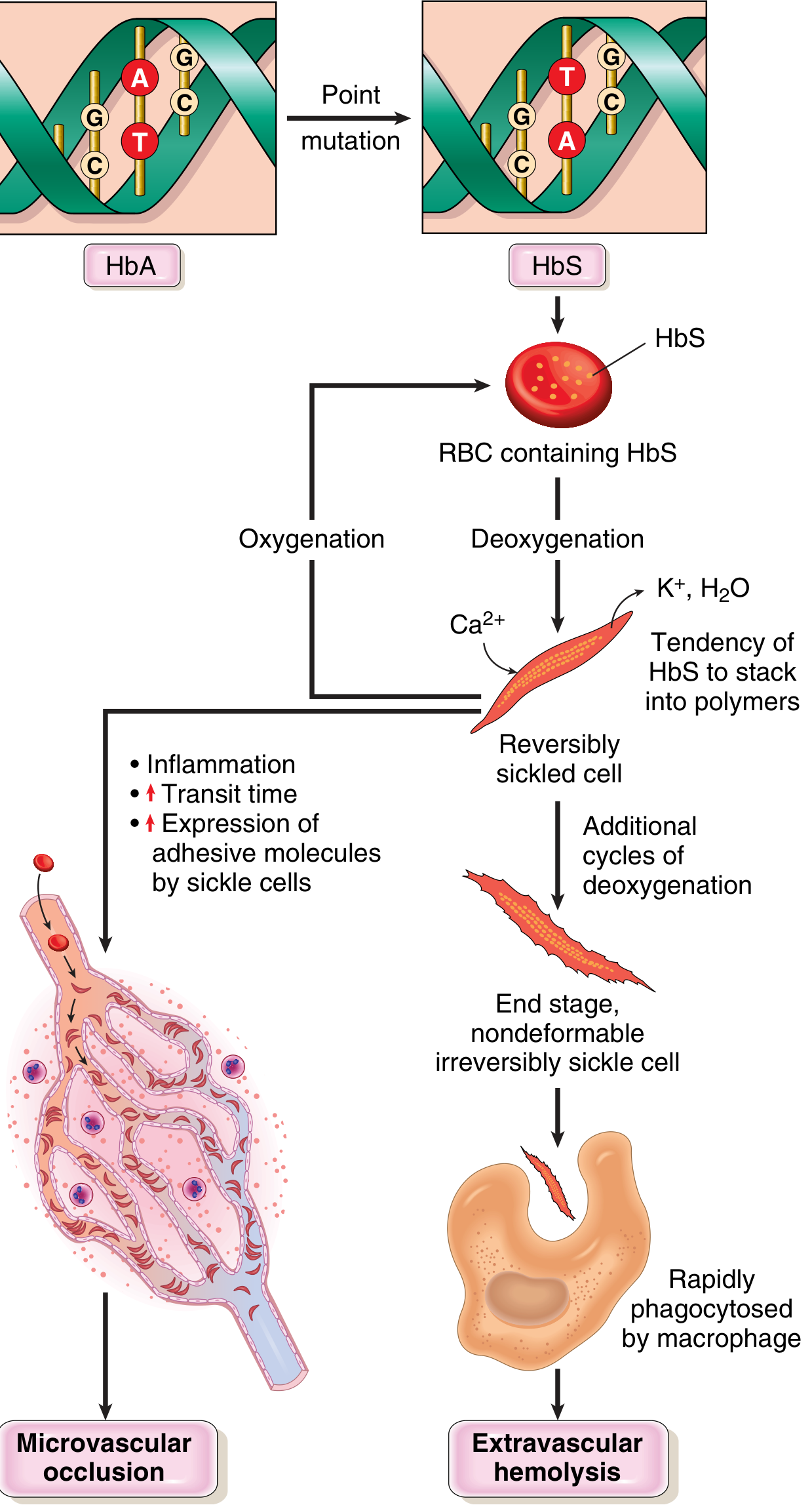
Point mutation (GAG→GTG) in HBB gene
                ↓
Glutamate → Valine substitution at β-globin position 6
                ↓
Under DEOXYGENATION: HbS forms long insoluble polymers
(intermolecular contacts via abnormal valine residue)
                ↓
Polymers distort RBC → SICKLE / CRESCENTIC shape
                ↓
       ┌────────────────────┬──────────────────────────────┐
       ↓                    ↓                              ↓
Re-oxygenation      Repeated sickling cycles         Ca²⁺ influx
       ↓                    ↓                              ↓
Reversible sickling  Membrane damage accumulates    K⁺ + H₂O efflux
                            ↓                              ↓
                    IRREVERSIBLY SICKLED CELLS       Cell dehydration
                            ↓                              ↓
             ┌──────────────┴──────────────────┐
             ↓                                 ↓
    Phagocytosed by macrophages        Obstruct microvasculature
    (extravascular haemolysis)          (vaso-occlusion)
             ↓                                 ↓
    HAEMOLYTIC ANAEMIA              ISCHAEMIA + INFARCTION
Three Key Factors That Determine Sickling
- Heterozygotes (HbAS): only ~40% is HbS; HbA retards polymerization → cells rarely sickle in vivo
- Homozygotes (HbSS): 100% HbS → severe sickling
- HbF (fetal Hb) interacts weakly with HbS → protects neonates from sickling (symptoms absent until HbF declines at 5-6 months of age)
- HbC (Lys at position 6) coexists in some patients → HbSC disease (milder than HbSS)
- Tissues with sluggish blood flow (spleen, bone marrow) most susceptible
- Inflammation increases adhesion of sickle RBCs to endothelium → prolongs transit time → increases sickling
- Triggers: infection, dehydration, acidosis, hypoxia, cold - all precipitate sickling crises
MORPHOLOGY
Peripheral Blood Smear
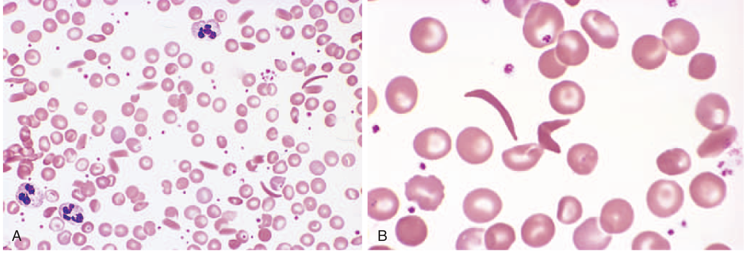
- Sickle cells (drepanocytes) - elongated, crescent/boat-shaped cells - pathognomonic
- Target cells (codocytes) - thin cells with central dense area
- Normoblasts (nucleated RBCs) - due to compensatory erythroid hyperplasia
- Polychromasia - reticulocytosis (compensatory)
- Howell-Jolly bodies - nuclear remnants due to autosplenectomy
- Anisocytosis and poikilocytosis
Bone Marrow
- Erythroid hyperplasia (compensatory) - increased normoblasts
- Expansion of marrow cavity into cortical bone → bone resorption
- "Crew-cut" appearance on skull X-ray (hair-on-end pattern) due to medullary expansion
- Extramedullary haematopoiesis in liver and spleen
Spleen
- Childhood: Moderate splenomegaly (up to 500 g) - red pulp congested with sickled RBCs
- Adulthood: Progressive infarction and fibrosis → "Autosplenectomy" - spleen reduced to a small fibrotic nubbin
- This is the hallmark gross finding in adults with sickle cell anaemia
Other Organs
- Heart, liver, renal tubules: Fatty changes due to hypoxia (both anaemia and vascular stasis)
- Bone: Avascular necrosis (particularly femoral head and humeral head); bone pain
- Kidneys: Infarction → haematuria; papillary necrosis; renal failure in late disease
- Pigment gallstones (cholelithiasis): Due to chronic haemolysis → excess bilirubin
- Skin: Leg ulcers over medial malleolus (due to microinfarcts + poor venous drainage)
- Priapism: Vascular obstruction in corpora cavernosa → fibrosis and erectile dysfunction
CLINICAL FEATURES
Onset
- Symptoms begin at 5-6 months of age (when HbF declines to adult levels)
- Diagnosed by newborn screening (haemoglobin electrophoresis)
Chronic Features
| System | Feature |
|---|---|
| Haematological | Chronic haemolytic anaemia (Hb 6-9 g/dL); jaundice; pallor; splenomegaly (childhood) |
| Musculoskeletal | Dactylitis (hand-foot syndrome) - earliest manifestation in infants; avascular necrosis of femoral/humeral heads; bone pain |
| Cardiovascular | Cardiomegaly (high-output cardiac failure due to anaemia) |
| Renal | Haematuria; papillary necrosis; inability to concentrate urine (isosthenuria) |
| Neurological | Stroke (most feared complication); seizures |
| Eyes | Proliferative retinopathy; vitreous haemorrhage |
| GI | Pigment gallstones; hepatomegaly; hepatic infarction |
| Skin | Chronic leg ulcers |
Sickle Cell Crises (Acute Episodes)
| Crisis Type | Mechanism | Features |
|---|---|---|
| Vaso-occlusive (Painful) Crisis | Most common; microvascular occlusion | Severe bone pain; abdominal pain; chest pain; triggered by infection, dehydration, cold |
| Acute Chest Syndrome | Lung infarction ± infection | Chest pain, fever, hypoxia, pulmonary infiltrates; most common cause of death |
| Aplastic Crisis | Parvovirus B19 infection of erythroid progenitors | Sudden severe drop in Hb; absent reticulocytes |
| Splenic Sequestration Crisis | Massive acute pooling of blood in spleen | Rapid splenomegaly; shock; occurs in children |
| Haemolytic Crisis | Accelerated haemolysis | Deepening jaundice; falling Hb |
LABORATORY DIAGNOSIS
| Test | Finding |
|---|---|
| Haemoglobin | Low: 6-9 g/dL |
| MCV | Normal (normocytic) |
| Reticulocyte count | Elevated (5-15%) |
| Peripheral smear | Sickle cells, target cells, Howell-Jolly bodies, nucleated RBCs |
| Serum bilirubin | Elevated indirect (unconjugated) - due to haemolysis |
| LDH | Elevated (haemolysis marker) |
| Serum haptoglobin | Decreased (consumed by free haemoglobin) |
| Sickling test (Solubility test) | Positive (Na metabisulphite reduces O₂ → sickle cells visible) |
| Haemoglobin electrophoresis | Gold standard - HbSS pattern (no HbA band; predominantly HbS + HbF) |
| Bone marrow | Erythroid hyperplasia |
| X-ray skull | "Crew-cut" / hair-on-end pattern |
COMPLICATIONS
- Stroke - most feared; occurs in ~11% of children; due to cerebrovascular sickling
- Acute chest syndrome - most common cause of death; pulmonary vaso-occlusion
- Autosplenectomy → increased susceptibility to encapsulated organisms (Streptococcus pneumoniae, Haemophilus influenzae, Salmonella) - reason why prophylactic penicillin and vaccinations are mandatory
- Salmonella osteomyelitis - classic association (vs. normal: Staphylococcus)
- Avascular necrosis of femoral and humeral heads
- Chronic renal failure - from repeated infarctions
- Pigment gallstones - from chronic haemolysis
- Aplastic crisis - from Parvovirus B19
- Priapism → erectile dysfunction
- Pulmonary hypertension - from chronic haemolysis and lung disease
- Retinopathy → blindness
SICKLE CELL TRAIT vs SICKLE CELL DISEASE
| Feature | Sickle Cell Trait (HbAS) | Sickle Cell Disease (HbSS) |
|---|---|---|
| HbS% | ~40% | ~85-90% |
| HbA% | ~60% | 0% |
| Anaemia | Absent | Moderate-severe |
| Sickling crises | Absent (rare, only extreme hypoxia) | Present, recurrent |
| Spleen | Normal | Autosplenectomy |
| Malaria protection | YES (selective advantage) | Partial |
| Life expectancy | Normal | Reduced (median ~50-60 years with treatment) |
TREATMENT (Brief, for completeness)
- Hydroxyurea - increases HbF production → reduces HbS polymerization; reduces frequency of crises (most important drug)
- Prophylactic penicillin + vaccinations (against S. pneumoniae, H. influenzae, meningococcus) due to functional asplenia
- Blood transfusions - for acute chest syndrome, stroke prevention
- Bone marrow / stem cell transplantation - only curative treatment
- Gene therapy - emerging (BCL11A gene editing to reactivate HbF)
MARKS BREAKDOWN (10/10)
| Section | Marks |
|---|---|
| Definition + Genetics + Epidemiology | 1.0 |
| Molecular basis (mutation, amino acid change) | 1.5 |
| Pathogenesis flow (sickling mechanism, 3 factors) | 2.0 |
| Morphology - smear, spleen, bone, other organs | 2.0 |
| Clinical features + 5 types of crises | 2.0 |
| Lab diagnosis (electrophoresis, smear, sickling test) | 1.0 |
| Complications (5+ named) | 0.5 |
| Total | 10 |
Source: Robbins & Kumar Basic Pathology; Robbins & Cotran Pathologic Basis of Disease - Chapter on Red Cell Disorders. Images directly from Robbins textbook.