
Explain being tumors of breast from ROBBINS AND COTRAN PATHOGENESIS OF DISEASE with flowchart diagrams and tables
Reading File
Now let me search the medical library for benign tumors of the breast from Robbins and Cotran.
Finding Sources
Finding Sources
Finding Sources
Finding Sources
Finding Sources
Finding Sources
Now let me read the relevant sections from Robbins Cotran on benign breast tumors and related disease spectrum:
Reading File
Finding Sources
Reading File
Reading File
Excellent! Now let me read the continuation of fibroadenoma section and also the phyllodes tumor and other benign breast tumor sections:
Reading File
Reading File
Excellent! Now let me get the risk table and the proliferative disease with atypia section, plus fetch the key images:
Reading File
Loading Image
Loading Image
Now let me get the risk stratification table and the phyllodes tumor image:
Reading File
Reading File
Loading Image
Reading File
I now have comprehensive content from Robbins & Cotran. Let me compile the full, detailed answer with flowcharts, tables, and textbook images.
Benign Tumors and Tumor-Like Lesions of the Breast
Robbins, Cotran & Kumar: Pathologic Basis of Disease
Overview: Breast Structure and Lesion Classification
The breast is composed of ducts, lobules, two epithelial cell types (luminal and myoepithelial), and two stromal compartments (intralobular and interlobular). Each element is a source of both benign and malignant disease.
BREAST ANATOMY → Sources of Disease
━━━━━━━━━━━━━━━━━━━━━━━━━━━━━━━━━━━━━━━━━━━━━━━━━━━━━━━━━━━━━━━━━━
BREAST PARENCHYMA
│
┌───────────────┴────────────────┐
EPITHELIAL CELLS STROMAL CELLS
│ │
┌─────┴──────┐ ┌──────┴──────┐
Luminal Myoepi- Intralobular Interlobular
cells thelial stroma stroma
│ cells │ │
│ Fibroadenoma Lipoma
│ Phyllodes Tumor Myofibro-
│ blastoma
│ Fibromatosis
│ Angiosarcoma
│
├─ Fibrocystic Changes (non-neoplastic)
├─ Proliferative Disease Without Atypia
├─ Proliferative Disease With Atypia
└─ Carcinoma (DCIS → Invasive)
━━━━━━━━━━━━━━━━━━━━━━━━━━━━━━━━━━━━━━━━━━━━━━━━━━━━━━━━━━━━━━━━━━
Key point: >90% of symptomatic breast lesions are benign. Most benign palpable masses are fibroadenomas or cysts.
FLOWCHART 1: Clinical Presentation of Breast Disease
Patient presents with breast complaint
│
┌──────────┼──────────────┬──────────────┐
▼ ▼ ▼ ▼
Lumpiness Pain Palpable Nipple
(diffuse (cyclic/ Mass Discharge
nodularity) non-cyclic) │ │
│ │ │ │
Fibrocystic Premenstrual ┌─┴──┐ ┌──┴───┐
Changes edema / Benign Malignant Milky Bloody/
ruptured (~95%) (~5% (galac- Serous
cyst ↑with torrhoea)(papilloma
age) /cancer)
│
┌──────────┴────────────┐
Round/oval, Irregular,
rubbery, mobile hard (scirrhous),
(Fibroadenoma, nonmobile
Cyst) (Carcinoma)
SECTION 1: Nonproliferative Breast Changes (Fibrocystic Changes)
Age group: 30-50 years | Cause: Hormonal fluctuations during menstruation | Risk: NO increased cancer risk
Three Principal Morphological Features:
FIBROCYSTIC CHANGES
━━━━━━━━━━━━━━━━━━━━━━━━━━━━━━━━━━━━━━━━
┌──────────────────────┐
│ FIBROCYSTIC CHANGES │
└──────────────────────┘
│
┌──────────────┼──────────────┐
▼ ▼ ▼
CYSTIC CHANGE FIBROSIS ADENOSIS
│ │ │
Dilation of Cyst rupture Increase in
lobules → → chronic acini per
larger cysts inflammation lobule
│
Lined by flat
or APOCRINE
cells
("Blue-dome Fibrosis = palpable
cysts") nodularity
│
Calcifications
common
━━━━━━━━━━━━━━━━━━━━━━━━━━━━━━━━━━━━━━━━
Apocrine Cysts (Fig. 23.5) - Cysts lined by apocrine cells with granular eosinophilic cytoplasm; blue-tinged turbid fluid; confirmed by FNA causing mass to disappear:
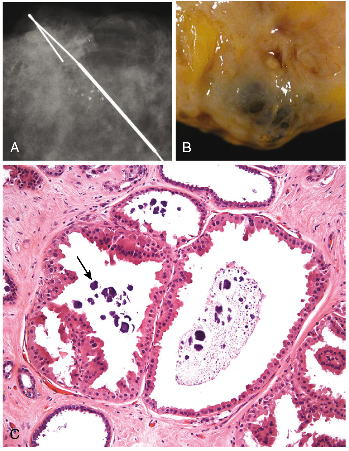
Special variant - Lactational Adenoma: Palpable mass in pregnant/lactating females; normal-appearing breast tissue with lactational changes; regresses after breastfeeding stops. Considered an exaggerated local response to gestational hormones rather than a true neoplasm.
SECTION 2: Proliferative Breast Disease Without Atypia
Risk: Small increase (~1.5-2x) in subsequent carcinoma in BOTH breasts | Usually detected as mammographic densities, calcifications, or incidental findings
Subtypes:
| Lesion | Morphology | Key Features | Clinical Presentation |
|---|---|---|---|
| Usual Ductal Hyperplasia (UDH) | Mixed luminal + myoepithelial cells filling ducts; irregular peripheral slit-like lumens | Mixed population (CK14-positive myoepithelial cells within mass) | Usually incidental |
| Sclerosing Adenosis | Increased acini, compressed by stromal fibrosis; "swirling" pattern with well-circumscribed outer border | May mimic invasive carcinoma; calcifications within lumens | Palpable mass, radiologic density, calcifications |
| Radial Scar (Complex Sclerosing Lesion) | Stellate lesion; entrapped glands in hyalinized elastotic stroma surrounded by radiating projections | Mimics invasive carcinoma radiologically + histologically. NOT caused by trauma/surgery. >1 cm = complex sclerosing lesion | Mammographic abnormality |
| Papilloma (Large Duct) | Multiple branching fibrovascular cores in dilated duct; UDH and apocrine metaplasia common | Solitary; central (lactiferous sinuses) | Nipple discharge - bloody (torsion/infarction) or serous; >80% produce discharge |
| Papilloma (Small Duct) | Same structure, peripheral ducts | Multiple; peripheral | Clinically occult; small palpable masses or mammographic densities |
PROLIFERATIVE DISEASE WITHOUT ATYPIA - RISK PATHWAY
━━━━━━━━━━━━━━━━━━━━━━━━━━━━━━━━━━━━━━━━━━━━━━━━━━━━━
Normal Breast → Fibrocystic Change (nonproliferative)
│
└─→ Proliferative Without Atypia ────────────────►
(UDH, Sclerosing Adenosis, RISK:
Radial Scar, Papilloma) ~1.5-2x
(both
breasts)
━━━━━━━━━━━━━━━━━━━━━━━━━━━━━━━━━━━━━━━━━━━━━━━━━━━━━
SECTION 3: Proliferative Breast Disease WITH Atypia (Precursor Lesions)
Risk: 4-5x increased risk of carcinoma | Regarded as precursors, not just risk markers
Flowchart: Low-Grade Neoplasia Pathway
MOLECULAR PROGRESSION - LOW GRADE PATHWAY
━━━━━━━━━━━━━━━━━━━━━━━━━━━━━━━━━━━━━━━━━━━━━━━━━━━━━━━━━━━━━━━━━━
Normal TDLU
│
▼
Columnar Cell Lesion (CCL)
- Clonal proliferation
- Earliest recognizable precursor
- Variably dilated acini
- Columnar epithelial cells
- Microcalcifications on mammography
│
▼
Flat Epithelial Atypia (FEA)
- CCL + CYTOLOGIC ATYPIA
- No architectural complexity (unlike ADH)
│
▼
Atypical Ductal Hyperplasia (ADH)
- Clonal; has SOME but NOT ALL features of
low-grade DCIS (found in ~10% of calcification biopsies)
OR
Atypical Lobular Hyperplasia (ALH)
- Incidental finding; found in <5% of biopsies
│
▼
Low-grade DCIS / LCIS
│
▼
Invasive Carcinoma (same morphologic/molecular features)
━━━━━━━━━━━━━━━━━━━━━━━━━━━━━━━━━━━━━━━━━━━━━━━━━━━━━━━━━━━━━━━━━━
SECTION 4: Relative Risk of Breast Cancer by Lesion Type
| Category | Lesion | Relative Risk vs. General Population |
|---|---|---|
| Nonproliferative | Fibrocystic changes, adenosis, mild hyperplasia | No increased risk (1x) |
| Proliferative without atypia | UDH, sclerosing adenosis, papilloma, radial scar | ~1.5-2x |
| Complex fibroadenoma | With cysts >0.3 cm, sclerosing adenosis, epithelial calcifications, papillary apocrine change | Slightly increased |
| Proliferative with atypia | Atypical ductal hyperplasia (ADH) | ~4-5x |
| Proliferative with atypia | Atypical lobular hyperplasia (ALH) | ~4-5x |
| LCIS / DCIS | Lobular/ductal carcinoma in situ | ~8-10x |
(Derived from Robbins, Cotran & Kumar Table 23.1 data)
SECTION 5: Fibroadenoma
The most common benign tumor of the female breast. Arises from intralobular stroma.
Molecular Pathogenesis:
FIBROADENOMA - PATHOGENESIS
━━━━━━━━━━━━━━━━━━━━━━━━━━━━━━━━━━━━━━━━━━━━━━━━━━━━━━━━━━━━━━━━━━
INTRALOBULAR STROMAL CELL
│
┌─────────────┴────────────┐
▼ ▼
MED12 mutation (~2/3) RARA mutation (~1/3)
(Mediator complex, (Retinoic acid receptor α;
regulates RNA Pol II estrogen target gene;
transcription) cooperates with ER)
│ │
└────────────┬─────────────┘
▼
Altered expression of sex hormone-regulated genes
→ stromal cell proliferation + survival
│
▼
FIBROADENOMA
━━━━━━━━━━━━━━━━━━━━━━━━━━━━━━━━━━━━━━━━━━━━━━━━━━━━━━━━━━━━━━━━━━
Note: Uterine leiomyoma also has MED12 mutations - both arise from hormone-responsive stromal cells.
Morphology:
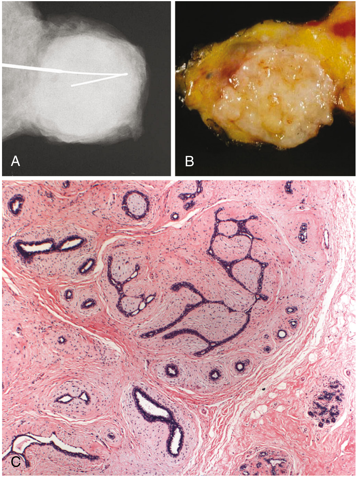
- Well-circumscribed, rubbery, gray-white nodule that bulges above surrounding tissue
- Slit-like spaces lined by epithelium
- Pericanalicular pattern: stroma surrounds patent ducts
- Intracanalicular pattern: stroma compresses/distorts ducts into cleft-like spaces
- In older women: densely hyalinized stroma + atrophic epithelium
Fig. 23.24 Fibroadenoma - (A) well-circumscribed mass on radiograph, (B) rubbery white gross specimen, (C) intralobular stroma compressing epithelium:
Subtypes of Fibroadenoma:
| Subtype | Features | Association |
|---|---|---|
| Sporadic | Standard morphology | MED12/RARA mutations |
| Myxoid | Myxoid stroma | Most sporadic; small proportion: Carney complex (PRKAR1A germline mutations, AD) |
| Complex | Cysts >0.3 cm, sclerosing adenosis, epithelial calcifications, or papillary apocrine change | Slightly increased cancer risk (likely due to co-existing at-risk lesions in surrounding breast) |
Clinical Features:
| Feature | Details |
|---|---|
| Age | Mainly 20s-30s |
| Number | Frequently multiple and bilateral |
| Hormonal response | Grow during pregnancy; regress after menopause; rapid growth/infarction in pregnancy may mimic carcinoma |
| Special association | ~50% of cyclosporin A recipients after renal transplant develop multiple bilateral fibroadenomas (regress after drug cessation) |
| Malignant potential | Extremely rare; no significant risk unless complex type |
SECTION 6: Phyllodes Tumor
Arises from intralobular stroma, like fibroadenoma, but much less common (~2.5% of fibroepithelial lesions).
Pathogenesis Comparison:
FIBROADENOMA vs. PHYLLODES TUMOR - MOLECULAR COMPARISON
━━━━━━━━━━━━━━━━━━━━━━━━━━━━━━━━━━━━━━━━━━━━━━━━━━━━━━━
Gene Fibroadenoma Phyllodes Tumor
─────────────────────────────────────────────────────────
MED12 ✓ (majority) ✓ (majority)
RARA ✓ (~1/3) ✓
TERT ✗ ✓ (additional)
TP53 ✗ ✓ (additional)
RB ✗ ✓ (additional)
─────────────────────────────────────────────────────────
→ Shared origin (intralobular stroma) BUT phyllodes has
additional genomic instability → more aggressive behavior
━━━━━━━━━━━━━━━━━━━━━━━━━━━━━━━━━━━━━━━━━━━━━━━━━━━━━━━
Grading:
PHYLLODES TUMOR GRADING
━━━━━━━━━━━━━━━━━━━━━━━━━━━━━━━━━━━━━━━━━━━━━━━━━━━━━━━━━━━━━━━━━━
BENIGN (~75%) BORDERLINE MALIGNANT (<25%)
│ │ │
Resembles More prominent Widely infiltrative;
fibroadenoma stromal atypia, difficult to distinguish
but stroma is cellularity, from sarcoma
more cellular + mitotic (marked stromal
mitotically activity overgrowth, few
active epithelial elements)
│ │ │
Occasional local Increased Hematogenous
recurrence recurrence metastasis in ~1/3
No metastasis (margin status Lymphatic spread
is key predictor) RARE
━━━━━━━━━━━━━━━━━━━━━━━━━━━━━━━━━━━━━━━━━━━━━━━━━━━━━━━━━━━━━━━━━━
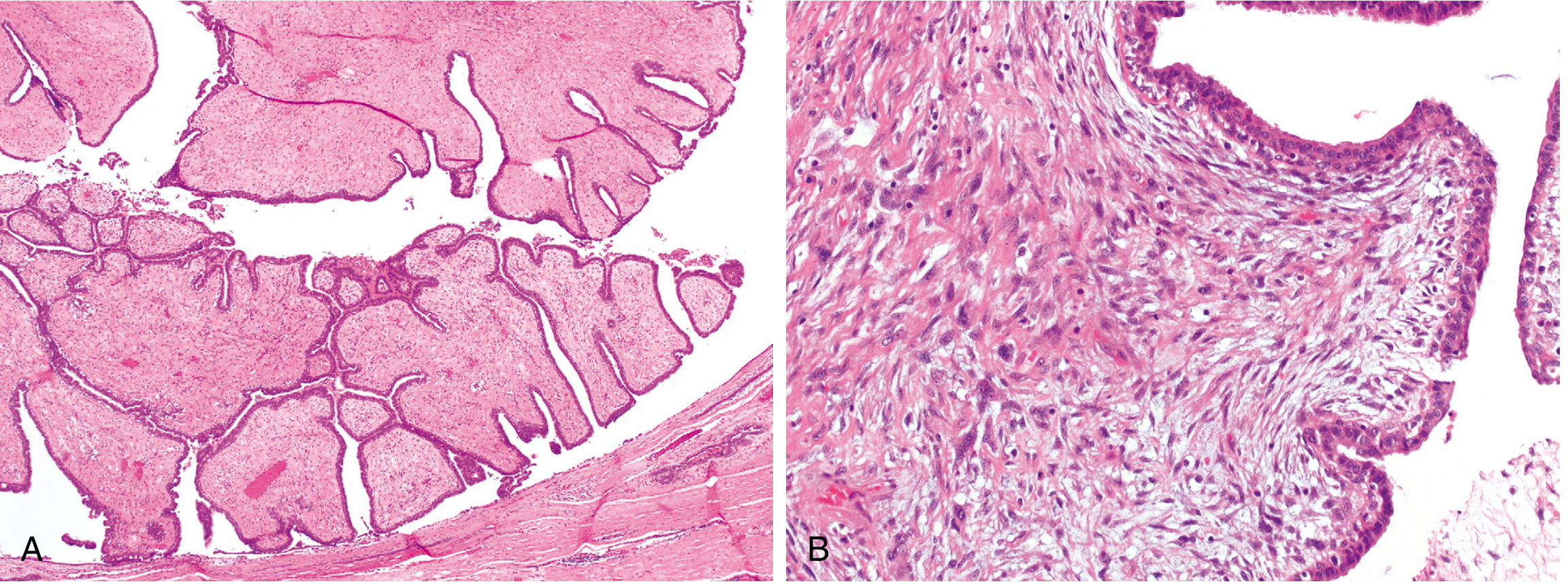
Fig. 23.25 Phyllodes Tumor - (A) leaf-like architecture at low power, (B) increased stromal cellularity, atypia and mitotic activity compared to fibroadenoma:
Key clinical points:
- Peak age: 6th decade (10-20 years later than fibroadenoma)
- "Phyllodes" = Greek for leaf-like (due to bulbous leaf-like projections into cystic spaces)
- Axillary lymph node dissection is contraindicated (lymphatic spread is rare regardless of grade)
- Only the neoplastic stromal component is present in metastases (not the epithelial component)
SECTION 7: Benign Lesions of Interlobular Stroma
These tumors consist of stromal cells only (no accompanying epithelial component).
| Tumor | Key Features |
|---|---|
| Myofibroblastoma | Composed of myofibroblasts; unique - only breast tumor equally common in both males and females |
| Lipoma | Palpable; fat-containing lesion on mammography |
| Fibromatosis | Clonal fibroblast/myofibroblast proliferation; irregular infiltrating mass; may involve muscle; locally aggressive but does NOT metastasize; associated with prior trauma/surgery, or FAP / hereditary desmoid syndrome / Gardner syndrome |
SECTION 8: Other Benign Breast Conditions (Inflammatory/Structural)
Gynecomastia (Male Breast)
The only benign lesion of any frequency in the male breast.
GYNECOMASTIA - PATHOGENESIS
━━━━━━━━━━━━━━━━━━━━━━━━━━━━━━━━━━━━━━━━━━━━━━━━━━━━━━━━━━
IMBALANCE: Estrogen ↑ / Androgen ↓
│
┌─────────────┼─────────────────┐
▼ ▼ ▼
PHYSIOLOGIC PATHOLOGIC DRUG-INDUCED
│ │ │
Puberty Cirrhosis Alcohol, Marijuana,
Old age (↓ estrogen Heroin, Antiretrovirals,
metabolism) Anabolic steroids
Klinefelter (47,XXY)
Leydig cell tumor
Sertoli cell tumor
━━━━━━━━━━━━━━━━━━━━━━━━━━━━━━━━━━━━━━━━━━━━━━━━━━━━━━━━━━
Morphology: Dense collagenous connective tissue +
epithelial hyperplasia of duct lining
(Lobule formation almost NEVER seen)
Duct Ectasia vs. Squamous Metaplasia of Lactiferous Ducts
| Feature | Duct Ectasia | Squamous Metaplasia of Lactiferous Ducts (Zuska Disease) |
|---|---|---|
| Age | 5th-6th decade, multiparous | Any age |
| Smoking | NOT associated | >90% are smokers |
| Presentation | Periareolar mass, thick white nipple secretions, occasional skin retraction | Painful erythematous subareolar mass; recurrent abscess; fistula tract |
| Morphology | Dilated ducts with inspissated secretions; lipid-laden macrophages; periductal lymphoplasmacytic inflammation; granulomas | Keratinizing squamous metaplasia extends into nipple ducts; keratin plugs → duct rupture → granulomatous inflammation |
| Key risk | May mimic invasive carcinoma clinically/radiologically | Recurrences common; may cause nipple inversion |
| Treatment | Observation | En bloc surgical removal of duct + fistula |
MASTER FLOWCHART: Benign Breast Disease - Overview and Cancer Risk
BENIGN BREAST DISEASE - RISK STRATIFICATION (ROBBINS)
━━━━━━━━━━━━━━━━━━━━━━━━━━━━━━━━━━━━━━━━━━━━━━━━━━━━━━━━━━━━━━━━━━━━━━━
FEMALE BREAST LESION
│
├─── INFLAMMATORY / STRUCTURAL (No cancer risk)
│ ├── Duct Ectasia
│ ├── Fat Necrosis
│ └── Squamous Metaplasia of Lactiferous Ducts (Zuska)
│
├─── NON-PROLIFERATIVE (No increased cancer risk)
│ └── Fibrocystic Changes
│ ├── Cysts (with apocrine metaplasia)
│ ├── Fibrosis
│ ├── Adenosis
│ └── Lactational Adenoma
│
├─── PROLIFERATIVE WITHOUT ATYPIA (RR ~1.5-2x)
│ ├── Usual Ductal Hyperplasia
│ ├── Sclerosing Adenosis
│ ├── Radial Scar / Complex Sclerosing Lesion
│ └── Papilloma (large duct / small duct)
│
├─── PROLIFERATIVE WITH ATYPIA (RR ~4-5x)
│ ├── Columnar Cell Lesions
│ ├── Flat Epithelial Atypia
│ ├── Atypical Ductal Hyperplasia (ADH)
│ └── Atypical Lobular Hyperplasia (ALH)
│ │
│ ▼
│ DCIS / LCIS (RR ~8-10x)
│ │
│ ▼
│ INVASIVE CARCINOMA
│
└─── STROMAL TUMORS (Neoplasms)
│
├─ INTRALOBULAR STROMA
│ ├── Fibroadenoma (BENIGN - most common)
│ └── Phyllodes Tumor (Benign/Borderline/Malignant)
│
└─ INTERLOBULAR STROMA
├── Myofibroblastoma (BENIGN)
├── Lipoma (BENIGN)
├── Fibromatosis (locally aggressive, no metastasis)
└── Angiosarcoma (MALIGNANT - rare)
━━━━━━━━━━━━━━━━━━━━━━━━━━━━━━━━━━━━━━━━━━━━━━━━━━━━━━━━━━━━━━━━━━━━━━━
QUICK COMPARISON TABLE: Major Benign Breast Tumors
| Feature | Fibroadenoma | Phyllodes Tumor | Papilloma (Large Duct) | Sclerosing Adenosis |
|---|---|---|---|---|
| Origin | Intralobular stroma | Intralobular stroma | Ductal epithelium | Lobular epithelium + stroma |
| Age | 20s-30s | 6th decade | Any | Reproductive age |
| Gross | Well-circumscribed, rubbery, gray-white | Variable size, leaf-like bulbous protrusions | Intraductal | Stellate/glandular |
| Key mutations | MED12, RARA | MED12, RARA, TERT, TP53, RB | - | - |
| Hormonal response | Yes (grows in pregnancy, regresses at menopause) | Less prominent | No | No |
| Key symptom | Palpable mobile mass | Palpable mass ± rapid growth | Nipple discharge (bloody/serous) | Palpable mass, calcifications |
| Cancer risk | None (slight if complex type) | 0% (benign) to ~33% metastasis (malignant) | Slight increase | Slight increase |
| Metastasis | No | Rare; only malignant type; hematogenous | No | No |
| LN dissection | N/A | Contraindicated (lymphatic spread rare) | N/A | N/A |
Source: Robbins, Cotran & Kumar Pathologic Basis of Disease, Chapter 23 - The Breast (pp. 945-985)
This is a shared conversation. Sign in to Orris to start your own chat.