TB pathology essay

Reading File
Finding Sources
Finding Sources
Reading File
Reading File
Finding Sources
Reading File
Finding Sources
Finding Sources
Reading File
Finding Sources
Reading File
Finding Sources
Loading Image
Loading Image
I now have comprehensive material. Let me compile the full essay.

Tuberculosis: Pathology

Based on Robbins, Cotran & Kumar Pathologic Basis of Disease (10th Ed.) and supporting textbooks.

1. Introduction and the Organism

Tuberculosis (TB) is a chronic granulomatous infectious disease caused by Mycobacterium tuberculosis, an obligate aerobic, non-spore-forming, non-motile bacillus. Its defining microbiological feature is a thick, waxy cell wall rich in mycolic acids, which accounts for its acid-fast staining property (Ziehl-Neelsen stain) and its resistance to desiccation, many disinfectants, and intracellular killing. The organism grows slowly (doubling time ~20 hours), making culture the definitive diagnostic method but a slow one. TB remains among the leading infectious causes of death worldwide, predominantly affecting the lungs but capable of infecting virtually any organ.

2. Pathogenesis

2.1 Entry and Initial Infection

Infection is almost invariably acquired by inhalation of airborne droplet nuclei expelled by a patient with active pulmonary TB. Droplet nuclei are small enough (<5 µm) to reach the alveoli. Once in the alveolus, M. tuberculosis is engulfed by alveolar macrophages via phagocytosis mediated by the mannose-binding lectin and the type 3 complement receptor (CR3) on the macrophage surface (Robbins, Cotran & Kumar, p. 351).

2.2 Intracellular Survival - The Phagosome Escape
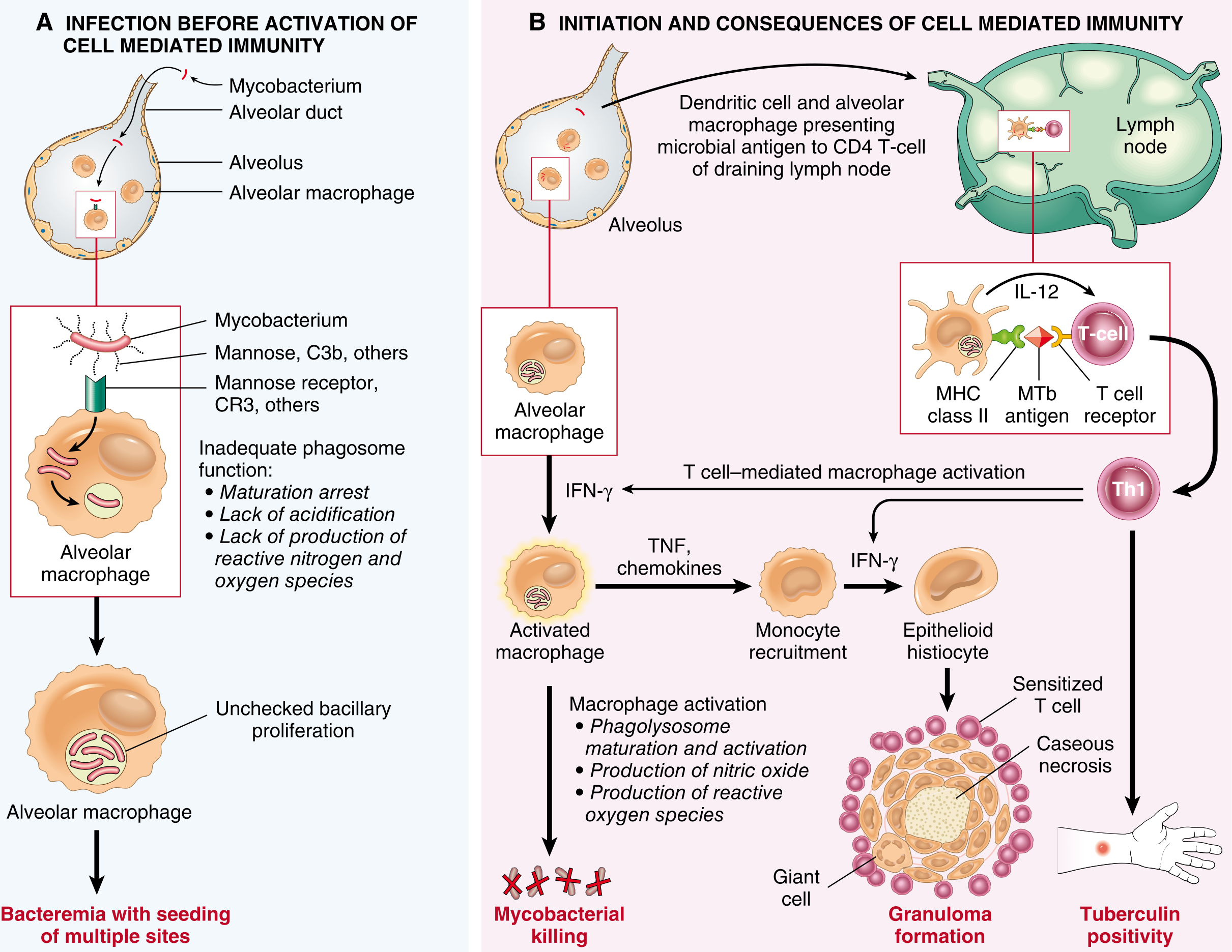
The bacterium's most important pathogenicity strategy is blocking phagolysosome fusion. M. tuberculosis recruits a host protein called coronin to the phagosomal membrane. Coronin activates the phosphatase calcineurin, which inhibits phagosome-lysosome fusion, preventing acidification and exposure to lysosomal enzymes. The result: unchecked bacillary proliferation within the macrophage. This leads to bacteremia with seeding of multiple sites even before adaptive immunity is activated (Robbins, p. 351).
Sequence of events in primary pulmonary tuberculosis - cellular immune response diagram
Fig. 8.26 - Sequence of events in primary pulmonary TB. (A) Early infection before T cell-mediated immunity: phagosome maturation arrest leads to unchecked bacillary proliferation and bacteremia. (B) After ~3 weeks, Th1 CD4+ cells produce IFN-γ, activating macrophages to kill bacteria and form granulomas. (Robbins, Cotran & Kumar)

2.3 Innate Immunity

Multiple pathogen-associated molecular patterns (PAMPs) of M. tuberculosis trigger innate receptors: mycobacterial lipoarabinomannan binds TLR2, and unmethylated CpG nucleotides bind TLR9. These interactions initiate cytokine production and prime the adaptive response.

2.4 Th1 Adaptive Response (~3 weeks post-infection)

Approximately three weeks after infection, a Th1 CD4+ T cell response is mounted. Dendritic cells, which have ingested mycobacteria and migrated to draining lymph nodes, present mycobacterial antigens via MHC class II to naïve T cells. IL-12 and IL-18 secreted by antigen-presenting cells drive Th1 differentiation.
Th1 cells produce IFN-γ, the pivotal mediator that activates macrophages to become bactericidal through three mechanisms:
  1. Stimulation of microbicidal nitric oxide and reactive oxygen species in the phagolysosome
  2. Mobilization of antimicrobial peptides (e.g., cathelicidin)
  3. Induction of autophagy, which sequesters and destroys intracellular bacteria
Activated macrophages also secrete TNF and chemokines, recruiting more monocytes to sustain the immune response. The critical importance of TNF in containing TB is demonstrated clinically by the increased risk of TB reactivation in patients receiving TNF antagonists for rheumatoid arthritis.

2.5 Granuloma Formation and Caseous Necrosis

IFN-γ-activated macrophages differentiate into epithelioid histiocytes that aggregate to form granulomas. Some epithelioid cells fuse to become Langhans-type multinucleated giant cells (nuclei arranged in a horseshoe or peripheral ring). The granuloma is enclosed within a fibroblastic rim with a peripheral cuff of lymphocytes.
The hallmark of TB granulomas - distinct from most other granulomatous diseases - is caseous necrosis: a central area of acellular, amorphous, cheese-like necrosis (from the Latin caseus, cheese). This is attributed to delayed-type hypersensitivity reactions (Type IV), tissue ischemia, and direct mycobacterial toxicity. Microscopically, caseous necrosis appears as an eosinophilic, granular, structureless zone with loss of tissue architecture.
In immunocompromised patients (e.g., HIV/AIDS), the typical granulomatous architecture fails to form; macrophages instead contain enormous numbers of bacilli without surrounding lymphocytic cuffing.

3. Morphology: Primary Tuberculosis

3.1 The Ghon Focus

Primary TB in adults almost always begins in the lungs. Inhaled bacilli implant in the distal airspaces of the lower part of the upper lobe or the upper part of the lower lobe, usually subpleurally. As sensitization develops over 3 weeks, a 1.0-1.5 cm area of gray-white consolidation appears - the Ghon focus (also called Ghon lesion). The center undergoes caseous necrosis.

3.2 The Ghon Complex

Bacilli, either free or within phagocytes, drain via lymphatics to regional hilar and paratracheal lymph nodes, which also undergo caseous necrosis. The combination of:
  • The parenchymal Ghon focus
  • Draining lymphangitis
  • Hilar/paratracheal lymphadenitis with caseation
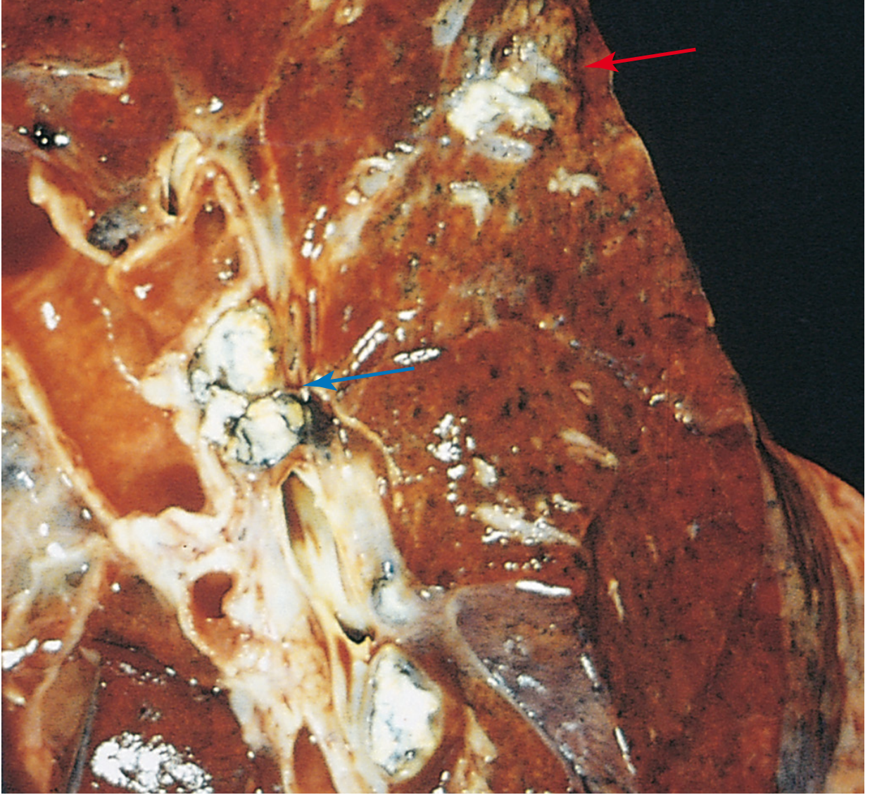
...is collectively called the Ghon complex (or Ranke complex when calcified).
Primary pulmonary tuberculosis Ghon complex - gross pathology specimen
Fig. 8.28 - Primary pulmonary tuberculosis, Ghon complex. The gray-white parenchymal focus is subpleural in the lower upper lobe (red arrow). Hilar lymph nodes with caseation visible on the left (blue arrow). (Robbins, Cotran & Kumar)

3.3 Outcome of Primary TB

In approximately 95% of immunocompetent individuals, cell-mediated immunity controls the infection. The Ghon complex undergoes progressive fibrosis and calcification (radiologically detectable). Viable organisms may remain dormant within these fibrocalcific lesions for decades - the basis of latent TB infection (LTBI). Despite early hematogenous seeding, no lesions develop at extra-pulmonary sites.
In the remaining 5%, or in immunocompromised hosts, the infection progresses to active disease.

4. Morphology: Secondary (Postprimary / Reactivation) Tuberculosis

Secondary TB arises in a previously sensitized host, either from:
  • Reactivation of latent bacilli (commonest, triggered by immunosuppression, malnutrition, renal failure, diabetes, HIV)
  • Exogenous reinfection with a new strain

4.1 Location and Initial Lesion

The hallmark location is the apex of the upper lobe (or superior segment of lower lobe), reflecting the high oxygen tension favored by M. tuberculosis. The initial lesion is a small (<2 cm) focus of consolidation, sharply circumscribed, firm, and gray-white to yellow, with variable central caseation and peripheral fibrosis.

4.2 Fibrocaseous Disease

In immunocompetent individuals the focus undergoes fibrous encapsulation - the body walls off the infection with a dense fibrous shell. Histologically, active lesions show characteristic coalescent tubercles with central caseation; acid-fast bacilli can be found by Ziehl-Neelsen staining during the early exudative and caseous phases.

4.3 Progressive Pulmonary TB

When immunity is inadequate, the apical lesion expands into adjacent lung and erodes into a bronchus. The caseous centre liquefies and is expectorated, leaving a ragged, irregular cavity poorly walled off by fibrous tissue. This cavity, with its aerobic internal environment, supports exponential bacillary proliferation and is the primary source of infectivity.
Key consequences of progressive disease:
  • Erosion of blood vessels within the cavity wall → hemoptysis (can be massive if a Rasmussen aneurysm forms)
  • Spread via airway → endobronchial, endotracheal, and laryngeal TB; contralateral lung seeding
  • Pleural involvement → serous pleural effusion, tuberculous empyema, or obliterative fibrous pleuritis
  • With treatment the cavities may become fibrotic and sterilized, though architectural distortion persists

5. Miliary Tuberculosis

When host defenses fail and bacilli enter the bloodstream, hematogenous dissemination occurs:
  • Miliary pulmonary TB: organisms draining through lymphatics enter the venous blood and circulate back to the lung, producing innumerable 2-mm yellow-white foci scattered through the lung parenchyma (resembling millet seeds, hence "miliary"). Individual lesions may coalesce to produce lobar consolidation.
  • Systemic miliary TB: dissemination via the systemic arterial circulation seeds virtually any organ. Organs classically involved include the liver, bone marrow, spleen, adrenals, meninges, kidneys, fallopian tubes, and epididymis (Robbins, p. 354).

6. Extrapulmonary TB

SitePathological Features
Lymph nodes (scrofula)Caseous granulomas in cervical nodes; most frequent form of extrapulmonary TB
Meninges (TB meningitis)Millet seed-sized tubercles on meningeal surfaces; subependymal granulomas; exudate in basal cisterns
KidneyCortical seeding during primary bacteremia; focal granulomas → coalescing cavities; calcification
Spine (Pott's disease)Granulomatous osteomyelitis of vertebral bodies; paravertebral "cold abscess" tracking along fascial planes
Adrenal glandsBilateral caseous destruction → Addison disease (historically an important cause)
IntestineTransverse ulcers over mucosal lymphoid aggregates (especially terminal ileum); healing → strictures; hyperplastic type (ileocaecal mass) mimics carcinoma
Fallopian tubes / EpididymisGranulomatous salpingitis/epididymitis; infertility
Skin (scrofuloderma)Direct extension from underlying lymphadenitis

7. Histological Summary

FeatureDescription
Tubercle (granuloma)Aggregate of epithelioid macrophages + Langhans giant cells + peripheral lymphocytes; enclosed in a fibroblastic rim
Caseous necrosisCentral eosinophilic, amorphous, cheese-like necrosis; structureless on H&E
Giant cellsLanghans type: nuclei arranged in horseshoe/peripheral ring pattern
Acid-fast bacilli (ZN stain)Beaded red rods on blue background; best found in early exudative/caseous phases
FibrocalcificationEnd-stage healed lesion; dystrophic calcification
Immunocompromised patternMany macrophages loaded with AFB; absent granulomas; "anergic" TB

8. Special Considerations

HIV co-infection: AIDS is the single greatest risk factor for progression from latent to active TB, due to loss of CD4+ Th1 cell function. HIV-positive patients often present with atypical patterns - lower lobe involvement, mediastinal lymphadenopathy without the classic apical infiltrate, and disseminated disease. Granuloma formation fails, and macrophages contain massive bacillary loads.
Drug resistance: Inadequate treatment selects for resistant mutants. Multi-drug resistant TB (MDR-TB, resistant to isoniazid and rifampicin) and extensively drug-resistant TB (XDR-TB) pose major public health challenges.
Immunological test (tuberculin/Mantoux): Intradermal PPD (purified protein derivative) elicits a Type IV delayed hypersensitivity reaction in sensitized individuals - induration at 48-72 hours. A positive test indicates sensitization, not necessarily active disease. It may be falsely negative in immunosuppression or anergy.

9. Summary

The pathology of TB is fundamentally the story of a host-pathogen arms race. M. tuberculosis survives in macrophages by blocking phagolysosome fusion; the host counters with Th1-mediated macrophage activation and granuloma formation. When immunity prevails, the infection is walled off as a fibrocalcific Ghon complex. When it fails, progressive necrosis, cavitation, and hematogenous spread produce the full spectrum of disease - from apical fibrocaseous cavities to miliary dissemination to extrapulmonary TB in virtually every organ system. The characteristic lesion throughout - the caseating granuloma - is both the mechanism of containment and, in excess, the cause of tissue destruction.

Primary sources: Robbins, Cotran & Kumar Pathologic Basis of Disease (10th Ed.); Bailey and Love's Short Practice of Surgery (28th Ed.); Sherris & Ryan's Medical Microbiology (8th Ed.)

For 2nd year exam give flow chart

Generating Image

A clean, detailed medical flowchart titled "TB Pathology - 2nd Year Exam Flowchart" showing the complete pathology of tuberculosis. The flowchart should flow top to bottom with clearly labeled boxes and arrows. Include these sequential steps and branches: START: Inhalation of M. tuberculosis droplet nuclei → Alveolar macrophage phagocytosis (via mannose receptor / CR3) Branch into two paths: LEFT PATH (Early - No immunity yet, <3 weeks): Coronin recruits to phagosome → Calcineurin activation → Phagolysosome fusion BLOCKED → Unchecked bacillary replication → Bacteremia + seeding of multiple organs RIGHT PATH (After 3 weeks - Adaptive immunity): Dendritic cells present antigen in lymph node → IL-12/IL-18 → Th1 CD4+ T cells → IFN-γ → Macrophage activation → NO + ROS + autophagy → Bacterial killing CENTRAL OUTCOME box: Granuloma Formation - Epithelioid histiocytes + Langhans giant cells + peripheral lymphocytes + fibroblastic rim → Caseous necrosis (central) Then branch into: BOX A - PRIMARY TB (95% immunocompetent): Ghon focus (1-1.5 cm, subpleural, lower upper lobe) → Lymphatic spread → Hilar lymphadenopathy → Ghon Complex → Fibrosis + Calcification = LATENT TB (dormant bacilli) BOX B - PROGRESSIVE/SECONDARY TB (5% or reactivation): Apical upper lobe location → Fibrocaseous lesion → Cavity formation → branching into: 1) Bronchial spread → endobronchial TB / contralateral lung, 2) Vascular erosion → Hemoptysis, 3) Pleural involvement → Effusion/Empyema, 4) Lymphatic/hematogenous → MILIARY TB MILIARY TB box → Systemic spread to: Liver, Spleen, Bone marrow, Meninges (TB meningitis), Kidney (renal TB), Spine (Pott disease), Adrenals (Addison disease), Fallopian tubes/Epididymis Bottom HISTOLOGY SUMMARY box: Tubercle = epithelioid cells + Langhans giant cells + caseous necrosis + lymphocytic cuff | ZN stain: acid-fast beaded red rods | Immunocompromised: no granuloma, macrophages loaded with AFB Use color coding: blue for pathogenesis steps, orange for primary TB, red for progressive/secondary TB, purple for miliary/extrapulmonary, green for histology. White background, clear readable fonts, professional medical diagram style.
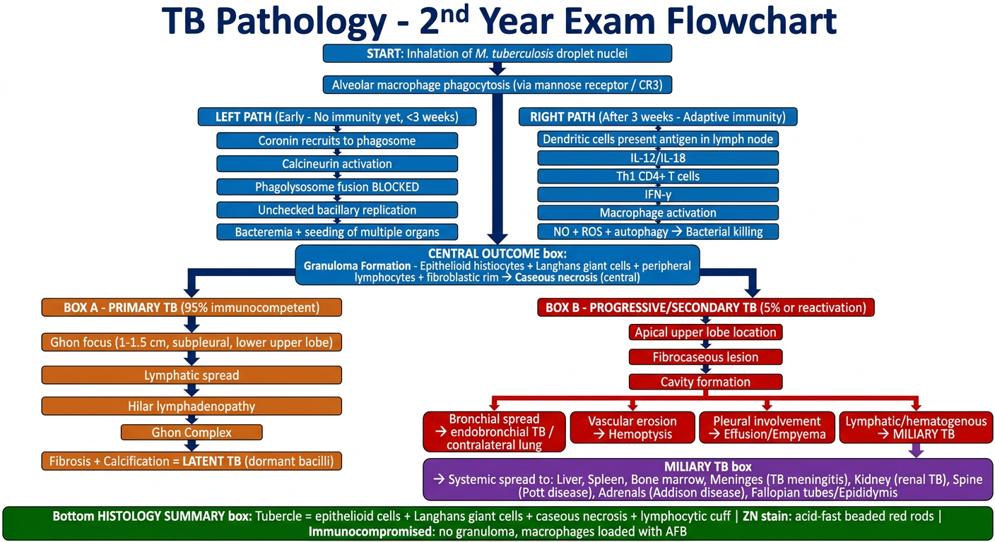
TB Pathology Flowchart for 2nd Year Exam
Here's a quick guide to reading the flowchart for exam purposes:
Top section (blue) - Pathogenesis: focus on the coronin/calcineurin mechanism for phagolysosome blockade, and the IFN-γ → macrophage activation axis.
Central box - The granuloma with caseous necrosis is THE defining lesion; examiners love asking what distinguishes TB granulomas from others (it's the caseation).
Orange branch - Primary TB: Ghon focus → Ghon complex → 95% heal with calcification = latent TB.
Red branch - Secondary/Reactivation TB: apical location, cavitation, then its three spread routes (airway, vascular, hematogenous).
Purple section - Miliary TB: know the organs in order - liver, bone marrow, spleen, adrenals, meninges, kidney, spine (Pott disease).
High-yield exam one-liners:
  • Ghon focus = subpleural, lower upper lobe, 1-1.5 cm
  • Ghon complex = focus + lymphangitis + hilar node caseation
  • Langhans giant cell = nuclei in horseshoe/peripheral ring
  • Pott disease = vertebral TB with "cold abscess"
  • ZN stain = beaded red rods on blue background
  • HIV co-infection = no granuloma, loads of AFB
This is a shared conversation. Sign in to Orris to start your own chat.