
I have dark neck and face then my whole body what to do
dark neck and body hyperpigmentation causes treatment acanthosis nigricans
acanthosis nigricans dark neck hyperpigmentation
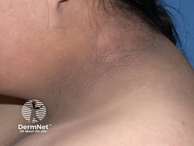
Clinical photography of the neck demonstrates acanthosis nigricans: hyperpigmented, velvety plaques within intertriginous regions of the cervical skin. The image shows a brown-to-dark brown, slightly pigmented area along the posterior and lateral neck folds, with smooth but thickened dermal-epidermal junction and papillomatosis consistent with acanthosis nigricans. The texture appears velvety on close inspection, and the surrounding skin remains comparatively lighter. There is no visible mucosal involvement or ulceration. The pattern is typically bilateral and symmetric, favoring the neck and adjacent flexural areas; extension to axillae or inguinal folds may be observed in some patients. This dermatologic finding is commonly associated with insulin resistance, obesity, type 2 diabetes mellitus, endocrine disorders, or, in rare adults, paraneoplastic syndromes. Clinically, acanthosis nigricans serves as a cutaneous sign prompting assessment of metabolic health, including fasting glucose, HbA1c, lipid profile, and weight management. The image is intended for educational purposes to illustrate morphological features such as hyperpigmentation, velvety texture, and intertriginous distribution; it aids differential diagnosis including frictional hyperpigmentation and postinflammatory changes. Potential uses include dermatology training, primary care screening, and research on metabolic disease correlations. Limitations: single-view photograph cannot assess texture depth or extent; clinical history crucial for confirming etiologies and guiding management decisions.
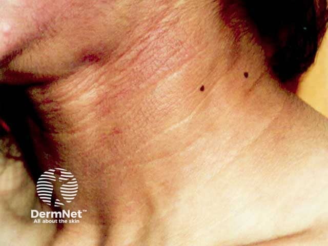
Clinical photograph of the neck illustrating acanthosis nigricans in a patient with acquired generalized lipodystrophy. The image shows pigmented, velvety plaques organized in intertriginous folds along the lateral neck and posterior cervical skin. The hyperpigmented bands are dark brown to slate-gray, with slight gloss and mild accentuation of skin creases. The lesion demonstrates epidermal thickening with papillomatosis and prominent acanthosis of the stratum spinosum; there is no overt induration or ulceration. This cutaneous marker is classically symmetric and may extend to other flexural areas, consistent with insulin resistance–related acanthosis nigricans. In the clinical context of generalized lipodystrophy, reduced adipose tissue storage and severe insulin resistance predispose to such pigmentary changes. The appearance is distinct from post-inflammatory hyperpigmentation and from malignant acanthosis nigricans, which may accompany paraneoplastic processes. The differential diagnosis includes benign physiologic/acanthosis nigricans in obesity, drug-induced variants, and paraneoplastic variants; however, in this case, the association with lipodystrophy implies a metabolic/endocrine etiology. Recognition of this sign prompts metabolic workup including fasting glucose, insulin, lipid panel, and assessment for diabetes risk. Clinically, it guides management toward glycemic control, weight management, and monitoring for metabolic syndrome complications. This sign is a visible cue for multidisciplinary care, including endocrinology and nutrition support.
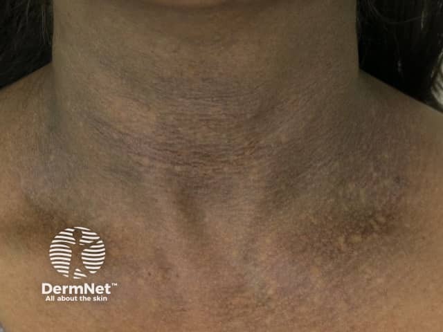
Clinical photography of the anterior neck reveals diffuse, symmetric hyperpigmentation with a thick, velvety, papillomatous texture classic for acanthosis nigricans. The plaques concentrate in neck flexures and along anterior cervical folds, extending laterally toward the upper chest. Color ranges from dark brown to slate-gray, with subtle variation between individual fold lines. The surface appears slightly creased and lichenified, but without overt ulceration, crusting, or scale. No acute erythema or edema is evident. In some regions, the pigmentation blends with adjacent normal-appearing skin, suggesting early or mild involvement. The pattern is typically bilateral and intertriginous, reflecting increased epidermal thickness and papillomatosis with dermal melanosis. Clinically, acanthosis nigricans is a cutaneous marker of insulin resistance, obesity, type 2 diabetes mellitus, endocrine disorders, or, less commonly, paraneoplastic processes. This image supports a differential that includes postinflammatory hyperpigmentation and frictional dermatitis in flexural areas, but the uniform texture and location strongly favor AN. The clinical significance lies in prompt metabolic screening and associated risk factor modification; management includes addressing underlying metabolic conditions, weight reduction, and consideration of dermatologic therapies for texture and pigment reduction. Educational utility includes teaching recognition of flexible-neck involvement and differentiation from other dermatoses in dermatology training and research.
Dark Skin on Neck, Face, and Body - What You Need to Know
What Does It Look Like?
Common Causes of Dark Skin (Especially Neck + Body)
| Cause | Key Features |
|---|---|
| Acanthosis Nigricans (most common) | Velvety, dirty-looking, dark skin in folds - neck, armpits, groin |
| Insulin resistance / Prediabetes / Diabetes | AN is a skin sign of high insulin levels |
| Obesity | Excess weight drives hyperinsulinemia |
| PCOS (in women) | Hormonal imbalance + insulin resistance |
| Thyroid disease | Can cause skin darkening |
| Sun exposure / Melasma | Affects face more than body |
| Nutritional deficiency (B12, folate) | Can cause diffuse darkening |
| Drug side effects | Glucocorticoids, nicotinic acid, estrogens |
| Rare: Malignancy | Rapid-onset AN in non-obese adults - warrants cancer workup |
What Acanthosis Nigricans Feels Like
- Skin feels velvety or rough to the touch
- Looks dirty or unwashed even after cleaning
- Most common on: back of neck, armpits, groin, inner thighs
- Can also affect: face, elbows, knees, around the belly button
- Sometimes accompanied by skin tags (acrochordons) in the armpits and groin
What You Should Do - Step by Step
- Fasting blood sugar + HbA1c (to check for diabetes)
- Fasting insulin level
- Lipid profile
- Thyroid function (TSH, T3, T4)
- For women: LH, FSH, testosterone (to rule out PCOS)
- Weight loss - even modest weight loss (5-10%) can visibly improve AN in overweight individuals
- Low-sugar, low-refined-carb diet to reduce insulin resistance
- Regular physical activity (30 min daily)
- Metformin - improves insulin sensitivity, may help the skin improve over time
- Topical treatments (used by dermatologists) - retinoids, salicylic acid, glycolic acid peels, calcipotriol, urea cream
- Laser treatment has been tried in some cases
- Gentle exfoliation (scrubbing too hard can worsen irritation)
- Moisturizing creams with urea or lactic acid can help soften the texture
- Sunscreen on the face to prevent further sun-induced darkening
Important Warning Signs - Go to the Doctor Urgently If:
- The darkening appeared very suddenly (weeks, not months)
- You are not overweight but still have AN
- You have unexplained weight loss, fatigue, or abdominal pain alongside the dark skin These could suggest an internal cancer (most commonly gastric), which is a rare but serious cause of AN.