
Which cell is responsible for immunity
White blood cells (leukocytes) are the primary cells responsible for immunity. The two main types are:
- Lymphocytes (B cells and T cells) - drive specific/adaptive immunity
- Phagocytes (neutrophils, macrophages, monocytes) - drive innate/non-specific immunity
Searching sources...
Reading File
Finding Sources
Finding Sources
Reading File
Reading File
Here is a complete, textbook-grounded answer:
Cells Responsible for Immunity
The lymphocyte is the principal cell responsible for immunity. Lymphocytes are a heterogeneous population with three major functional types, each serving a distinct role.
1. T Lymphocytes (T Cells) - Cell-Mediated Immunity
T cells are responsible for cell-mediated immunity. They are characterized by the surface markers CD2, CD3, CD5, CD7, and T-cell receptors (TCRs). They have a long lifespan and handle:
- Delayed hypersensitivity reactions
- Graft rejection and graft-versus-host reactions
- Defense against intracellular organisms (e.g., Mycobacterium tuberculosis, Brucella)
- Defense against tumor cells
- Producing cytokines: IL-2 (T cell proliferation), IL-3, IL-4, IL-5, IL-6, lymphotoxin, and migratory inhibitory factor
T cells make up the majority (~80-90%) of circulating lymphocytes and have lifespans of months to years.
2. B Lymphocytes (B Cells) - Humoral (Antibody-Mediated) Immunity
B cells are characterized by CD9, CD19, CD20 markers and surface IgM/IgD. They are responsible for humoral immunity by:
- Recognizing antigens
- Transforming into plasma cells, which produce large quantities of antibodies (immunoglobulins)
- Generating memory B cells with a long lifespan
B cells make up only 10-20% of circulating lymphocytes.
3. Natural Killer (NK) Cells - Innate Cytotoxic Immunity
NK cells are non-T, non-B lymphocytes expressing CD16a, CD56, and CD94. They:
- Are not antigen-specific - no T-cell receptor, no immunoglobulin
- Destroy virus-infected cells and some tumor cells via a cytotoxic mechanism
- Act as a rapid first-line defense (part of innate immunity)
4. Other Key Immune Cells (Innate Immunity)
Beyond lymphocytes, these cells also contribute to immunity:
| Cell | Role |
|---|---|
| Neutrophils | First responders; phagocytose bacteria |
| Macrophages | Phagocytosis, antigen presentation, cytokine secretion |
| Dendritic cells | Antigen presentation to T cells (link innate to adaptive) |
| Eosinophils | Defense against parasites, allergic reactions |
| Mast cells/Basophils | Allergic and inflammatory responses |
| Plasma cells | Derived from B cells; antibody factories |
Summary
| Cell | Immunity Type | Key Function |
|---|---|---|
| T cells | Adaptive - Cellular | Kill infected/tumor cells, regulate immune response |
| B cells / Plasma cells | Adaptive - Humoral | Produce antibodies |
| NK cells | Innate - Cytotoxic | Kill virus-infected and tumor cells without prior sensitization |
| Neutrophils/Macrophages | Innate | Phagocytosis, inflammation |
The figure below shows a large granular lymphocyte (NK/T cell type) as seen on peripheral blood smear:
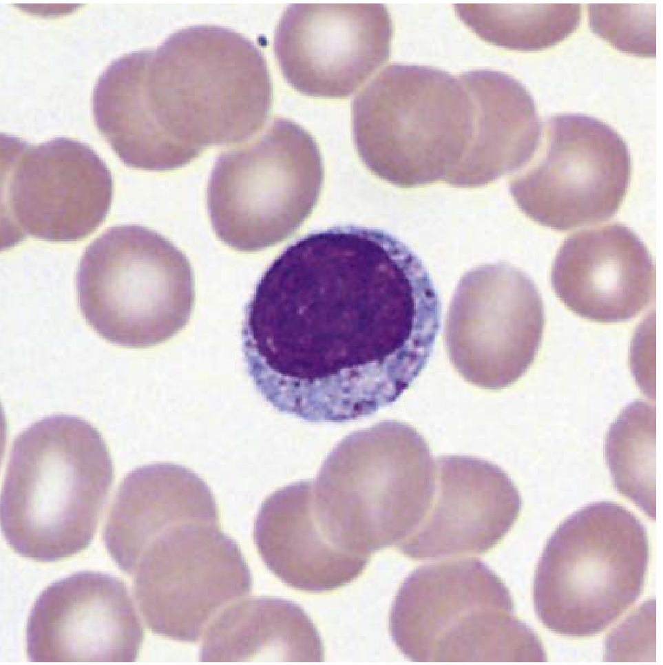
Large granular lymphocyte with azurophilic cytoplasmic granules (Wright-Giemsa, 1000x)
Sources:
- Histology: A Text and Atlas with Correlated Cell and Molecular Biology, p. 529-530
- Henry's Clinical Diagnosis and Management by Laboratory Methods, p. 2417-2426
Antigen and antibody
An antigen is any substance that triggers an immune response, while an antibody is a protein produced by B cells/plasma cells that specifically binds to that antigen to neutralize or destroy it.
Searching sources...
Finding Sources
Finding Sources
Reading File
Reading File
Finding Sources
Reading File
Reading File
Finding Sources
Finding Sources
Loading Image
Reading File
Reading File
Finding Sources
Reading File
Loading Image
Here is a complete, textbook-grounded explanation of antigens and antibodies:
Antigen and Antibody
ANTIGEN
Definition
"An antigen is any substance that can induce a specific immune response."
- Histology: A Text and Atlas, p. 1153
More specifically: an antigen is usually a large, structurally complex foreign molecule - such as a protein, polysaccharide, or glycolipid - that is recognized by cells of the immune system. It does not have to be pathogenic in origin; it simply needs to be "recognized as foreign."
Antigen vs. Immunogen vs. Hapten
| Term | Meaning |
|---|---|
| Antigen | Any substance recognized by the immune system |
| Immunogen | An antigen that can actually trigger an immune response (all immunogens are antigens, but not vice versa) |
| Hapten | A small molecule (usually <20 kDa) that cannot trigger immunity alone but can if bound to a larger carrier protein (e.g., penicillin) |
Epitopes (Antigenic Determinants)
Each antigen has multiple epitopes - specific regions/sites that are recognized by the immune system. A single antigen can contain many different epitopes, and copies of the same epitope make it multivalent, greatly increasing antibody binding strength.
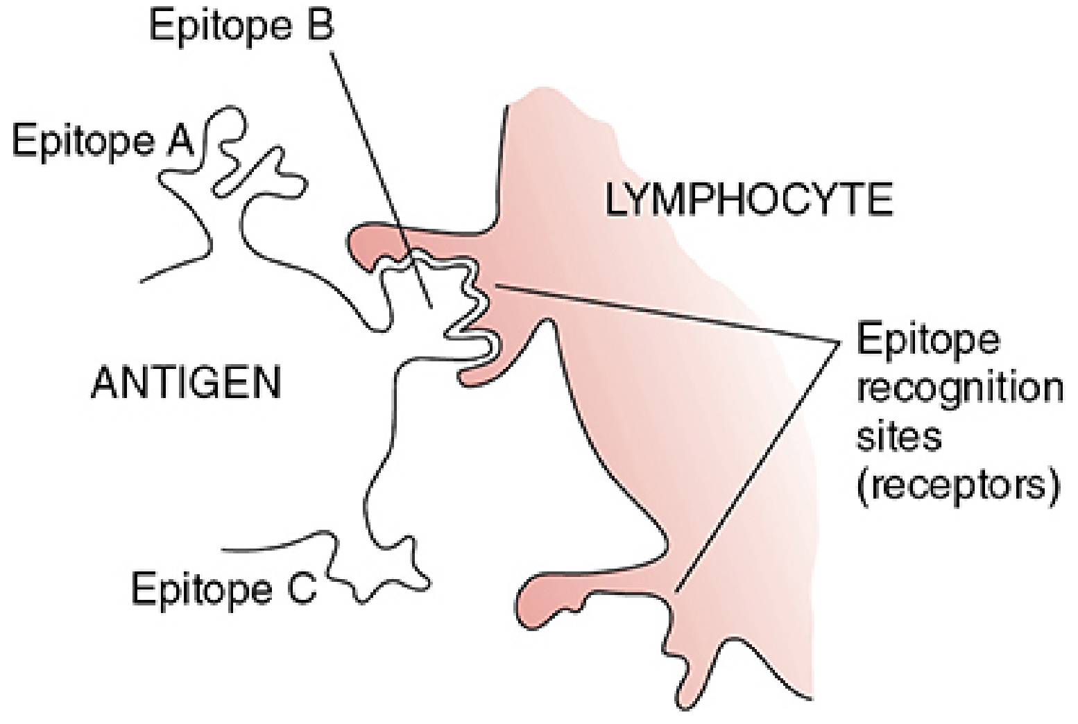
Types of Antigens
- T-dependent antigens (e.g., proteins): require T-cell help to stimulate B cells; produce high-affinity antibody + memory cells
- T-independent antigens (e.g., polysaccharides): stimulate B cells directly without T cells; produce lower-affinity antibody, no memory cells - reason why polysaccharide vaccines don't work in children under 2 years
ANTIBODY (Immunoglobulin)
Definition
Antibodies are glycoproteins produced by plasma cells (differentiated B cells) that specifically bind to the antigen that triggered their production. They belong to the immunoglobulin (Ig) family.
Structure of an Antibody (IgG model)
The basic unit is a Y-shaped tetramer consisting of:
- 2 heavy (H) chains - define the antibody class
- 2 light (L) chains - either kappa (κ) or lambda (λ) type
- Chains are connected by disulfide bonds
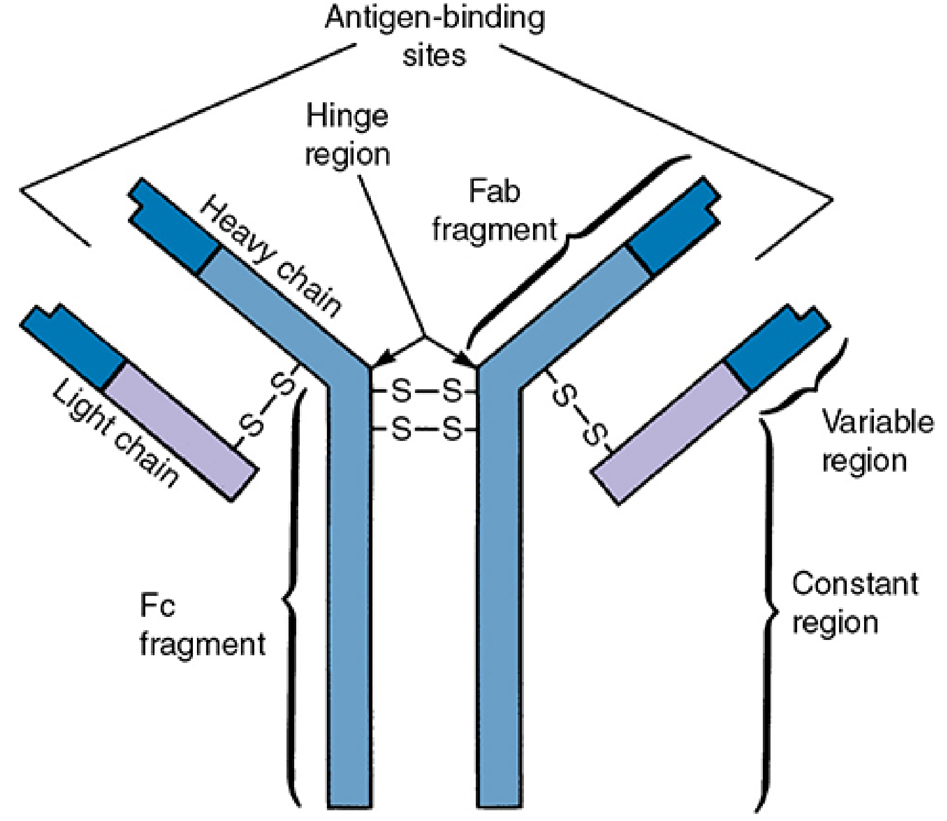
Two Functional Regions:
| Region | Fragment | Function |
|---|---|---|
| Variable region (VH/VL) | Fab - antigen-binding fragment | Binds to the specific antigen (2 identical sites per IgG) |
| Constant region (CH) | Fc - crystallizable fragment | Binds complement, interacts with macrophages/NK cells/mast cells, controls transport across placenta/mucosa |
Papain digestion yields:
- 2 Fab fragments - each with 1 antigen-binding site
- 1 Fc fragment - effector functions
The 5 Classes of Antibodies (Isotypes)
Each class is defined by its heavy chain type:
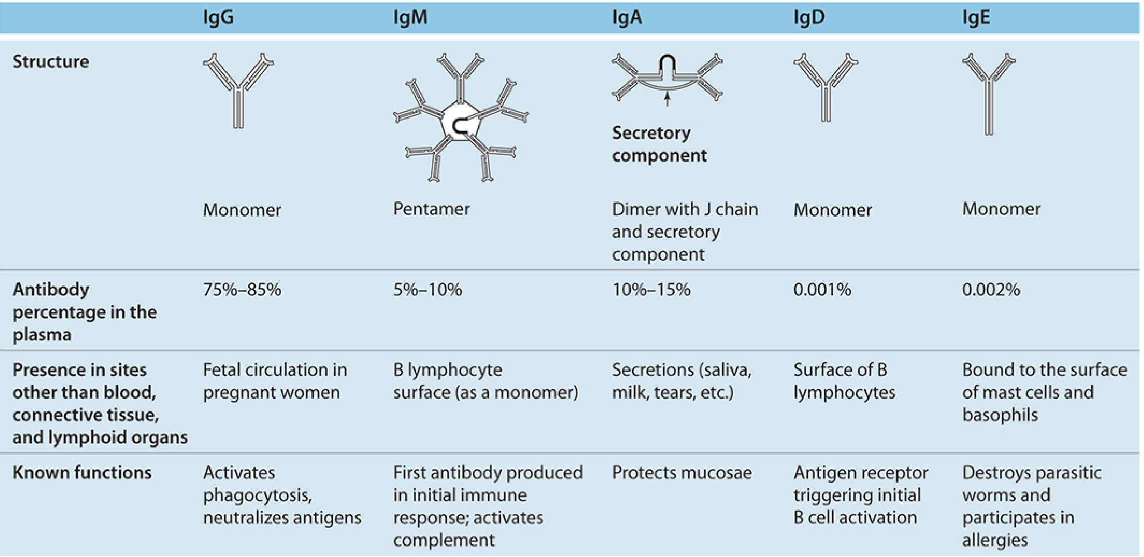
| Class | Heavy Chain | Structure | % in Blood | Key Functions |
|---|---|---|---|---|
| IgG | γ (gamma) | Monomer | 75-85% | Most abundant; neutralizes antigens; activates phagocytosis; crosses the placenta (passive immunity to fetus) |
| IgM | μ (mu) | Pentamer | 5-10% | First antibody produced in any immune response; most effective at activating complement |
| IgA | α (alpha) | Dimer (secretory) | 10-15% | Protects mucosae; found in saliva, milk, tears, mucus; secretory immunity |
| IgD | δ (delta) | Monomer | 0.001% | Acts as antigen receptor on B cell surface; triggers initial B cell activation |
| IgE | ε (epsilon) | Monomer | 0.002% | Bound to mast cells/basophils; triggers allergic reactions; defense against parasitic worms |
Antigen-Antibody Interaction
The binding between antigen and antibody is reversible and non-covalent, involving:
- Hydrophobic bonds
- Hydrogen bonds
- Van der Waals forces
- Ionic interactions
These forces only work when the antigen fits closely into the complementary binding site of the antibody - like a lock and key.
Key concepts:
- Affinity - tightness of binding at a single site (IgG has 2 sites)
- Avidity - total binding strength of all sites together; IgM (10 sites as pentamer) has far greater avidity than IgG despite similar per-site affinity
- Affinity maturation - as the immune response progresses, antibody affinity increases over time
Antigen-Antibody: Summary Comparison
| Feature | Antigen | Antibody |
|---|---|---|
| Nature | Foreign substance (protein, polysaccharide, etc.) | Glycoprotein (immunoglobulin) |
| Produced by | External source / pathogen | Plasma cells (from B lymphocytes) |
| Role | Triggers immune response | Neutralizes / eliminates antigen |
| Specific region | Epitope (on antigen) | Paratope (combining site on antibody) |
| Size | Usually large (>10 kDa for immunogens) | ~150 kDa (IgG) to ~900 kDa (IgM pentamer) |
Sources:
- Histology: A Text and Atlas with Correlated Cell and Molecular Biology, p. 1153-1154
- Sherris & Ryan's Medical Microbiology, 8th Ed., p. 77-87
- Junqueira's Basic Histology, 17th Ed., p. 671-672
- Henry's Clinical Diagnosis and Management by Laboratory Methods, p. 1087
This is a shared conversation. Sign in to Orris to start your own chat.