
A 25-year-old male presented with fever, body ache & retro-orbital pain for 5 days. On examination, skin rashes were found over the whole body, a) What is your probable diagnosis? b) How would you proceed to confirm the diagnosis in such a case in the laboratory? c) How to prevent such a condition?
dengue fever skin rash maculopapular
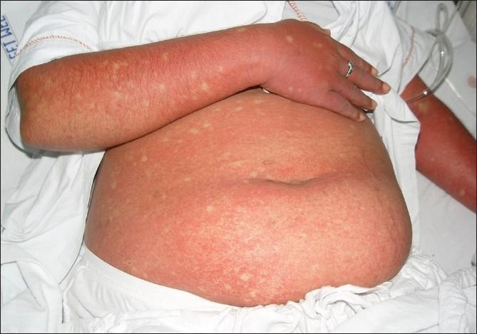
This clinical photograph displays a patient's abdomen and arm presenting a generalized, confluent erythematous rash, a characteristic cutaneous manifestation of Dengue Fever. The exanthem is marked by widespread, intense redness resulting from capillary dilatation and coalesced maculopapular lesions. A diagnostic hallmark visible here is the presence of 'islands of sparing'—small, rounded, or oval-shaped patches of normal-colored skin scattered throughout the erythematous background, often described as 'white islands in a sea of red'. These spared areas are randomly distributed across the affected trunk and limbs. The image serves as a key educational reference for recognizing the secondary rash phase of Dengue viral infection, typically occurring 3 to 6 days after the onset of fever. The visual presentation is essential for clinical diagnosis and differentiation from other viral exanthems, highlighting the specific immunologic response patterns seen in Dengue Fever (DF) and Dengue Hemorrhagic Fever (DHF).
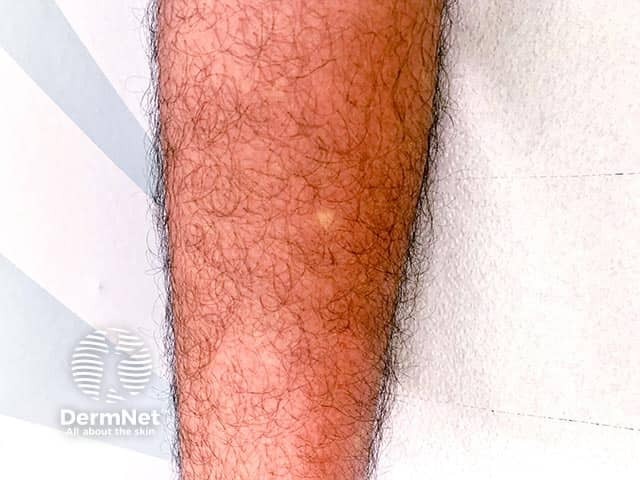
This is a high-resolution clinical photograph of the lower leg illustrating a diffuse febrile maculopapular eruption in the setting of suspected dengue fever. The image shows a broad area of coalescing erythema with patches of preserved or pale skin appearing as white islands within the red background, often described as white islands in a sea of red. The rash spans a substantial portion of the exposed leg surface and appears non-vesicular, with fine surface capillary blanching evident under light. There is no visible mucosal involvement or purpura in this view. The patient reports a 5-day history of fever, severe myalgias/body aches, and backache, compatible with the febrile phase of dengue. Morphology is consistent with a maculopapular eruption commonly seen in flavivirus infections; pattern may be confluent on extremities with interspersed hypopigmented or ischemic-looking islands. In the clinical context of dengue, such eruptions typically appear during the acute febrile period and may fade with defervescence. This image is valuable for dermatology and infectious disease education, aiding recognition of dengue-associated rash patterns, differentiation from other viral exanthems ( chikungunya, Zika ), and consideration of differential diagnoses and appropriate laboratory testing. NS1 antigen, RT-PCR, serology. It supports teaching on cutaneous manifestations of dengue.
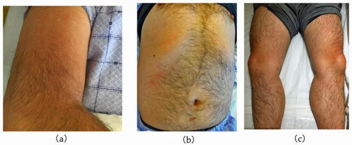
A series of three clinical photographs demonstrating cutaneous manifestations in a patient with dengue fever. (a) Right upper arm showing confluent, blanchable erythema with a diffuse distribution. (b) Anterior trunk (abdomen and chest) displaying a patchy, mottled erythematous rash interspersed with areas of normal skin tone, characteristic of the 'islands of white in a sea of red' pattern often seen in viral exanthems. (c) Lower limbs showing localized, more intense erythema focused around the knees and pretibial regions. The visual findings illustrate the secondary maculopapular rash typical of the recovery or late febrile phase of dengue virus infection. These images serve as an educational resource for identifying the clinical presentation of viral hemorrhagic fever rashes and their variable morphology across different anatomical regions, including the trunk, extremities, and periarticular areas.
Probable Diagnosis: Dengue Fever (Classical Dengue / "Breakbone Fever")
a) Probable Diagnosis
| Feature | This Patient |
|---|---|
| Acute febrile illness (5 days) | ✓ |
| Retro-orbital pain (on eye movement/pressure) | ✓ |
| Myalgia / body ache ("breakbone fever") | ✓ |
| Widespread skin rash | ✓ |
| Age group (healthy young adult) | ✓ (highest risk group) |
b) Laboratory Confirmation
Direct Virological Tests (Days 1-7, during viremic phase)
- Detects non-structural protein 1 (NS1) of dengue virus
- Positive from day 1 of fever up to day 6
- Commercial rapid kits give results in minutes; ELISA takes ~1 day
- Does not differentiate between serotypes
- Best early-phase test - can detect cases from day 1
- Detects viral RNA genome (positive days 1-7 of illness)
- High specificity; identifies serotype
- Turnaround: 1-2 days
- Specimen: acute phase serum (days 1-5)
- Gold standard but impractical - takes 1 week or more
- Specimen: acute serum/plasma, buffy coat (within first 6 days)
- Done only in reference/research labs
Serological Tests (Best after Day 5)
- IgM detectable from day 3-5 of illness onset
- 99% of patients have IgM by day 10
- IgM peaks at 2 weeks, then declines over 2-3 months
- Can cross-react with Zika and other flaviviruses
- Can detect both primary and secondary infections
- IgG persists for life
- A fourfold rise in IgG titers between acute (days 1-5) and convalescent sera (day >15) confirms recent infection
- HIA titre ≥1:1280 on single sample is supportive
- Distinguishes primary infection (high IgM:IgG ratio) from secondary infection (high IgG, lower IgM)
Haematological Parameters (Mandatory Monitoring)
- Complete Blood Count (CBC): Leukopenia (WBC ≤5000/mm³) and thrombocytopenia (platelets <150,000/mm³) strongly support dengue
- Haematocrit: Rising haematocrit (≥5-10%) suggests plasma leakage - flags progression to DHF
- Tourniquet test (Rumple-Leede test): ≥20 petechiae per 2.5 cm² is positive
Summary Table (Day of Illness)
| Days of Fever | Recommended Test | Method | Time to Result |
|---|---|---|---|
| Day 1-5 | NS1 Antigen | Rapid kit / ELISA | Minutes / 1 day |
| Day 1-7 | RT-PCR | Molecular | 1-2 days |
| Day 5+ | IgM ELISA | Serology | 1-2 days |
| Day 1-5 + Day 15-21 | IgG seroconversion | ELISA / HIA | 1-2 days |
Practical approach for this patient (day 5): Collect a single serum sample and test NS1 antigen + IgM antibody simultaneously - this combination identifies ≥90% of primary and secondary dengue cases.
- Park's Textbook of Preventive and Social Medicine, p. 292-294
- Red Book 2021, Report of the Committee on Infectious Diseases, p. 522-523
c) Prevention
1. Personal Protection (Host)
- Wear full-sleeve clothing (shirts and trousers) during peak mosquito activity hours (dawn and dusk)
- Use mosquito repellents (creams, liquids, coils, mats containing DEET or picaridin)
- Use bed nets (especially for infants and children sleeping during daytime, when Aedes is active)
- Isolate febrile patients under bed nets during the first few days of illness to prevent mosquito-to-human transmission
2. Vector Control (Aedes aegypti elimination)
- Empty, clean, or cover all water storage containers weekly ("weekly dry day")
- Remove or overturn items that collect rainwater (tyres, flower pots, coconut shells, discarded containers)
- Manage roof tops, porticos, and sunshades that collect water
- Properly cover overhead tanks and water drums
- Larviciding: Temephos (Abate) in water containers that cannot be emptied
- Adulticiding (fogging): Pyrethroid sprays during outbreaks to kill adult mosquitoes (malathion, deltamethrin)
- Introduction of Bacillus thuringiensis israelensis (Bti) - a larvicide
- Larvivorous fish (Gambusia, Poecilia) in large water bodies
3. Vaccination
- Tetravalent, live attenuated vaccine against all 4 serotypes (DENV 1-4)
- Schedule: 3 injections of 0.5 ml subcutaneously, at 6-month intervals
- Indicated for individuals 9-45 years living in dengue-endemic areas
- Recommended only for seropositive individuals (prior dengue exposure) - risk of severe disease in seronegative vaccinees
- Contraindicated in: immunocompromised individuals, HIV infection, pregnant/breastfeeding women
4. Surveillance and Public Health Measures
-
Early case notification and reporting to health authorities
-
Entomological surveillance (monitoring Aedes density)
-
Laboratory confirmation at sentinel surveillance hospitals
-
IEC (Information, Education, and Communication) campaigns for community awareness
-
Inter-sectoral coordination (urban development, education, transport)
-
Park's Textbook of Preventive and Social Medicine, p. 298-299