
Give me imp points from robbins textbook of pathology on pleomorphic adenoma
pleomorphic adenoma histology salivary gland
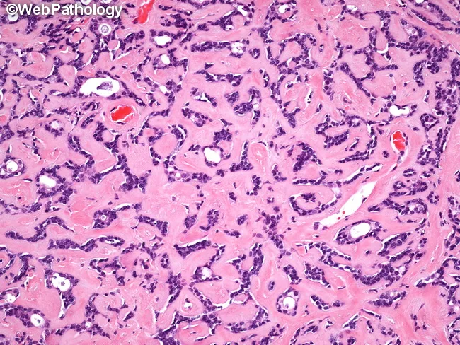
Pleomorphic adenoma of salivary gland on hematoxylin and eosin stained histology. This micrograph highlights abundant hyalinized eosinophilic matrix reminiscent of basement membrane material surrounding and intermingling with the tumor epithelium. The epithelial component forms tubular and acinar structures, composed of uniform cuboidal to columnar cells with basophilic nuclei and inconspicuous nucleoli. These duct- and acinus-like elements are variably compressed by the dense eosinophilic stroma, yielding a characteristic admixture of epithelial elements embedded within a predominantly hyalinized, fibrous background. The matrix material resembles basement membrane and may create a pseudo-cartilaginous appearance in other views, but in this field it constrains glandular architecture rather than producing well-developed cartilaginous differentiation. The overall pattern is typical of benign salivary gland neoplasia: a biphasic pattern with epithelial ductal elements and a myoepithelial/mesenchymal stroma, here dominantly hyalinized. The image is diagnostic of pleomorphic adenoma when correlated with clinical presentation (slow-growing, painless parotid mass) and radiologic assessment; however it also bears resemblance to basal cell adenoma in its basaloid epithelial proliferation. Clinically relevant features include the potential for local recurrence after incomplete excision if capsule breached. This myxoid-like matrix and tubular/acinar epithelial proliferation are pertinent for pathologic confirmation, surgical planning, and differential diagnosis in head and neck oncology.
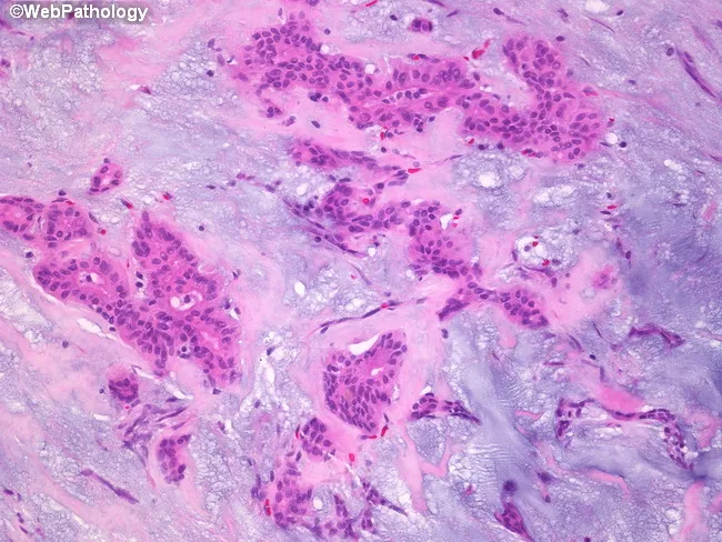
Pleomorphic adenoma histology, parotid salivary gland, examined by bright-field microscopy on Hematoxylin and Eosin stained sections. Imaging modality: Light microscopy; technique: Hematoxylin and Eosin staining. The sample reveals a classic biphasic neoplasm comprising epithelial ductal structures and myoepithelial cells embedded in an abundant stromal backdrop. Epithelial components form cords, ducts, and small island formations lined by cuboidal to columnar cells with eosinophilic cytoplasm and hyperchromatic nuclei. Intermixed myoepithelial cells display plasmacytoid or spindle morphologies, often surrounding epithelial elements. The stroma is a prominent feature, ranging from basophilic, myxoid to more eosinophilic, hyalinized areas. The myxoid matrix is rich in glycosaminoglycans and imparts a loose, gelatinous appearance, while hyalinized zones contribute dense eosinophilic bands. Overall architecture is often well circumscribed with minimal pleomorphism and low mitotic activity, consistent with benign behavior. Clinical significance includes differentiation from malignant salivary tumors; prognosis is favorable with complete surgical excision. Differential considerations include mucoepidermoid carcinoma, adenoid cystic carcinoma, basal cell adenoma, and Warthin tumor. This image is useful for educational purposes in pathology, otolaryngology, and head-and-neck surgery, illustrating quintessential pleomorphic adenoma features: epithelial and myoepithelial proliferation in a chondromyxoid stroma, with duct-like structures and a variable stromal composition. This histologic pattern aids diagnosis and education.
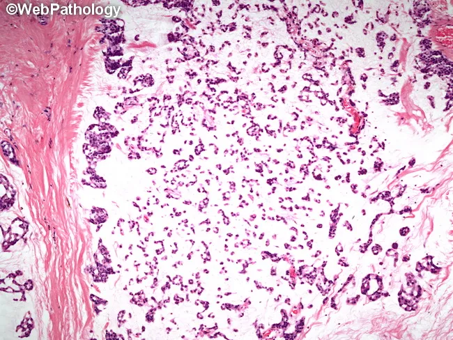
Histology image of a salivary gland tumor demonstrating classic pleomorphic adenoma (mixed tumor). Prepared as a hematoxylin and eosin stained slide and evaluated under light microscopy. The specimen represents salivary gland tissue in the parotid region. Epithelial elements are organized into small nests and rudimentary tubular structures, reflecting ductal differentiation. These epithelial components are embedded in a substantial myxoid background with scattered chondromyxoid areas. Myoepithelial cells contribute to the cellular admixture, accounting for variable cell shapes and occasional plasmacytoid appearances. The stroma ranges from loose, myxoid to more fibrous or hyaline areas, producing a heterogeneous, biphasic architecture typical of pleomorphic adenoma. The overall pattern is benign-appearing with well-demarcated margins in this field; however, the lesion is known for potential recurrence if incompletely excised. Pleomorphic adenoma is a benign salivary gland neoplasm featuring epithelial and myoepithelial components within mucoid and cartilaginous stroma. Diagnostic significance rests on recognizing the biphasic morphology and chondromyxoid matrix, which helps distinguish from malignant mimics such as mucoepidermoid carcinoma or adenoid cystic carcinoma. Clinically, these findings correlate with a slow-growing, painless parotid mass in middle-aged adults. This image is valuable for education, differential diagnosis practice, and radiology-pathology correlation in salivary gland tumors. for clinical education and research.
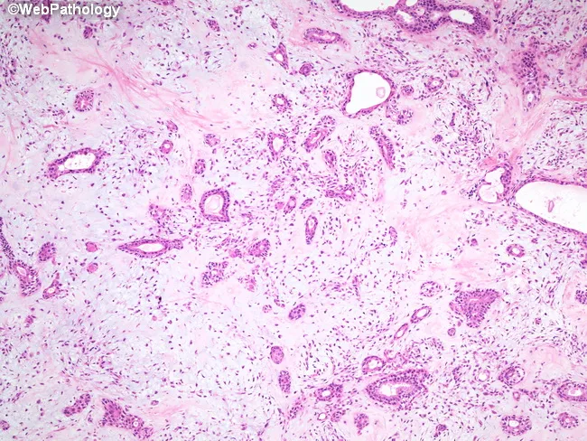
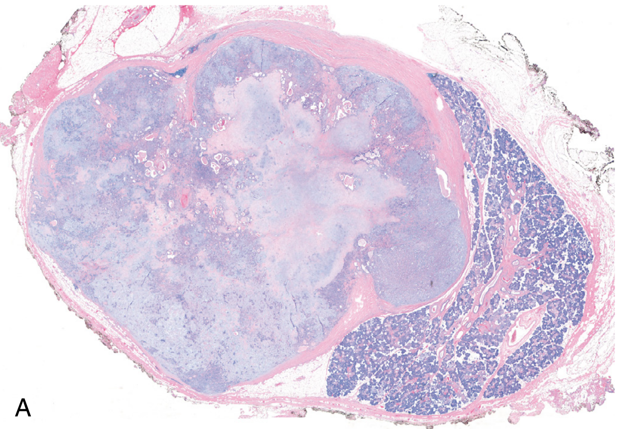
Low-power brightfield histology of a salivary gland tumor stained with hematoxylin and eosin reveals the classical pleomorphic adenoma pattern. The lesion is encapsulated within the parotid region, presenting as a benign, slow-growing mass. The epithelial component forms duct-like and acinar structures lined by a single layer of cuboidal to columnar cells, while an outer layer of neoplastic myoepithelial cells displays plasmacytoid, spindled, or stellate morphologies. The stroma is variably myxoid to chondroid, producing a heterogeneous, often cartilaginous appearance and imparting the 'pleomorphic' descriptor. Cellular atypia and mitotic activity are absent or minimal, consistent with benign behavior. The lesion demonstrates biphasic architecture with a mosaic of epithelial nests, cords, and tubules embedded in an abundant extracellular matrix rich in mucopolysaccharides and chondroid material; occasional hyalinization may be seen. Clinically, this histology corresponds to a pleomorphic adenoma, the most common benign salivary gland neoplasm, characteristically presenting in adults with a painless, mobile mass. Differential diagnoses include mucoepidermoid carcinoma, adenoid cystic carcinoma, and basal cell adenoma; however, the combination of ductal epithelial cells with myoepithelial myxochondroid stroma supports benign pleomorphic adenoma. The diagnostic significance lies in confirming a benign, surgically curable process requiring complete excision to minimize recurrence. Correlation with clinical exam advised today.
Pleomorphic Adenoma - Robbins Pathology
Overview / Definition
Epidemiology & Location
- 65-80% arise in the parotid gland
- 10% in the submandibular gland
- Remainder in minor salivary glands (including sublingual)
- The smaller the gland, the greater the proportion of malignant tumors - parotid ~15-30% malignant, sublingual ~70-90% malignant
- Occur predominantly in adults; only ~5% before age 16
Pathogenesis / Molecular Biology
- Radiation exposure increases risk
- All tumor elements (including apparently mesenchymal ones) are hypothesized to be of myoepithelial or ductal reserve cell (stem cell) origin
- Many cases have chromosomal rearrangements causing overexpression of PLAG1, a transcription factor that promotes expression of genes increasing cell growth (including growth factor receptor signaling pathways)
- HMGA2 gene mutations (encodes a DNA-binding protein) are associated with cases lacking PLAG1 overexpression
Morphology (Gross)
- Rounded, well-demarcated masses, usually <6 cm in greatest dimension
- Encapsulated (circumscribed), but capsule may be incomplete - especially on the palate
- Incomplete capsule allows expansile growth with tongue-like protrusions into surrounding tissue - leads to recurrences if merely enucleated
- Cut surface: gray-white with myxoid and blue translucent chondroid areas
Morphology (Histology)
- Epithelial elements: ductal or myoepithelial cells arranged as ducts, acini, irregular tubules, strands, or sheets
- These cells are dispersed within a mesenchymal-like background containing:
- Loose myxoid tissue
- Islands of cartilage (chondroid) - the blue translucent areas grossly
- Rarely, foci of bone
- Duct formation: cuboidal to columnar cells with an underlying layer of deeply chromatic, small myoepithelial cells
- Islands of well-differentiated squamous epithelium may be present
- No epithelial dysplasia or mitotic activity in most cases
- Tumor behaves similarly regardless of whether epithelial or mesenchymal elements predominate
Clinical Features
- Painless, slow-growing, mobile, discrete mass in the parotid or submandibular area, or buccal cavity
- May be present for months to years before seeking attention
- Not clinically reliable to differentiate benign from malignant - requires biopsy/surgical resection for definitive diagnosis
Treatment & Recurrence
| Procedure | Recurrence Rate |
|---|---|
| Simple enucleation | ~25% recurrence |
| Parotidectomy | ~4% recurrence |
Malignant Transformation - Carcinoma Ex Pleomorphic Adenoma
| Duration of Tumor | Risk of Malignant Transformation |
|---|---|
| < 5 years | ~2% |
| > 15 years | ~10% |
- Cancers are usually adenocarcinoma or undifferentiated carcinoma
- Highly infiltrative - may completely replace the precursor lesion (making the diagnosis difficult)
- Among the most aggressive salivary gland neoplasms
- Mortality: 30-50% at 5 years
Key Points to Remember (Summary Table)
| Feature | Detail |
|---|---|
| Most common salivary gland tumor | Yes - 60% of parotid tumors |
| Cell types | Ductal epithelial + myoepithelial + mesenchymal |
| Alternate name | Mixed tumor |
| Key gene | PLAG1 (overexpressed), HMGA2 (mutated) |
| Gross | Encapsulated, gray-white, myxoid + chondroid areas |
| Histology hallmark | Morphologic heterogeneity (NOT nuclear pleomorphism) |
| Cartilage in tumor | Yes - chondroid areas are characteristic |
| Mitoses/dysplasia | Absent in benign form |
| Recurrence after enucleation | ~25% |
| Recurrence after parotidectomy | ~4% |
| Malignant transformation | 2% (<5 yrs) → 10% (>15 yrs) |
| Malignant type | Carcinoma ex pleomorphic adenoma |