
Make 200 most probable High yeild que for neetpg 2026 , also analyse last 5 year que , new updates and must conclude recall based image based que of last 5 years
Which subject(s) should these 200 high-yield NEET PG 2026 questions cover? · What format do you want the output in?
NEET PG 2024 2025 2026 high yield topics recalled questions analysis all subjects
NEET PG 2025 image based questions recalled anatomy pathology radiology
https://www.medexamsprep.com/blog/neet-pg-2025-paper-analysi…
NEET PG 2026 - 200 Most Probable High-Yield Questions
Based on: Last 5-Year PYQ Analysis (2022-2026) + Recent Updates + Recall-Based Image Questions
TREND ANALYSIS: NEET PG 2022-2026
| Subject | 2025 Qs | Weightage | Trend |
|---|---|---|---|
| Medicine (incl. Derm, Psych) | 30 | 15% | Highest - clinical vignettes |
| OBGYN | 20 | 10% | Very high - PPH, ANC, fertility |
| Surgery | 18 | 9% | High - trauma, GI, oncology |
| PSM | 16 | 8% | Consistent - programs, statistics |
| Pharmacology | 15 | 7.5% | Rising - clinical drug use |
| Biochemistry | 15 | 7.5% | Rising - metabolic integration |
| Pathology | 12 | 6% | Stable - IHC, chromosomal |
| Microbiology | 12 | 6% | Stable - clinical context |
| Anatomy | 9 | 4.5% | Moderate - radiological focus |
| Dermatology | 8 | 4% | Rising - image-heavy |
| Forensic Medicine | 6 | 3% | Stable |
| Orthopaedics | 5 | 2.5% | Stable |
| Ophthalmology | 5 | 2.5% | Stable |
| ENT | 5 | 2.5% | Stable |
| Paediatrics | 5 | 2.5% | Stable |
| Radiology | 5 | 2.5% | Rising |
| Physiology | 5 | 2.5% | Integrated |
| Anaesthesia | 4 | 2% | Stable |
- ~50% image-based (doubled from 2022)
- ~70% clinical vignette format
- ~30% repeats from PYQs
- Basic sciences now mostly integrated into clinical scenarios
- Drug of choice tested 15-20 times per paper
PART 1 - 200 HIGH-YIELD MCQs WITH ANSWERS
ANATOMY (9 Questions)
PHYSIOLOGY (5 Questions)
BIOCHEMISTRY (15 Questions)
PHARMACOLOGY (15 Questions)
PATHOLOGY (12 Questions)
MICROBIOLOGY (12 Questions)
GENERAL MEDICINE (30 Questions)
OBSTETRICS & GYNAECOLOGY (20 Questions)
SURGERY (18 Questions)
PSM / COMMUNITY MEDICINE (16 Questions)
PAEDIATRICS (5 Questions)
ORTHOPAEDICS (5 Questions)
OPHTHALMOLOGY (5 Questions)
ENT (5 Questions)
DERMATOLOGY (8 Questions)
FORENSIC MEDICINE (6 Questions)
RADIOLOGY (5 Questions)
ANAESTHESIA (4 Questions)
ADDITIONAL HIGH-YIELD CLINICAL INTEGRATORS (4 Questions completing 200)
| Feature | Bacterial | Viral | TB |
|---|---|---|---|
| Appearance | Turbid/purulent | Clear | Cobweb clot |
| Cells | PMN (100-10,000) | Lymphocytes | Lymphocytes |
| Protein | Very high (>100) | Normal/slight ↑ | High (100-500) |
| Glucose | Very low (<45) | Normal | Low (very) |
| Opening pressure | High | Normal/mild ↑ | Elevated |
PART 2 - LAST 5-YEAR RECALLED IMAGE-BASED QUESTIONS (2022-2026)
ANATOMY - Image Recalls
SURGERY - Image Recalls
PATHOLOGY - Image Recalls
MICROBIOLOGY - Image Recalls
MEDICINE / CARDIOLOGY - Image Recalls
DERMATOLOGY - Image Recalls
RADIOLOGY - Image Recalls
QUICK REVISION: 2025-2026 NEW UPDATES TO KNOW
SUBJECT-WISE PRIORITY FOR NEET PG 2026
| Priority | Subject | Focus |
|---|---|---|
| ★★★★★ | Medicine | Clinical vignettes, ECGs, emergencies, rheumatology |
| ★★★★★ | OBGYN | Obstetric emergencies, infertility, gynecological malignancies |
| ★★★★★ | Surgery | Trauma, GI surgery, oncology, hernia classification |
| ★★★★ | PSM | Biostatistics, national programs, epidemiology |
| ★★★★ | Pharmacology | Drug of choice, drug interactions, newer drugs |
| ★★★★ | Pathology | IHC, chromosomal translocations, histology images |
| ★★★★ | Microbiology | Clinical context, special stains, serology |
| ★★★ | Biochemistry | Enzyme deficiencies, metabolic pathways |
| ★★★ | Dermatology | Image-heavy, bullous diseases, photosensitivity |
| ★★★ | Radiology | X-ray signs, CT patterns, named signs |
| ★★ | Anatomy | Neuroanatomy, radiological, embryology |
| ★★ | Forensic | Medico-legal, thanatology basics |
| ★★ | Ortho | X-ray diagnosis, named conditions |
| ★★ | Ophthalmology | Retinal conditions, glaucoma, congenital |
| ★★ | ENT | Audiometry, ear conditions, neck masses |
| ★★ | Paediatrics | Growth milestones, congenital conditions |
| ★ | Physiology | Integrated into other subjects |
| ★ | Anaesthesia | Specific drugs, monitoring, complications |
Image based que with ans from last 5 year, Recall based
NEET PG 2021 2022 2023 2024 2025 recall image based questions with answers all subjects
Reed Sternberg cell owl eye histology Hodgkin lymphoma
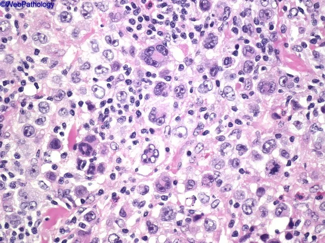
This high-power light micrograph of a lymph node biopsy, stained with Hematoxylin and Eosin, demonstrates classical Hodgkin lymphoma histology. The tissue is densely cellular with a polymorphic inflammatory background comprising small lymphocytes, eosinophils, plasma cells, and histiocytes. Scattered among the background are large binucleated Reed-Sternberg (RS) cells exhibiting prominent, mirror-image eosinophilic nucleoli and ample cytoplasm, giving an owl's eye appearance. Mononuclear RS variants with similar nuclear features are present, reflecting heterogeneity within the malignant cell population. The RS cells may appear with delicate nuclear membranes and occasional lobulation. The surrounding milieu often includes reactive T-lymphocytes and occasional eosinophils attracted by cytokines produced by RS cells. The overall architecture lacks well-formed nodules, and the background shows a mixed inflammatory infiltrate, characteristic of classical Hodgkin lymphoma subtypes such as mixed cellularity. Important differential considerations include infectious or benign reactive processes, but the presence of RS cells with classic morphology supports a diagnosis of classical HL. Clinically, these findings correlate with nodal enlargement and B symptoms in many patients, and histology guides staging and therapy decisions, including chemotherapeutic regimens such as ABVD and potential radiotherapy in select cases. Correlation with immunohistochemistry and clinical data improves diagnostic confidence and treatment planning for patient care.
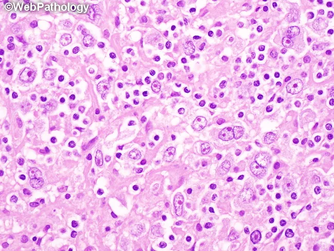
This histopathology image depicts a hematoxylin and eosin (H&E) stained lymph node biopsy section examined under bright-field light microscopy. The predominant nodular sclerosis pattern features fibrous bands dividing the paracortex into discrete nodules. Within the nodules, numerous Reed-Sternberg (RS) cells and Hodgkin cells are evident; RS cells frequently display binucleation with prominent eosinophilic nucleoli and an 'owl's eye' appearance. A lacunar variant of RS cells may be present, characterized by abundant pale cytoplasm and retraction artifacts in formalin-fixed tissue. Background inflammatory cells are relatively sparse but include small lymphocytes and occasional eosinophils; overall lymphoid architecture is disrupted by collagenous septa. The appearance is characteristic of classic Hodgkin lymphoma, nodular sclerosis subtype, which often presents with mediastinal or nodal involvement. Diagnostic significance lies in identifying RS/Hodgkin cells amid a fibrotic nodular framework, supporting a diagnosis of cHL and guiding therapy decisions. Clinically, this image is relevant for educational purposes, hematopathology training, differential diagnosis discussions with reactive lymphadenopathy, NLPHL distinctions, and confirmation of HL subtypes. Potential diagnoses to consider include nodular sclerosis cHL versus NLPHL, other B-cell lymphomas; immunophenotypic studies (CD30+, CD15+, PAX5 weak) and staging (PET-CT) commonly accompany histology for treatment planning.
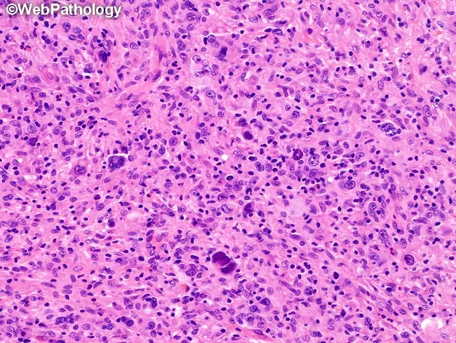
Histopathology image: Lymph node biopsy stained with Hematoxylin and Eosin (H&E) examined under light microscopy, high magnification. The sample displays lymphoreticular tissue composed of a dense inflammatory background with numerous small lymphocytes and occasional histiocytes. Scattered large atypical cells with prominent nucleoli are identifiable, consistent with Reed–Sternberg (RS) cells and their variants. In this Grade II presentation of nodular sclerosis classical Hodgkin lymphoma (cHL), numerous, bizarre, anaplastic RS cells are seen within a milieu that lacks substantial lymphocyte depletion. The architectural pattern may include nodularity interspersed by fibrous bands, a characteristic of nodular sclerosis, although the image emphasizes individual RS cells and a reactive infiltrate. The RS cells resemble owl-eye nucleoli within a large cytoplasm; immunophenotype typically CD30+, CD15+, CD20−/variable, PAX5 weak, EBV status variable. The observed features correspond to a higher-grade cHL subset, yet grading is primarily a research construct and not essential for routine clinical management. Clinically, this histology correlates with aggressive behavior in some nodal HL cases but usually responds to standard chemotherapy regimens. The image is useful for education about HL morphology, RS cell pleomorphism, differential diagnosis (anaplastic large cell lymphoma, NLPHL, reactive lymphoid hyperplasia) and research correlations in HL grading strategies.
spondylolisthesis lumbar spine X-ray anterior vertebral slip
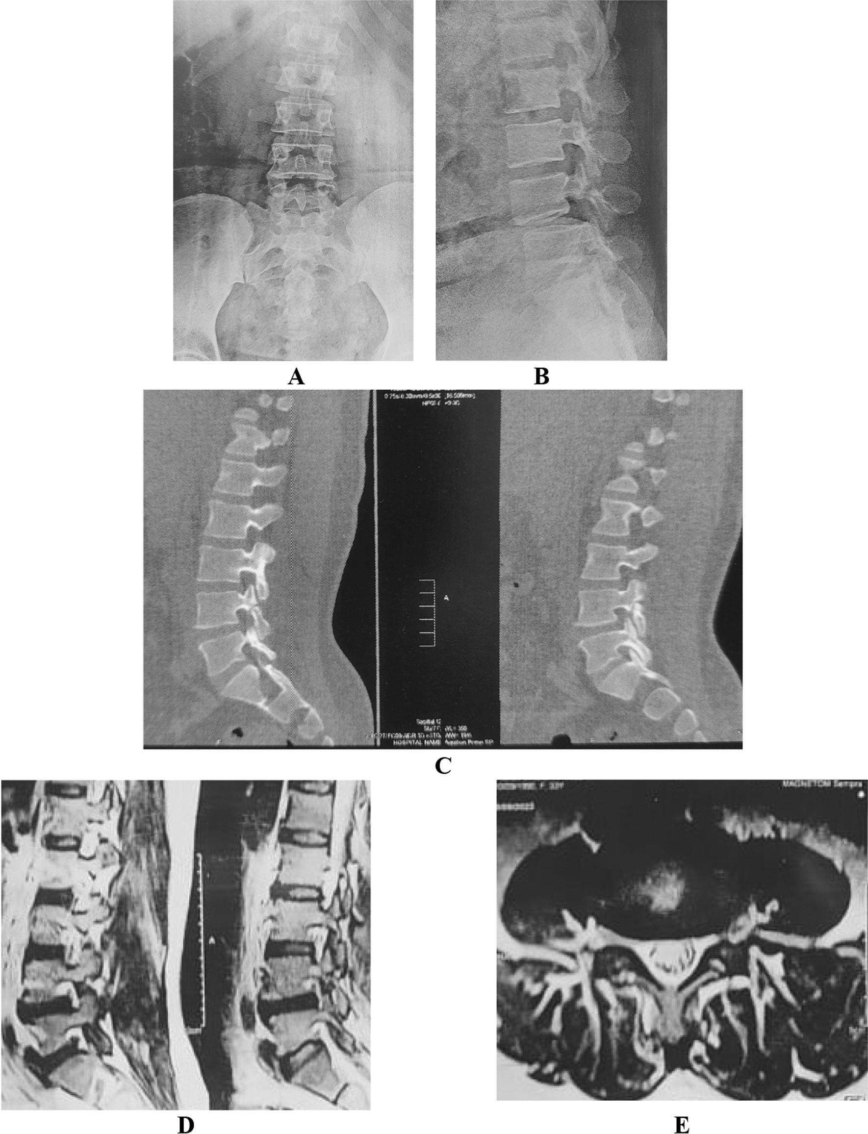
Multi-modal diagnostic imaging of the lumbar spine showcasing a case of lytic spondylolisthesis scheduled for MIS-TLIF. Panel A (Anterior-Posterior X-ray) and B (Lateral X-ray) demonstrate lumbar spinal alignment with a visible grade I or II anterolisthesis at the L4-L5 level. Panel C consists of sagittal CT reconstructions highlighting a fractured pars interarticularis (spondylolysis) at the affected level, contributing to the instability. Panel D (Sagittal MRI) and E (Axial MRI) provide soft tissue detail, showing the relationship between the slipped vertebral body, the intervertebral disc, and the neural elements within the spinal canal. The MRI axial cut (E) specifically illustrates the status of the spinal canal and foraminal space at the level of the slip. Collectively, these images represent the preoperative workup for a minimally invasive transforaminal lumbar interbody fusion (MIS-TLIF) to address spondylolytic spondylolisthesis.
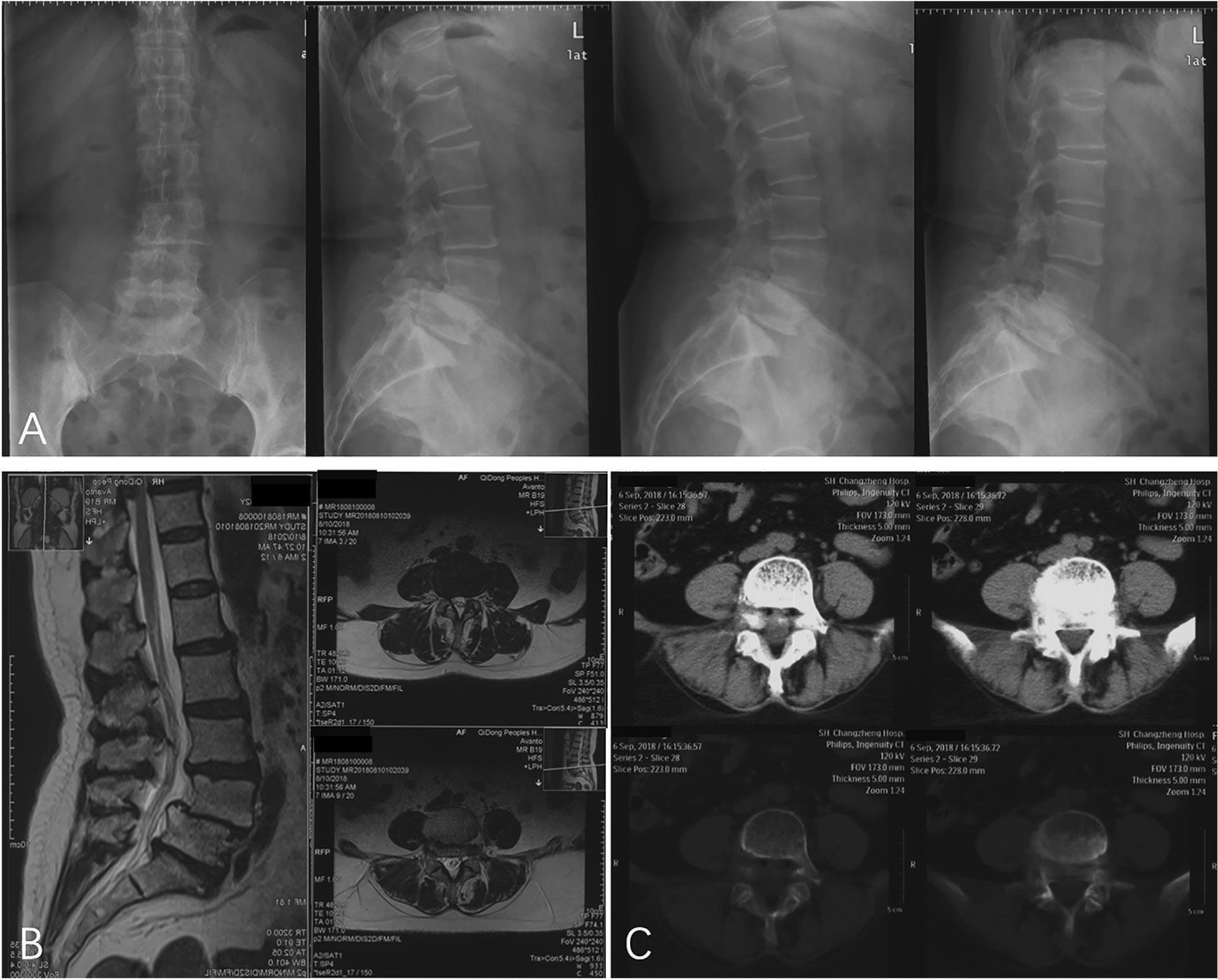
This composite figure displays multi-modal preoperative diagnostic imaging of the lumbar spine, focusing on the L4-L5 segment. Panel A consists of plain radiographs in anterior-posterior and lateral views (neutral, flexion, and extension), demonstrating Grade I degenerative anterolisthesis of the L4 vertebral body relative to L5. Key features include vertebral malalignment, intervertebral disc space narrowing, and osteophyte formation. Panel B presents sagittal and axial T2-weighted MRI scans, highlighting soft tissue pathologies such as segmental spinal canal stenosis and compression of the spinal cord and nerve roots associated with the slip. Panel C shows axial CT scans in bone and soft tissue windows, providing detailed visualization of the vertebral bodies and facet joints while confirming the absence of isthmic fractures, classifying the condition as degenerative rather than spondylolytic spondylolisthesis. This comparative imaging set illustrates the respective strengths of X-ray for alignment, MRI for neural elements and soft tissue, and CT for cortical bone integrity in the evaluation of lumbar degenerative diseases.
aspergillus septate hyphae acute angle branching histopathology
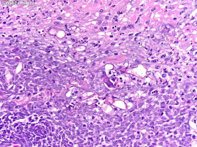
High-magnification light microscopy of a necrotic tissue biopsy stained with hematoxylin and eosin (H&E) reveals broad, nonseptate fungal hyphae with irregular, often wide-angle branching consistent with Mucorales (mucormycosis). The specimen shows extensive focal necrosis with loss of parenchymal architecture, irregular stromal disruption, and sparse neutrophilic and lymphohistiocytic inflammatory infiltrate. Within necrotic zones, thick-walled hyphae measuring approximately 5–20 μm in diameter traverse the tissue, displaying paucity of septa and occasional right-angle or variable-angle branching. The background demonstrates vascular congestion and occasional hyaline fungal elements; occasional sporangia are not clearly visible. The appearance is highly suggestive of invasive zygomycosis in an immunocompromised host or poorly controlled diabetes. Clinically important features include the need for rapid antifungal therapy (liposomal amphotericin B or amphotericin B), urgent surgical debridement, and correction of underlying acidosis or immunosuppression. Differential diagnosis includes Aspergillus species (septate hyphae with acute-angle branching), Fusarium, and other mould infections; however, septation and branching patterns help distinguish. The observation supports a diagnosis of fungal invasion in necrotic tissue, correlating with systemic signs of infection. In microbiology, culture and molecular assays can confirm genus. Utility: educational reference for histopathology, diagnostic pathology, and fungal infection management.
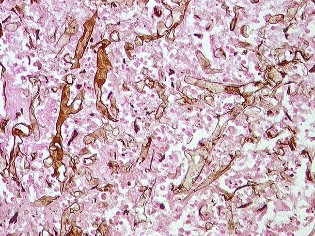
Imaging modality with technique details: Grocott methenamine silver stain applied to tissue biopsy. Light microscopy. The image shows a granulating necrotic focus with scattered, irregular thick fungal hyphae visible against a pale background. Hyphae are broad and variably wide, with intermittent septation and irregular, snake-like contours. The stain accentuates fungal cell walls in dark pigment (black/brown) on the eosinophilic necrotic tissue and inflammatory debris. The surrounding tissue demonstrates necrosis with sparse inflammatory cells. The morphological pattern is compatible with invasive molds; presence of hyphae within necrotic debris suggests tissue invasion. Distinguishing features include broad, nonseptate or pauci-septate hyphae with wide branching, a classic clue for Mucorales (mucormycosis); in contrast, Aspergillus would typically show narrow, septate hyphae with acute angle branching. Clinical correlation is essential given risk factors such as diabetes, neutropenia, or immunosuppression. The diagnostic significance lies in early recognition of invasive fungal infection to guide prompt antifungal therapy liposomal amphotericin B and surgical debridement where appropriate. Potential differential diagnoses include mucormycosis, aspergillosis, and other zygomycetes; PCR and culture should be performed for species-level identification. This image is a valuable educational resource for histopathology, infectious disease, and pathology training; courtesy of Dr. Katharina Glatz, University of Basel, Switzerland. Thank you.
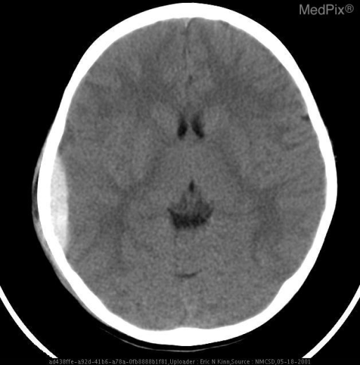
epidural hematoma biconvex lenticular CT brain
Tetralogy of Fallot boot shaped heart X-ray coeur en sabot
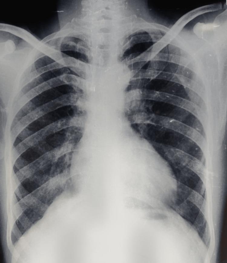
A posteroanterior (PA) chest X-ray of an adult male demonstrating classic radiological features associated with Tetralogy of Fallot (TOF). The cardiac silhouette exhibits mild cardiomegaly and a characteristic 'boot-shaped' heart (coeur en sabot). This morphology is defined by an upturned, rounded cardiac apex, indicating right ventricular hypertrophy, and a concave pulmonary artery segment (pulmonary conus). The lung fields appear relatively oligemic, reflecting decreased pulmonary blood flow typical of right-to-left shunting or pulmonary stenosis. No acute pulmonary infiltrates or pleural effusions are visible. The costophrenic angles are clear. This diagnostic image is intended for medical students and clinicians to identify visual markers of cyanotic congenital heart disease and right ventricular pressure overload in an adult clinical presentation.
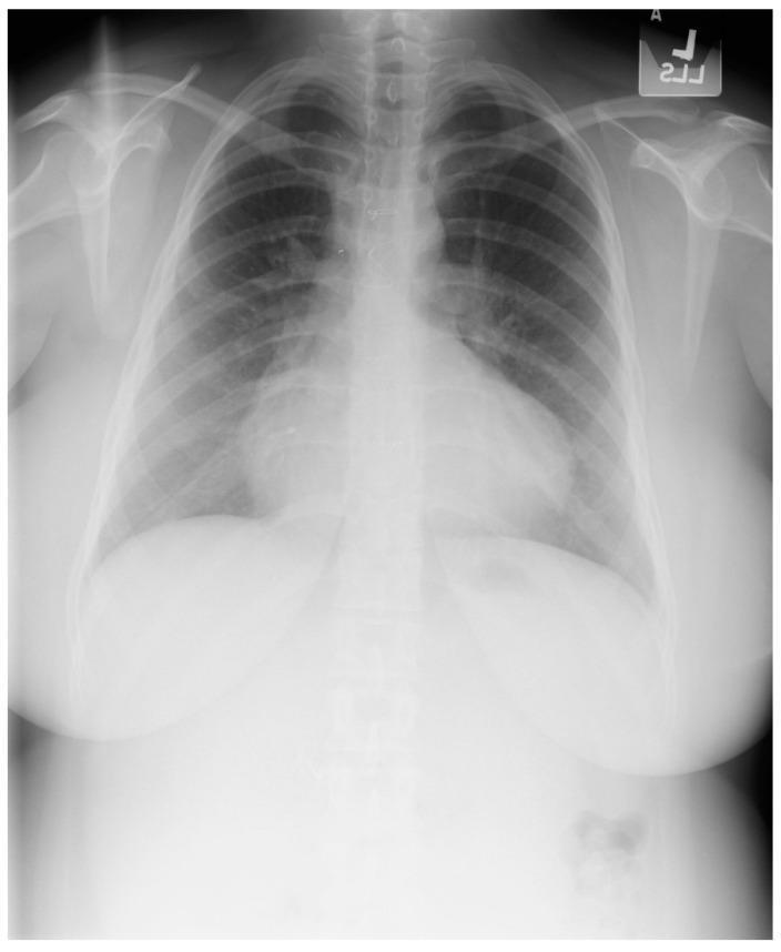
This diagnostic posteroanterior (PA) chest X-ray demonstrates the characteristic findings of Tetralogy of Fallot in a 21-year-old female patient. The image shows a well-centered view with adequate inspiratory effort. The primary finding is an increased cardiothoracic ratio with a distinctive 'boot-shaped' heart, also known as 'coeur en sabot.' This morphology is characterized by an upturned cardiac apex, resulting from significant right ventricular hypertrophy, and a concave pulmonary artery segment. The right heart border is notably prominent, indicating right atrial enlargement. Despite the history of surgical repair (including VSD closure and RV outflow tract reconstruction), these structural changes remain visible. The pulmonary vasculature appears normal, and the lung fields are clear without evidence of opacities, focal lesions, or pleural effusions. Sternal wires and surgical markers from prior cardiac procedures may be visible in the midline. The image serves as a classic educational example of chronic cardiac remodeling in congenital heart disease.
Congo red amyloid apple green birefringence polarized light
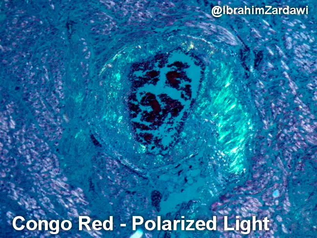
Imaging modality and technique: Light microscopy of Congo red-stained cardiac tissue examined under polarized light. Primary subject: Cardiac amyloidosis with amyloid deposition in the vascular wall. Specimen type: Cardiac tissue biopsy specimen. Imaging perspective: Polarized-light evaluation of Congo red–stained sections. Visual features: Irregular, eosinophilic, amorphous extracellular deposits confined to small- to mid-sized vessel walls within the myocardium; deposits appear as dense, salmon-pink material on routine brightfield and exhibit characteristic apple-green birefringence under polarized illumination. Diagnostic significance: Congo red positivity with apple-green birefringence confirms amyloid deposition in heart tissue, supporting a diagnosis of systemic amyloidosis with cardiac involvement. This finding is clinically relevant because myocardial amyloid deposition can cause restrictive cardiomyopathy with diastolic dysfunction and heart failure; identification guides systemic workup for AL (light-chain) vs ATTR (transthyretin) etiologies, and informs prognosis and treatment strategies. Differential considerations: Non-amyloid eosinophilic deposits or pigmentations would lack Congo red positivity and birefringence; however, the combination of Congo red staining and birefringence is consistent with amyloidosis. Clinical correlation: Correlate with echocardiography, cardiac MRI, serum free light chains, immunohistochemistry, and genetic testing as indicated.
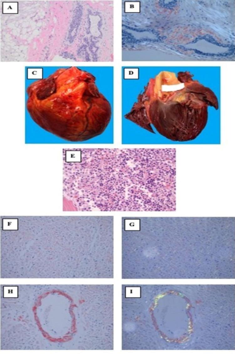
A multi-panel figure illustrating diagnostic findings of systemic AL amyloidosis through microscopy and gross pathology. Panels A and B show a breast biopsy; Congo red staining (A) reveals pink amorphous stromal amyloid deposits, which demonstrate characteristic apple-green birefringence under polarized light (B). Panels C and D are gross photographs of an autopsied heart showing significant cardiomegaly and a thickened left ventricular myocardium, indicative of restrictive cardiomyopathy. Panel E displays a bone marrow microscopic section with hypercellularity and an abnormal plasma cell infiltrate. Panels F through I show myocardial autopsy sections; Congo red stain identifies widespread peri-myocytic (F) and vascular (H) pink amyloid deposits. Corresponding polarized light images (G, I) confirm these as amyloid through apple-green birefringence within the cardiac interstitium and vessel walls. This composite figure serves as an educational resource for identifying multisystemic amyloid infiltration and the underlying plasma cell dyscrasia.
psoriasis silvery white plaques extensor elbow skin clinical
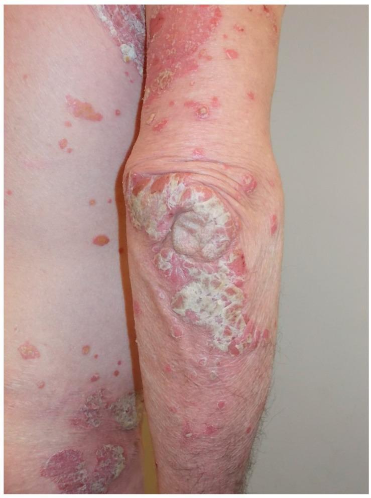
This clinical photograph displays a lateral view of a patient's upper extremity and torso demonstrating classic features of plaque psoriasis. The focal point is the extensor surface of the elbow, which exhibits large, confluent, well-demarcated erythematous plaques. These plaques are characterized by a thick, micaceous, silvery-white scale, which is most prominent on the joint surface. Scattered across the surrounding forearm, upper arm, and lateral trunk are numerous smaller, discrete erythematous papules and plaques of varying sizes, representing a guttate or eruptive pattern. Some of these peripheral lesions show early scale formation, while others appear as simple erythematous macules. The distribution follows a typical pattern for chronic plaque-type psoriasis, emphasizing involvement of extensor surfaces. This image is an educational tool for dermatology, illustrating primary skin lesions including plaques, papules, and scales, and highlighting the hallmark morphological features used in the clinical diagnosis of psoriatic disease.
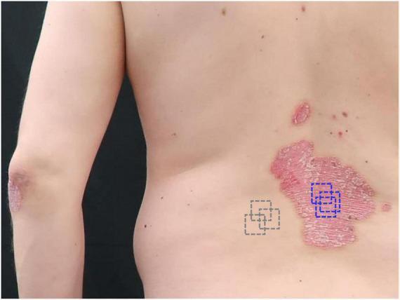
A clinical photograph of a 45-year-old male patient demonstrating moderate-to-severe plaque psoriasis on the lower back and elbow. The image shows a large, well-demarcated, erythematous plaque on the trunk characterized by thick, silvery-white micaceous scaling and a dry, fissured surface. Smaller satellite plaques are visible superior and lateral to the primary lesion. A similar thick, scaly plaque is noted on the extensor surface of the left elbow. The image includes annotations for clinical research monitoring: blue dashed squares delineate the target lesional site on the primary plaque for optical coherence tomography (OCT) scanning, while gray dashed squares indicate a perilesional control site on normal-appearing skin. This visual demonstrates the classic morphology of psoriasis vulgaris used for long-term therapeutic monitoring, with a reported PASI score of 10.8 and BSA of 10%.
intussusception target doughnut sign ultrasound pediatric
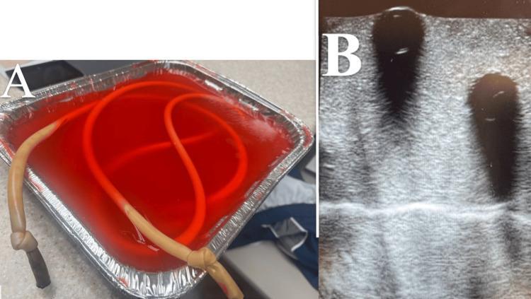
This composite image illustrates the development of a low-cost, high-fidelity ultrasound training phantom for pediatric intussusception. Panel A shows a clinical photograph of a benchtop model consisting of silicone tubing submerged in a red-tinted gelatin and psyllium fiber matrix within a foil container. The tubing is coiled and invaginated to simulate loops of bowel and intussusception pathology. Panel B displays a diagnostic ultrasound image obtained from this phantom. The ultrasound reveals significant acoustic shadowing—characterized by dark vertical bands—emanating from the simulated bowel loops. This shadowing is an artifact likely caused by trapped air or dense material within the tubing, which prevents clear visualization of the classic 'target sign' or 'doughnut sign' typically sought in diagnostic imaging for intussusception. The model serves as an educational tool for emergency medicine and pediatric residents to practice bedside ultrasound techniques and recognize imaging pitfalls such as bowel gas artifacts.
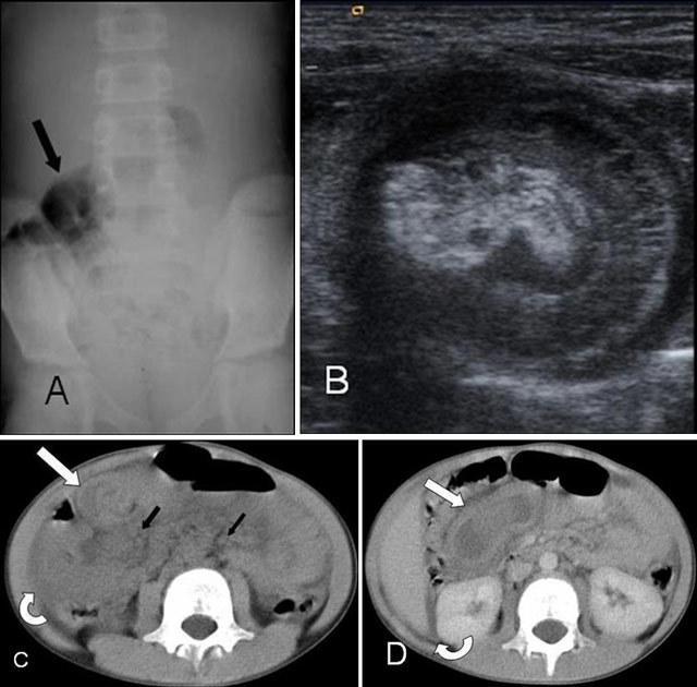
Multi-modal clinical imaging of a nine-year-old pediatric patient with Henoch-Schönlein purpura (HSP) and ileoileal intussusception. Panel A (Plain Abdominal X-ray) shows focal bowel distention with gas in the right lower quadrant (black arrow). Panel B (Abdominal Ultrasound) demonstrates the classic 'target sign' or 'doughnut sign' in cross-section, indicative of intestinal invagination. Panels C and D (Axial CT, non-contrast and contrast-enhanced) reveal the site of intussusception with thickened intestinal walls forming a target sign (white arrow). Associated findings include enlarged para-aortic lymph nodes (black arrows) and free peritoneal fluid (curved arrow) around the liver and intestinal loops. These images illustrate the diagnostic workup for acute abdominal pain in children with systemic vasculitis, highlighting the transition from non-specific radiography to definitive cross-sectional imaging (US and CT) for identifying mechanical complications like intussusception and associated lymphadenopathy.
atrial flutter ECG sawtooth flutter waves rate 150
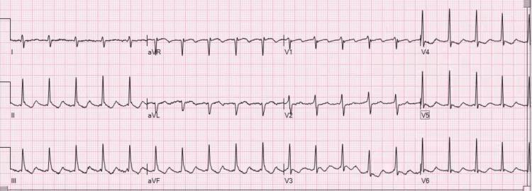
Diagnostic Image: This 12-lead electrocardiogram (ECG) demonstrates atrial flutter with a 2:1 atrioventricular (AV) conduction ratio. The tracing shows highly regular atrial activity with a characteristic sawtooth morphology. In the inferior leads (II, III, and aVF), the flutter waves are distinctly positive (upright), while in lead V1, the flutter waves exhibit a negative (inverted) polarity. This specific polarity pattern—upright flutter waves in the inferior leads and inverted in V1—is diagnostic of clockwise typical atrial flutter (atypical by counter-clockwise standards). The QRS complexes are regular and narrow, occurring at approximately half the frequency of the atrial rate (atrial rate ~300 bpm; ventricular rate ~150 bpm). In the precordial leads V4-V6, the QRS and T-wave morphologies are preserved, although discrete P waves are replaced by the undulating flutter baseline. The image illustrates a classic supraventricular tachycardia frequently encountered in cardiology and emergency medicine, serving as an educational example of macro-reentrant atrial rhythms.
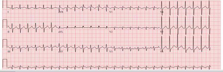
A 12-lead electrocardiogram (ECG) recorded at a standard paper speed of 25 mm/s, demonstrating a narrow complex supraventricular tachycardia. The primary finding is atrial flutter with a 2:1 atrioventricular (AV) conduction ratio, resulting in a ventricular rate of approximately 150 beats per minute. Distinctive 'sawtooth' flutter waves (F-waves) are most prominent in the inferior leads (II, III, and aVF), characterized by negative deflections without an intervening isoelectric baseline. In the precordial leads V1-V3, the flutter waves appear more upright or biphasic. The QRS complexes are regular and maintain a consistent morphology and narrow duration, indicating normal ventricular depolarization following the 2:1 conduction from the atria. This tracing is a classic educational example used to distinguish atrial flutter from sinus tachycardia or atrial fibrillation in the context of regular narrow-complex tachycardia.
NEET PG 2021-2025: RECALL-BASED IMAGE QUESTIONS WITH ANSWERS
PATHOLOGY IMAGE QUESTIONS
IMG-PATH-01 | NEET PG 2023 & 2025 (Repeated Concept)
- Langhans giant cells
- Reed-Sternberg cells
- Touton giant cells
- Aschoff cells
- Diagnosis = Classical Hodgkin Lymphoma
- Immunohistochemistry: CD15+, CD30+, PAX5 (weak), CD20-
- Most common subtype = Nodular Sclerosis (young females, mediastinal mass)
- Treatment: ABVD chemotherapy ± radiation
IMG-PATH-02 | NEET PG 2022 & 2024 (Frequently repeated)
- Glycogen storage disease - PAS stain
- Amyloidosis - Congo red with apple-green birefringence
- Iron deposition - Prussian blue
- Lipofuscinosis
- Congo red stain + apple-green birefringence = AMYLOID (only finding that confirms)
- AL amyloid (light chain) = most common systemic; associated with myeloma
- AA amyloid = chronic inflammation (TB, RA, IBD)
- ATTR amyloid = cardiac (restrictive cardiomyopathy in elderly)
- Serum amyloid P scan = whole body amyloid burden
IMG-PATH-03 | NEET PG 2024
- Aspergillus fumigatus
- Candida albicans
- Mucor / Rhizopus (Mucormycosis)
- Cryptococcus neoformans
| Feature | Aspergillus | Mucor/Rhizopus |
|---|---|---|
| Hyphae | Narrow, septate | Broad, non-septate (ribbon-like) |
| Branching | Acute angle (45°) | Right angle (90°) |
| Septae | Present (regular) | Absent or rare |
- Mucormycosis: angioinvasive; rhinocerebral form = commonest; associated with DKA + neutropenia
- Treatment: liposomal amphotericin B + surgical debridement
- Aspergillus: septate, 45° = commonest opportunistic mold in immunocompromised
IMG-PATH-04 | NEET PG 2023
- Glioblastoma multiforme
- Oligodendroglioma
- Ependymoma
- Pilocytic astrocytoma
- WHO Grade II/III; frontal lobe most common
- Molecular marker: 1p/19q codeletion (diagnostic + better prognosis)
- IDH mutation usually present
- Best prognosis among diffuse gliomas
IMG-PATH-05 | NEET PG 2024
- Goblet cells in normal bowel
- Signet ring cells - Diffuse gastric adenocarcinoma (Linitis plastica)
- Clara cells in lung
- Chief cells in stomach
- Linitis plastica = diffuse infiltrating gastric carcinoma; "leather bottle stomach" on barium
- Associated with CDH1 (E-cadherin) mutation - hereditary diffuse gastric cancer
- Krukenberg tumor = bilateral ovarian mets from gastric signet ring carcinoma
- Diffuse type: not associated with H. pylori (unlike intestinal type)
IMG-PATH-06 | NEET PG 2023
- Grade I (Pilocytic astrocytoma)
- Grade II (Low-grade astrocytoma)
- Grade III (Anaplastic astrocytoma)
- Grade IV (Glioblastoma multiforme - GBM)
- Most common primary brain tumor in ADULTS
- Butterfly glioma: crosses corpus callosum
- MGMT promoter methylation = better response to temozolomide
- IDH wildtype = worse prognosis
- Treatment: Surgery + Temozolomide + Radiation (Stupp protocol)
RADIOLOGY IMAGE QUESTIONS
IMG-RAD-01 | NEET PG 2025
- Spondylolisthesis
- Spondylolysis
- Vertebral compression fracture
- Degenerative disc disease
- Spondylolysis = pars interarticularis FRACTURE only, no slippage ("Scottie dog with collar" on oblique X-ray)
- Spondylolisthesis = actual forward SLIP of vertebra
- Meyerding grading: Grade I (<25%), II (25-50%), III (50-75%), IV (>75%), V = spondyloptosis
IMG-RAD-02 | NEET PG 2024
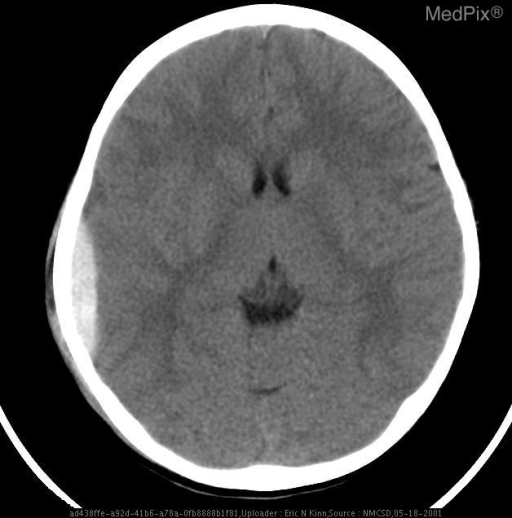
- Subdural hematoma - bridging veins
- Epidural hematoma - middle meningeal artery
- Subarachnoid hemorrhage - circle of Willis aneurysm
- Intracerebral hemorrhage - lenticulostriate artery
| Feature | EDH | SDH |
|---|---|---|
| Shape | Biconvex (lens) | Crescent |
| Suture crossing | NO | YES |
| Vessel | MMA (artery) | Bridging veins |
| Lucid interval | YES (classic) | Rare |
| CT density | Hyperdense (acute) | Variable |
| Age group | Young trauma | Elderly, alcoholics |
IMG-RAD-03 | NEET PG 2023 & Repeated 2024

- Transposition of Great Arteries
- Truncus arteriosus
- Tetralogy of Fallot
- Total anomalous pulmonary venous drainage
- Pulmonary stenosis (outflow obstruction)
- Right ventricular hypertrophy
- Overiding aorta
- Ventricular septal defect
- Egg... no that's TGA
- "Squatting" relieves hypercyanotic spells (Tet spells) - increases SVR, reduces R→L shunt
- Pink TOF: if PS is mild, can be acyanotic
- Treatment: Blalock-Taussig shunt (palliation) then total correction
IMG-RAD-04 | NEET PG 2024
- Epidural hematoma
- Subdural hematoma
- Subarachnoid hemorrhage
- Hypertensive intracerebral bleed
- Most common cause = ruptured Berry (saccular) aneurysm at Circle of Willis (ACoA most common)
- "Thunderclap headache" = worst headache of life, sudden onset
- "Xanthochromia" on CSF (yellow pigment) = confirms SAH when CT is negative
- CT sensitivity: 98% in first 12 hours; drops to 50% by day 7
- Vasospasm (days 4-10) = nimodipine to prevent
IMG-RAD-05 | NEET PG 2024
- Appendicitis - "target sign"
- Intussusception - air enema reduction
- Volvulus
- Hirschsprung disease
- Most common: ileocolic (ileum telescopes into colon)
- Age: 6 months - 2 years (peak)
- Classic triad: colicky abdominal pain + currant-jelly stool + sausage-shaped mass (RIF)
- Lead point in adults: Meckel's diverticulum, polyp, lymphoma
- Treatment: Air/barium/saline enema (diagnostic + therapeutic) if no perforation; surgery if failed or perforated
IMG-RAD-06 | NEET PG 2022
- Caecal volvulus - "kidney-bean" sign
- Sigmoid volvulus - "coffee-bean sign"
- Large bowel obstruction - multiple fluid levels
- Toxic megacolon
- Most common volvulus in India = Sigmoid (due to high-fiber diet + redundant sigmoid)
- X-ray: "coffee-bean" or "bent inner tube"
- Treatment: Flexible sigmoidoscopy decompression (if viable bowel) → elective sigmoid resection
- If gangrene/perforation: emergency Hartmann's procedure
IMG-RAD-07 | NEET PG 2023
- Lymphoma - mediastinal
- Thymoma - associated with Myasthenia gravis
- Teratoma - anterior mediastinum
- Pancoast tumor
- Thymoma most common anterior mediastinal tumor in adults
- 30% of thymoma patients have Myasthenia gravis
- 10-15% of MG patients have thymoma
- Staging: Masaoka classification
- Treatment: Thymectomy (improves MG even without thymoma)
CARDIOLOGY ECG IMAGE QUESTIONS
IMG-ECG-01 | NEET PG 2024-25 (Both years)
- Atrial fibrillation - rate 150 bpm - amiodarone
- Atrial flutter with 2:1 block - rate 150 bpm - diltiazem
- SVT - rate 150 bpm - adenosine
- Sinus tachycardia
- Atrial flutter: atrial rate 300 bpm; usually 2:1 block → ventricular rate 150 bpm
- DISTINGUISH: AF = irregularly irregular + no P waves (coarse fibrillation baseline)
- Rate control: Diltiazem (first line), beta-blockers; NOT amlodipine/nifedipine
- DO NOT give diltiazem/verapamil if HFrEF (negative inotropy)
- Cardioversion: DC shock 50-100J (flutter is very responsive)
IMG-ECG-02 | NEET PG 2023
- NSTEMI - IV heparin + early angiography
- Anterior STEMI (LAD occlusion) - primary PCI within 90 min
- Right heart strain (PE) - anticoagulation
- Brugada syndrome - ICD implant
- ST elevation + chest pain = STEMI until proven otherwise
- Time is muscle: Door-to-balloon <90 min (PCI available) or thrombolytics <30 min (no PCI)
- Anterior (V1-V4) = LAD; Inferior (II, III, aVF) = RCA/LCx; Lateral (I, aVL, V5-V6) = LCx
- Reciprocal changes = confirm STEMI (not pericarditis which is diffuse, saddle-shaped)
OBSTETRICS & GYNAECOLOGY IMAGE QUESTIONS
IMG-OBG-01 | NEET PG 2025
- Burns-Marshall technique
- Lovset's maneuver
- Mauriceau-Smellie-Veit maneuver
- Prague maneuver
| Step | Maneuver |
|---|---|
| Deliver trunk | Spontaneous/assisted |
| Deliver shoulders | Lovset's maneuver (trunk rotation) |
| Deliver aftercoming head | Mauriceau-Smellie-Veit (jaw-flexion-shoulder) |
| If head stuck | Burn-Marshall (pendulum swing) |
IMG-OBG-02 | NEET PG 2025
- Sacrospinous ligament
- Mackenrodt's (transverse cervical / cardinal) ligament
- Broad ligament
- Round ligament
- Uterovaginal prolapse primary support = Mackenrodt's ligament (Level I support - DeLancey)
- Level I = cardinal + uterosacral ligaments (apical support)
- Level II = ATFP, paravaginal attachments (mid-vaginal)
- Level III = perineal body, external anal sphincter (distal)
IMG-OBG-03 | NEET PG 2025
- Endometrial carcinoma
- Uterine leiomyoma (fibroid)
- Adenomyosis
- Endometrial polyp
- Most common benign uterine tumor
- Hormone-sensitive (grow in reproductive years, regress after menopause)
- GnRH analogues (leuprolide) = preoperative reduction
- Types: Submucosal (most symptomatic - heavy bleeding); Subserosal (pedunculated); Intramural
IMG-OBG-04 | NEET PG 2023
- Endometrial polyp - hysteroscopy
- Endometrial carcinoma - endometrial biopsy
- Cervical carcinoma - colposcopy
- Uterine sarcoma - CT scan
- Most common gynecological malignancy in DEVELOPED countries
- Most common presenting symptom = postmenopausal bleeding
- Risk factors: unopposed estrogen, obesity, nulliparity, diabetes, HTN, tamoxifen, Lynch syndrome
- Staging: FIGO surgical staging (2023 updated); Grade 3 = worst
- DOC investigation: Endometrial biopsy (Pipelle); D&C if biopsy inadequate
SURGERY IMAGE QUESTIONS
IMG-SURG-01 | NEET PG 2025
- Intersphincteric
- Suprasphincteric
- Extrasphincteric
- Transsphincteric
| Type | Path | Frequency | Treatment |
|---|---|---|---|
| Intersphincteric | Between sphincters | ~70% (most common) | Fistulotomy |
| Transsphincteric | Through both sphincters | ~25% | Seton procedure |
| Suprasphincteric | Above puborectalis | ~5% | Complex repair |
| Extrasphincteric | Outside all sphincters | ~1% (least common) | Stoma ± repair |
IMG-SURG-02 | NEET PG 2025
- Fasciotomy for compartment syndrome
- Escharotomy for circumferential burns
- Amputation
- Skin grafting
- Escharotomy = incision through burn eschar (not into fascia = fasciotomy)
- Indicated for circumferential full-thickness (3rd degree) burns of limbs or chest
- Chest escharotomy → improves respiratory compliance
- Limb escharotomy → prevents compartment syndrome from swelling
IMG-SURG-03 | NEET PG 2025
- Surgeon's knot
- Square knot
- Granny's knot
- Slip knot
- Square knot = two single throws in OPPOSITE directions (most secure, standard)
- Granny knot = two single throws in SAME direction (unreliable, slips)
- Surgeon's knot = double loop first + single loop second (used in wet/slippery tissues)
- Slip knot = adjustable (used in ligatures)
IMG-SURG-04 | NEET PG 2024
- Large bowel obstruction
- Small bowel obstruction (SBO)
- Sigmoid volvulus
- Ileus
- SBO vs LBO on X-ray:
- SBO: central + dilated loops + valvulae conniventes (complete across lumen) + "string of pearls"
- LBO: peripheral + haustral folds (incomplete across lumen) + large loop
- Most common cause SBO = adhesions (post-surgery); LBO = carcinoma colon
- Treatment: NGT decompression + fluid resuscitation; surgery if strangulation
IMG-SURG-05 | NEET PG 2022
- Ewing's sarcoma - onion-peel periosteal reaction
- Osteosarcoma - Codman's triangle = elevated periosteum with reactive new bone
- Giant cell tumor - soap-bubble epiphysis
- Chondrosarcoma - ring-and-arc calcification
| Tumor | X-ray sign | Location | Age |
|---|---|---|---|
| Osteosarcoma | Sunburst + Codman's | Distal femur/Prox tibia metaphysis | 10-25 yrs |
| Ewing's sarcoma | Onion-skin/laminated | Diaphysis (flat bones also) | 5-15 yrs |
| GCT | Soap bubble (eccentric) | Epiphysis (distal femur) | 20-40 yrs |
| Aneurysmal BC | Soap bubble (expansile) | Metaphysis | Adolescents |
| Chondrosarcoma | Ring-and-arc calcification | Pelvis/prox femur | 40-70 yrs |
DERMATOLOGY IMAGE QUESTIONS
IMG-DERM-01 | NEET PG 2025 & 2023 (Most repeated)
- Lichen planus - Wickham's striae
- Psoriasis - Auspitz sign (pinpoint bleeding)
- Pityriasis rosea - Herald patch
- Seborrheic dermatitis - greasy yellow scales
- Psoriasis: T-cell mediated; HLA-Cw6 association
- Grattage test: candle-grease scrapings → Auspitz sign
- Histology: Munro's microabscesses (PMNs in parakeratotic SC) + suprapapillary plate thinning
- Koebner phenomenon: lesions appear on traumatized skin
- Scalp psoriasis: most common site; nail pitting = most common nail change
IMG-DERM-02 | NEET PG 2024
- Pemphigus vulgaris - Nikolsky positive - intraepidermal
- Bullous pemphigoid - Nikolsky NEGATIVE - subepidermal
- Dermatitis herpetiformis - Nikolsky negative
- Epidermolysis bullosa
| Feature | Pemphigus vulgaris | Bullous pemphigoid |
|---|---|---|
| Blister | Flaccid, fragile | Tense, turgid |
| Nikolsky | POSITIVE | NEGATIVE |
| Oral involvement | YES (first) | Rare/mild |
| Split | Intraepidermal (suprabasal) | Subepidermal |
| Antibody | Anti-desmoglein 1&3 | Anti-BP180, BP230 |
| Age | Middle-aged | Elderly |
| DIF | IgG intercellular "fish-net" | Linear IgG at DEJ |
IMG-DERM-03 | NEET PG 2023
- Lupus erythematosus - ANA testing
- Lupus vulgaris (cutaneous TB) - skin biopsy + AFB culture/CBNAAT
- Lepromatous leprosy - slit-skin smear
- Rosacea - clinical diagnosis
- Most common form of cutaneous TB in India = Lupus vulgaris
- Face > neck > trunk
- Histology: Epithelioid granulomas WITHOUT caseation (or minimal)
- Complication: scar carcinoma (SCC in chronic lesions)
- Treatment: standard anti-TB drugs (rifampicin-based)
IMG-DERM-04 | NEET PG 2022
- Lepromatous leprosy (LL) - Lepromin negative
- Tuberculoid leprosy (TT) - Lepromin STRONGLY positive
- Borderline lepromatous (BL) - weakly positive
- Indeterminate leprosy
| Feature | Tuberculoid (TT) | Lepromatous (LL) |
|---|---|---|
| Lesions | Few (1-5) | Numerous |
| Margins | Well defined | Ill defined |
| Sensation | Absent (anesthetic) | Relatively intact initially |
| Nerve thickening | Yes (few nerves) | Yes (many nerves) |
| Lepromin | Strongly positive | Negative |
| AFB | Absent/few (paucibacillary) | Numerous (multibacillary) |
| Immunity | High CMI | Low CMI, high humoral |
| Treatment | 6 months (PB) | 12 months (MB) |
MICROBIOLOGY / INFECTIOUS DISEASE IMAGE QUESTIONS
IMG-MICRO-01 | NEET PG 2022
- Aspergillus - voriconazole
- Candida - fluconazole
- Cryptococcus neoformans - amphotericin B + flucytosine
- Mucor - liposomal amphotericin B
- Capsular antigen test (Cryptococcal antigen/CrAg) = best diagnostic test
- India ink = classic but less sensitive
- Treatment: Induction (amphotericin B + flucytosine 2 weeks) → Consolidation (fluconazole 8 weeks) → Maintenance (fluconazole lifelong)
- Raised ICP = therapeutic lumbar puncture
IMG-MICRO-02 | NEET PG 2024
- P. vivax - CQ sensitive
- P. falciparum - chloroquine resistance common
- P. malariae - quartan malaria
- P. ovale - similar to vivax
- Multiple rings per RBC = P. falciparum (only species that does this)
- Banana/crescent-shaped gametocytes = ONLY P. falciparum
- "Appliqué forms" (rings at edge of RBC)
- Maurer's clefts (not Schuffner's dots)
| Feature | P. falciparum | P. vivax |
|---|---|---|
| Rings/RBC | Multiple | Single |
| Gametocyte | Banana/crescent | Round |
| RBC size | Normal | Enlarged |
| Dots | Maurer's clefts | Schüffner's dots |
| Fever | Subtertian (36-48h) | Tertian (48h) |
| Hypnozoites | NO | YES (relapses) |
| Severe malaria | YES | Less common |
IMG-MICRO-03 | NEET PG 2025
- X-linked dominant
- Mitochondrial inheritance
- Autosomal dominant
- X-linked recessive
- MELAS = Mitochondrial Encephalomyopathy, Lactic Acidosis, Stroke-like episodes
- MERRF = Myoclonic Epilepsy with Ragged Red Fibers
- Kearns-Sayre syndrome = PEO + retinitis pigmentosa + cardiac block
- Leber's hereditary optic neuropathy (LHON)
- All from MATERNAL inheritance; heteroplasmy accounts for variable expressivity
OPHTHALMOLOGY IMAGE QUESTIONS
IMG-OPH-01 | NEET PG 2022 & 2023
- Grade II hypertensive retinopathy - AV nipping
- Grade III - flame hemorrhages + exudates
- Grade IV - papilledema + star pattern = Hypertensive emergency
- Central retinal vein occlusion
- Grade I: Arteriolar narrowing + silver wiring
- Grade II: AV nipping (Gunn's sign) + copper wiring
- Grade III: Flame hemorrhages + soft exudates (cotton-wool spots) + hard exudates
- Grade IV: Grade III + Papilledema + macular star
IMG-OPH-02 | NEET PG 2023
- Congenital cataract
- Retinoblastoma
- Persistent hyperplastic primary vitreous (PHPV)
- Coat's disease
- Retinoblastoma: most common intraocular malignancy in children (<5 years)
- Leukocoria (60%) + strabismus (20%) = most common presentations
- RB1 gene (13q14) = tumor suppressor; "2-hit hypothesis" (Knudson)
- Bilateral = hereditary (germline mutation); Unilateral = usually somatic
- Trilateral RB = bilateral retinoblastoma + pinealoblastoma
ENT IMAGE QUESTIONS
IMG-ENT-01 | NEET PG 2025
- Sensorineural hearing loss
- Otosclerosis
- Presbycusis
- Noise-induced hearing loss
- Otosclerosis: abnormal remodeling of otic capsule (bony cochlea)
- Autosomal dominant; more common in females; bilateral in 70%
- Tympanogram: "As" type (reduced compliance of fixed stapes)
- Treatment: Stapedectomy / stapedotomy (gold standard); hearing aid also effective
- Fluoride therapy (sodium fluoride): can slow progression
IMG-ENT-02 | NEET PG 2024
- Thyroid nodule
- Branchial cyst
- Carotid body tumor (Paraganglioma) - Lyre's sign
- Cervical lymph node
- Paraganglioma (chemodectoma) at carotid bifurcation
- Pulsatile, mobile side-to-side but NOT up-down (Fontaine's sign)
- 10% rule: 10% bilateral, 10% malignant, 10% familial
- Familial: SDH gene mutations (succinate dehydrogenase)
- Treatment: surgical resection (preop embolization for large tumors)
FORENSIC MEDICINE IMAGE QUESTIONS
IMG-FM-01 | NEET PG 2024
- Cadaveric spasm - indicates ante-mortem violent activity
- Pugilistic attitude - due to heat coagulation of muscles post-mortem (NOT self-defense)
- Rigor mortis in fighting position
- Decomposition artefact
- Pugilistic/boxer's attitude = HEAT ARTIFACT (protein coagulation shortens flexor muscles more than extensors)
- Does NOT indicate the person was fighting or in self-defense
- Important medico-legal point: examiners must NOT misinterpret as defense injuries
- Cadaveric spasm = instantaneous rigor at moment of death (violent emotion/exertion); DOES indicate ante-mortem activity
IMG-FM-02 | NEET PG 2022
- Contact wound - star-shaped laceration + burning + soot
- Close-range shot (<45 cm) - blackening + tattooing
- Medium range (45-90 cm) - tattooing only (no blackening)
- Long range (>90 cm) - only entry wound, no secondary effects
- Soot + burning + tattooing = Close range (<45 cm)
- Tattooing only = Medium range (45-90 cm)
- Entry wound only, no secondary effects = Long range (>90 cm)
- Entry wound: small, inverted margins, abrasion collar
- Exit wound: large, everted (exploded outward), no abrasion collar
- Tattooing (stippling) = unburned powder grains embedded in skin = CANNOT be washed off
- Soiling = soot deposits on skin = CAN be washed off
- Contact wound: weapon pressed against skin = "impression mark" + gas explosion inside skin = cross-shaped laceration
ANATOMY IMAGE QUESTIONS
IMG-ANAT-01 | NEET PG 2025
- CN IX
- CN X
- CN XI
- CN XII
- CN IX (Glossopharyngeal)
- CN X (Vagus)
- CN XI (Accessory spinal)
- Inferior petrosal sinus (anteriorly)
- CN XII = exits through HYPOGLOSSAL CANAL (separate foramen)
IMG-ANAT-02 | NEET PG 2022-23
- Portal vein
- Gastroduodenal artery
- Common bile duct
- IVC
- POSTERIOR to D1: gastroduodenal artery + portal vein + CBD (hepatoduodenal ligament)
- INFERIOR to D1: pancreatic head
- "Hot Cross Bun" pneumonic: GDA + CBD + PV form the "holy trinity" posterior to D1
PAEDIATRICS IMAGE QUESTIONS
IMG-PAED-01 | NEET PG 2023
- Osteogenesis imperfecta - multiple fractures
- Nutritional rickets (Vit D deficiency) - low Ca, low PO4, HIGH ALP, HIGH PTH
- Scurvy - subperiosteal hemorrhage + Pelkan spurs
- Lead poisoning - dense metaphyseal bands
- Widening of physis (growth plate)
- Fraying (irregular zone of provisional calcification)
- Cupping + splaying of metaphysis
- Periosteal reaction
- Long bone bowing (genu varum in ambulatory children)
- Craniotabes (in infants)
- "Rosary" at costochondral junctions
IMG-PAED-02 | NEET PG 2024
- Tetralogy of Fallot - boot-shaped
- Transposition of Great Arteries (TGA) - "egg on its side"
- Total anomalous pulmonary venous drainage - "snowman" sign
- Truncus arteriosus - enlarged heart
- TGA: Aorta arises from RV (anterior); PA from LV (posterior) = parallel circuits
- Neonate depends on mixing via PDA/ASD/VSD for survival
- Emergency: Prostaglandin E1 (keep PDA open) + Balloon Atrial Septostomy (Rashkind)
- Definitive: Arterial switch operation (Jatene procedure) within first 2 weeks
RAPID-FIRE IMAGE SIGNS TESTED IN NEET PG (2021-2025)
| Sign | Image Description | Diagnosis | Year Tested |
|---|---|---|---|
| Boot-shaped heart (Coeur en sabot) | CXR: upturned apex + concave PA bay | Tetralogy of Fallot | 2022, 2023 |
| Egg on its side | CXR: narrow mediastinum + oval heart | TGA | 2024 |
| Coffee-bean sign | AXR: omega loop pointing to RUQ | Sigmoid volvulus | 2022 |
| String of pearls | AXR: small air bubbles in SB | SBO | 2024 |
| Target/doughnut sign | USG abdomen: concentric rings in child | Intussusception | 2024 |
| Sunburst + Codman's triangle | X-ray: metaphysis of long bone | Osteosarcoma | 2022 |
| Onion-skin periosteum | X-ray: diaphysis of long bone | Ewing's sarcoma | 2023 |
| Soap bubble (epiphysis) | X-ray: eccentric lytic lesion | GCT / Aneurysmal BC | 2023 |
| Lyre/goblet sign | Carotid angiogram: splayed bifurcation | Carotid body tumor | 2025 |
| Carhart's notch | Audiogram: dip at 2000 Hz BC | Otosclerosis | 2025 |
| Sawtooth waves | ECG at 150 bpm | Atrial flutter 2:1 | 2024, 2025 |
| Apple-green birefringence | Congo red under polarized light | Amyloidosis | 2022, 2024 |
| Owl-eye cells | Lymph node H&E | Hodgkin lymphoma | 2023, 2025 |
| Pseudopalisading necrosis | Brain biopsy H&E | GBM | 2023 |
| Fried egg cells | Brain biopsy H&E | Oligodendroglioma | 2023 |
| Apple-jelly nodules | Diascopy of facial plaque | Lupus vulgaris | 2023 |
| Banana gametocyte | Blood smear Giemsa | P. falciparum | 2024 |
| Encapsulated yeast India ink | CSF prep | Cryptococcus | 2022 |
| Pugilistic attitude | Burns victim X-ray | Heat artifact (NOT defense) | 2024 |
| Leukocoria | White pupillary reflex photo | Retinoblastoma | 2023 |
| Macular star + papilledema | Fundus photo | Grade IV HTN retinopathy | 2022, 2023 |
| Tense bullae (elderly) | Clinical skin photo | Bullous pemphigoid | 2024 |
| Flaccid bullae + oral erosions | Clinical photo, middle-aged | Pemphigus vulgaris | 2023 |
| Silver plaques extensor | Clinical skin photo | Psoriasis | 2023, 2025 |
Give me 5 year microbiology question with ans of neet pg
NEET PG 2021 2022 2023 2024 2025 microbiology recall questions answers
microbiology medical education question recall
https://www.diginerve.com/blogs/neet-pg-microbiology-previou…
NEET PG Microbiology: 5-Year Recall-Based Questions with Answers
Coverage: NEET PG 2021-2025
NEET PG 2025 Microbiology Recall
Answer: Bacillus anthracis.
McFadyean reaction demonstrates the polypeptide capsule as a purple halo.
Answer: Lipoteichoic acid.
Also remember: M protein is the major antiphagocytic virulence factor and is linked to rheumatic fever.
Answer: Enterobius vermicularis.
Diagnosis: cellulose tape or Scotch-tape test, not routine stool microscopy.
Answer: Histoplasmosis due to Histoplasma capsulatum.
Histoplasma is dimorphic and appears as intracellular yeasts in macrophages in tissue.
Answer: Weil disease, severe leptospirosis.
Caused by Leptospira interrogans.
Answer: Aspergillus.
Aspergillus: narrow septate hyphae, acute-angle branching. Mucor: broad pauci-septate hyphae, right-angle branching.
Answer: Gram-positive, branching filamentous bacterium that is weakly acid-fast.
Modified Ziehl-Neelsen stain is used; it is aerobic and can cause pulmonary/CNS infection in immunocompromised hosts.
Answer: Paraffin bait technique.
Answer: Terminal complement deficiency, C5-C9.
Deficiency of the membrane attack complex predisposes to recurrent Neisseria infections.
Answer: Rifampicin.
Single-dose ciprofloxacin or ceftriaxone are alternatives depending on clinical context.
Answer: Anti-HBs antibody.
Vaccination pattern: anti-HBs positive alone, with anti-HBc negative.
Answer: Within 2-8 weeks.
Delay is generally longer in TB meningitis because of risk from CNS IRIS.
NEET PG 2024 Microbiology Recall
Answer: Plasmodium falciparum.
Banana/crescent gametocytes are characteristic.
Answer: Mucormycosis caused by Mucorales.
Treatment principle: liposomal amphotericin B, urgent surgical debridement, and correction of metabolic derangement.
Answer: Cryptococcus neoformans.
The capsule gives a clear halo against dark background. Cryptococcal antigen testing is more sensitive.
Answer: Mycoplasma pneumoniae.
No cell wall, hence no Gram stain visualization and intrinsic resistance to beta-lactams.
Answer: Rickettsial infection.
It detects cross-reacting antibodies to Proteus OX strains. It is an older, nonspecific test.
Answer: Malassezia furfur causing pityriasis versicolor.
Answer: Clostridioides difficile.
Its toxin B is cytotoxic; toxin A is enterotoxic.
Answer: VDRL or RPR.
Confirm with a treponemal test such as TPHA/TPPA.
NEET PG 2023 Microbiology Recall
Answer: Enveloped single-stranded RNA virus, measles virus.
Answer: Oral candidiasis due to Candida albicans.
Answer: Genital herpes, usually HSV-2.
HSV-1 is increasingly implicated too.
Answer: Sandfly.
Visceral leishmaniasis or kala-azar due to Leishmania donovani, transmitted by female Phlebotomus sandfly.
Answer: Enterobius vermicularis infection.
Answer: Viral meningitis.
Answer: Blocking release of inhibitory neurotransmitters GABA and glycine.
It cleaves synaptobrevin, preventing inhibitory interneuron neurotransmitter release.
Answer: Cryptococcal antigen detection in CSF or serum.
India ink is classic but less sensitive.
NEET PG 2022 Microbiology Recall
Answer: Brugia malayi.
Answer: Wuchereria bancrofti.
Answer: Histamine-producing bacteria such as Morganella morganii, Klebsiella, and some Pseudomonas species.
Older recall keys may state Pseudomonas aeruginosa. The mechanism is bacterial conversion of fish histidine to histamine.
Answer: Cefotetan.
It is clinically important in cystic fibrosis and chronic granulomatous disease.
Answer: Measles.
Answer: Cryptococcus neoformans.
Answer: Mucormycosis.
Answer: Enterobius vermicularis.
NEET PG 2021 Recall Themes
Answer: Vibrio cholerae toxin ADP-ribosylates Gs alpha, causing increased cAMP and chloride-water secretion.
Answer: Tuberculin skin test or interferon-gamma release assay.
Neither distinguishes latent from active TB by itself.
Answer: Entamoeba histolytica.
Diagnostic trophozoites may contain ingested RBCs.
Answer: Cytomegalovirus infection.
Answer: Mumps virus, an enveloped negative-sense single-stranded RNA paramyxovirus.
Answer: Past natural infection with immunity.
Answer: Hippocampal pyramidal cells and Purkinje cells of cerebellum in rabies.
Answer: Yersinia pestis.
Staining may be demonstrated by Wayson or Giemsa stain.
High-Yield One-Liner Revision Table
| Topic | Recall answer |
|---|---|
| McFadyean reaction | Bacillus anthracis capsule |
| Narrow-based budding yeast | Histoplasma capsulatum |
| Broad nonseptate 90° hyphae | Mucor/Rhizopus |
| Septate 45° hyphae | Aspergillus |
| India ink halo | Cryptococcus neoformans |
| Pseudohyphae + budding yeast | Candida albicans |
| Weakly acid-fast branching filament | Nocardia |
| Paraffin bait technique | Nocardia |
| Banana-shaped gametocyte | P. falciparum |
| Kinetoplast in macrophage | Leishmania donovani |
| Sheathed microfilaria with two tail nuclei | Brugia malayi |
| Perianal pruritus + planoconvex egg | Enterobius vermicularis |
| Koplik spots | Measles |
| Cold agglutinins, atypical pneumonia | Mycoplasma pneumoniae |
| Terminal complement C5-C9 deficiency | Recurrent Neisseria |
| Anti-HBs alone | Hepatitis B vaccination immunity |
| Weil-Felix | Rickettsial infection |
| “Spaghetti and meatballs” | Malassezia furfur |
| Apple-jelly? | Lupus vulgaris, cutaneous TB |
| Tetanospasmin | Blocks GABA and glycine release |
Important correction for exams
Quick answer
NEET PG Microbiology: 5-Year Recall-Based Questions with Answers
2021-2025
NEET PG 2021 Microbiology Recalls
Answer: Sandfly
Diagnosis: Visceral leishmaniasis (Kala-azar), caused by Leishmania donovani.
Answer: Enveloped single-stranded RNA virus
Organism: Measles virus, Paramyxoviridae.
Answer: Genital herpes
Organism: Usually HSV-2, although HSV-1 can also cause it.
Answer: Enterobius vermicularis
Key point: D-shaped or planoconvex eggs; diagnose using early-morning Scotch tape test.
Answer: Terminal complement components C5-C9
Key point: Membrane attack complex deficiency predisposes to recurrent Neisseria infection.
NEET PG 2022 Microbiology Recalls
Answer: Brugia malayi
Trap:
- Wuchereria bancrofti: tail tip free of nuclei
- Brugia malayi: two terminal nuclei
- Loa loa: nuclei extend to tail tip
Answer: Gram-positive, branching filamentous bacilli that are weakly acid-fast on modified Ziehl-Neelsen staining.
Answer: Oral candidiasis caused by Candida albicans.
Answer: Histamine-producing bacteria, classically Morganella morganii; some recall sources list Pseudomonas aeruginosa among implicated organisms.
Exam concept: Improperly stored fish leads to bacterial histidine decarboxylation and histamine toxicity.
Answer: Cefotetan
Clinical association: Opportunistic infection in cystic fibrosis and chronic granulomatous disease.
Answer: Cryptococcus neoformans
Key point: Cryptococcal antigen testing is more sensitive than India ink.
NEET PG 2023 Microbiology Recalls
Answer: Mycoplasma pneumoniae
Key point: It lacks a cell wall, so beta-lactams are ineffective.
Answer: Blocks release of inhibitory neurotransmitters, GABA and glycine, from inhibitory interneurons.
Answer: Rickettsial infections
Key point: It detects cross-reacting antibodies against Proteus OX strains.
Answer: Lupus vulgaris, a form of cutaneous tuberculosis.
Answer: Mucor/Rhizopus, causing mucormycosis.
Association: Diabetic ketoacidosis, neutropenia, iron overload.
Answer: Clostridioides difficile
Key point: Toxin A is enterotoxin and toxin B is cytotoxin.
NEET PG 2024 Microbiology Recalls
Answer: Plasmodium falciparum
Answer: Weil disease due to Leptospira interrogans.
Answer: Aspergillus species
Contrast: Mucor is broad, aseptate/pauciseptate, and branches at right angles.
Answer: Histoplasmosis due to Histoplasma capsulatum
Key point: Dimorphic fungus, mould at 25°C and yeast at 37°C.
Answer: Epstein-Barr virus
Diagnosis: Oral hairy leukoplakia.
Answer: Molluscum contagiosum
Inclusion: Henderson-Patterson bodies.
NEET PG 2025 Microbiology Recalls
Answer: Bacillus anthracis
Key point: McFadyean reaction demonstrates the capsule of anthrax bacilli.
Answer: Lipoteichoic acid
Trap: M protein is the major antiphagocytic virulence factor and is linked to rheumatic fever.
Answer: Enterobius vermicularis
Answer: Paraffin bait technique
Also remember: It is weakly acid-fast on modified ZN stain.
Answer: Septate hyphae with acute-angle, approximately 45°, dichotomous branching.
Answer: Anti-HBs antibody
Pattern after vaccination: Anti-HBs positive, anti-HBc negative.
Answer: Early-morning perianal cellophane tape test.
Answer: Immune Reconstitution Inflammatory Syndrome, IRIS.
Answer: Cryptococcal meningitis caused by Cryptococcus neoformans.
Must-Revise Microbiology Images from the Last 5 Years
| Image / finding | Answer |
|---|---|
| Broad aseptate, right-angle branching hyphae | Mucormycosis |
| Septate, acute-angle branching hyphae | Aspergillosis |
| India ink halo around yeast | Cryptococcus neoformans |
| Banana-shaped gametocyte | P. falciparum |
| Multiple ring forms in one RBC | P. falciparum |
| Planoconvex/D-shaped egg on tape test | Enterobius vermicularis |
| Macrophages with LD bodies and kinetoplast | Visceral leishmaniasis |
| Budding yeast + pseudohyphae | Candida albicans |
| Apple-jelly nodules on diascopy | Lupus vulgaris |
| McFadyean reaction | Bacillus anthracis |
| Umbilicated papules with Henderson-Patterson bodies | Molluscum contagiosum |
| Koplik spots + descending rash | Measles |
Highest-Yield Repeated Topics
- Fungal morphology: Aspergillus vs Mucor, Cryptococcus, Candida, Histoplasma
- Parasite images: malaria smear, microfilariae, Enterobius, Leishmania
- Stains and tests: Modified ZN for Nocardia, India ink, McFadyean reaction, Weil-Felix
- Virology: HBV serology, measles, EBV, HIV-related infections
- Clinical bacteriology: anthrax, leptospirosis, GAS virulence factors, C. difficile
- Opportunistic infections in HIV: Cryptococcus, candidiasis, PCP, toxoplasmosis, CMV
All important qe of anatomy in neet pg last 10 year
Which Anatomy PYQ module should I give first, with recall-style questions and answers?
NEET PG 2026 - 200 Most Probable High-Yield Questions
Based on: Last 5-Year PYQ Analysis (2022-2026) + Recent Updates + Recall-Based Image Questions
TREND ANALYSIS: NEET PG 2022-2026
| Subject | 2025 Qs | Weightage | Trend |
|---|---|---|---|
| Medicine (incl. Derm, Psych) | 30 | 15% | Highest - clinical vignettes |
| OBGYN | 20 | 10% | Very high - PPH, ANC, fertility |
| Surgery | 18 | 9% | High - trauma, GI, oncology |
| PSM | 16 | 8% | Consistent - programs, statistics |
| Pharmacology | 15 | 7.5% | Rising - clinical drug use |
| Biochemistry | 15 | 7.5% | Rising - metabolic integration |
| Pathology | 12 | 6% | Stable - IHC, chromosomal |
| Microbiology | 12 | 6% | Stable - clinical context |
| Anatomy | 9 | 4.5% | Moderate - radiological focus |
| Dermatology | 8 | 4% | Rising - image-heavy |
| Forensic Medicine | 6 | 3% | Stable |
| Orthopaedics | 5 | 2.5% | Stable |
| Ophthalmology | 5 | 2.5% | Stable |
| ENT | 5 | 2.5% | Stable |
| Paediatrics | 5 | 2.5% | Stable |
| Radiology | 5 | 2.5% | Rising |
| Physiology | 5 | 2.5% | Integrated |
| Anaesthesia | 4 | 2% | Stable |
- ~50% image-based (doubled from 2022)
- ~70% clinical vignette format
- ~30% repeats from PYQs
- Basic sciences now mostly integrated into clinical scenarios
- Drug of choice tested 15-20 times per paper
PART 1 - 200 HIGH-YIELD MCQs WITH ANSWERS
ANATOMY (9 Questions)
PHYSIOLOGY (5 Questions)
BIOCHEMISTRY (15 Questions)
PHARMACOLOGY (15 Questions)
PATHOLOGY (12 Questions)
MICROBIOLOGY (12 Questions)
GENERAL MEDICINE (30 Questions)
OBSTETRICS & GYNAECOLOGY (20 Questions)
SURGERY (18 Questions)
PSM / COMMUNITY MEDICINE (16 Questions)
PAEDIATRICS (5 Questions)
ORTHOPAEDICS (5 Questions)
OPHTHALMOLOGY (5 Questions)
ENT (5 Questions)
DERMATOLOGY (8 Questions)
FORENSIC MEDICINE (6 Questions)
RADIOLOGY (5 Questions)
ANAESTHESIA (4 Questions)
ADDITIONAL HIGH-YIELD CLINICAL INTEGRATORS (4 Questions completing 200)
| Feature | Bacterial | Viral | TB |
|---|---|---|---|
| Appearance | Turbid/purulent | Clear | Cobweb clot |
| Cells | PMN (100-10,000) | Lymphocytes | Lymphocytes |
| Protein | Very high (>100) | Normal/slight ↑ | High (100-500) |
| Glucose | Very low (<45) | Normal | Low (very) |
| Opening pressure | High | Normal/mild ↑ | Elevated |
PART 2 - LAST 5-YEAR RECALLED IMAGE-BASED QUESTIONS (2022-2026)
ANATOMY - Image Recalls
SURGERY - Image Recalls
PATHOLOGY - Image Recalls
MICROBIOLOGY - Image Recalls
MEDICINE / CARDIOLOGY - Image Recalls
DERMATOLOGY - Image Recalls
RADIOLOGY - Image Recalls
QUICK REVISION: 2025-2026 NEW UPDATES TO KNOW
SUBJECT-WISE PRIORITY FOR NEET PG 2026
| Priority | Subject | Focus |
|---|---|---|
| ★★★★★ | Medicine | Clinical vignettes, ECGs, emergencies, rheumatology |
| ★★★★★ | OBGYN | Obstetric emergencies, infertility, gynecological malignancies |
| ★★★★★ | Surgery | Trauma, GI surgery, oncology, hernia classification |
| ★★★★ | PSM | Biostatistics, national programs, epidemiology |
| ★★★★ | Pharmacology | Drug of choice, drug interactions, newer drugs |
| ★★★★ | Pathology | IHC, chromosomal translocations, histology images |
| ★★★★ | Microbiology | Clinical context, special stains, serology |
| ★★★ | Biochemistry | Enzyme deficiencies, metabolic pathways |
| ★★★ | Dermatology | Image-heavy, bullous diseases, photosensitivity |
| ★★★ | Radiology | X-ray signs, CT patterns, named signs |
| ★★ | Anatomy | Neuroanatomy, radiological, embryology |
| ★★ | Forensic | Medico-legal, thanatology basics |
| ★★ | Ortho | X-ray diagnosis, named conditions |
| ★★ | Ophthalmology | Retinal conditions, glaucoma, congenital |
| ★★ | ENT | Audiometry, ear conditions, neck masses |
| ★★ | Paediatrics | Growth milestones, congenital conditions |
| ★ | Physiology | Integrated into other subjects |
| ★ | Anaesthesia | Specific drugs, monitoring, complications |
NEET PG 2021-2025: RECALL-BASED IMAGE QUESTIONS WITH ANSWERS
PATHOLOGY IMAGE QUESTIONS
IMG-PATH-01 | NEET PG 2023 & 2025 (Repeated Concept)
- Langhans giant cells
- Reed-Sternberg cells
- Touton giant cells
- Aschoff cells
- Diagnosis = Classical Hodgkin Lymphoma
- Immunohistochemistry: CD15+, CD30+, PAX5 (weak), CD20-
- Most common subtype = Nodular Sclerosis (young females, mediastinal mass)
- Treatment: ABVD chemotherapy ± radiation
IMG-PATH-02 | NEET PG 2022 & 2024 (Frequently repeated)
- Glycogen storage disease - PAS stain
- Amyloidosis - Congo red with apple-green birefringence
- Iron deposition - Prussian blue
- Lipofuscinosis
- Congo red stain + apple-green birefringence = AMYLOID (only finding that confirms)
- AL amyloid (light chain) = most common systemic; associated with myeloma
- AA amyloid = chronic inflammation (TB, RA, IBD)
- ATTR amyloid = cardiac (restrictive cardiomyopathy in elderly)
- Serum amyloid P scan = whole body amyloid burden
IMG-PATH-03 | NEET PG 2024
- Aspergillus fumigatus
- Candida albicans
- Mucor / Rhizopus (Mucormycosis)
- Cryptococcus neoformans
| Feature | Aspergillus | Mucor/Rhizopus |
|---|---|---|
| Hyphae | Narrow, septate | Broad, non-septate (ribbon-like) |
| Branching | Acute angle (45°) | Right angle (90°) |
| Septae | Present (regular) | Absent or rare |
- Mucormycosis: angioinvasive; rhinocerebral form = commonest; associated with DKA + neutropenia
- Treatment: liposomal amphotericin B + surgical debridement
- Aspergillus: septate, 45° = commonest opportunistic mold in immunocompromised
IMG-PATH-04 | NEET PG 2023
- Glioblastoma multiforme
- Oligodendroglioma
- Ependymoma
- Pilocytic astrocytoma
- WHO Grade II/III; frontal lobe most common
- Molecular marker: 1p/19q codeletion (diagnostic + better prognosis)
- IDH mutation usually present
- Best prognosis among diffuse gliomas
IMG-PATH-05 | NEET PG 2024
- Goblet cells in normal bowel
- Signet ring cells - Diffuse gastric adenocarcinoma (Linitis plastica)
- Clara cells in lung
- Chief cells in stomach
- Linitis plastica = diffuse infiltrating gastric carcinoma; "leather bottle stomach" on barium
- Associated with CDH1 (E-cadherin) mutation - hereditary diffuse gastric cancer
- Krukenberg tumor = bilateral ovarian mets from gastric signet ring carcinoma
- Diffuse type: not associated with H. pylori (unlike intestinal type)
IMG-PATH-06 | NEET PG 2023
- Grade I (Pilocytic astrocytoma)
- Grade II (Low-grade astrocytoma)
- Grade III (Anaplastic astrocytoma)
- Grade IV (Glioblastoma multiforme - GBM)
- Most common primary brain tumor in ADULTS
- Butterfly glioma: crosses corpus callosum
- MGMT promoter methylation = better response to temozolomide
- IDH wildtype = worse prognosis
- Treatment: Surgery + Temozolomide + Radiation (Stupp protocol)
RADIOLOGY IMAGE QUESTIONS
IMG-RAD-01 | NEET PG 2025
- Spondylolisthesis
- Spondylolysis
- Vertebral compression fracture
- Degenerative disc disease
- Spondylolysis = pars interarticularis FRACTURE only, no slippage ("Scottie dog with collar" on oblique X-ray)
- Spondylolisthesis = actual forward SLIP of vertebra
- Meyerding grading: Grade I (<25%), II (25-50%), III (50-75%), IV (>75%), V = spondyloptosis
IMG-RAD-02 | NEET PG 2024
- Subdural hematoma - bridging veins
- Epidural hematoma - middle meningeal artery
- Subarachnoid hemorrhage - circle of Willis aneurysm
- Intracerebral hemorrhage - lenticulostriate artery
| Feature | EDH | SDH |
|---|---|---|
| Shape | Biconvex (lens) | Crescent |
| Suture crossing | NO | YES |
| Vessel | MMA (artery) | Bridging veins |
| Lucid interval | YES (classic) | Rare |
| CT density | Hyperdense (acute) | Variable |
| Age group | Young trauma | Elderly, alcoholics |
IMG-RAD-03 | NEET PG 2023 & Repeated 2024
- Transposition of Great Arteries
- Truncus arteriosus
- Tetralogy of Fallot
- Total anomalous pulmonary venous drainage
- Pulmonary stenosis (outflow obstruction)
- Right ventricular hypertrophy
- Overiding aorta
- Ventricular septal defect
- Egg... no that's TGA
- "Squatting" relieves hypercyanotic spells (Tet spells) - increases SVR, reduces R→L shunt
- Pink TOF: if PS is mild, can be acyanotic
- Treatment: Blalock-Taussig shunt (palliation) then total correction
IMG-RAD-04 | NEET PG 2024
- Epidural hematoma
- Subdural hematoma
- Subarachnoid hemorrhage
- Hypertensive intracerebral bleed
- Most common cause = ruptured Berry (saccular) aneurysm at Circle of Willis (ACoA most common)
- "Thunderclap headache" = worst headache of life, sudden onset
- "Xanthochromia" on CSF (yellow pigment) = confirms SAH when CT is negative
- CT sensitivity: 98% in first 12 hours; drops to 50% by day 7
- Vasospasm (days 4-10) = nimodipine to prevent
IMG-RAD-05 | NEET PG 2024
- Appendicitis - "target sign"
- Intussusception - air enema reduction
- Volvulus
- Hirschsprung disease
- Most common: ileocolic (ileum telescopes into colon)
- Age: 6 months - 2 years (peak)
- Classic triad: colicky abdominal pain + currant-jelly stool + sausage-shaped mass (RIF)
- Lead point in adults: Meckel's diverticulum, polyp, lymphoma
- Treatment: Air/barium/saline enema (diagnostic + therapeutic) if no perforation; surgery if failed or perforated
IMG-RAD-06 | NEET PG 2022
- Caecal volvulus - "kidney-bean" sign
- Sigmoid volvulus - "coffee-bean sign"
- Large bowel obstruction - multiple fluid levels
- Toxic megacolon
- Most common volvulus in India = Sigmoid (due to high-fiber diet + redundant sigmoid)
- X-ray: "coffee-bean" or "bent inner tube"
- Treatment: Flexible sigmoidoscopy decompression (if viable bowel) → elective sigmoid resection
- If gangrene/perforation: emergency Hartmann's procedure
IMG-RAD-07 | NEET PG 2023
- Lymphoma - mediastinal
- Thymoma - associated with Myasthenia gravis
- Teratoma - anterior mediastinum
- Pancoast tumor
- Thymoma most common anterior mediastinal tumor in adults
- 30% of thymoma patients have Myasthenia gravis
- 10-15% of MG patients have thymoma
- Staging: Masaoka classification
- Treatment: Thymectomy (improves MG even without thymoma)
CARDIOLOGY ECG IMAGE QUESTIONS
IMG-ECG-01 | NEET PG 2024-25 (Both years)
- Atrial fibrillation - rate 150 bpm - amiodarone
- Atrial flutter with 2:1 block - rate 150 bpm - diltiazem
- SVT - rate 150 bpm - adenosine
- Sinus tachycardia
- Atrial flutter: atrial rate 300 bpm; usually 2:1 block → ventricular rate 150 bpm
- DISTINGUISH: AF = irregularly irregular + no P waves (coarse fibrillation baseline)
- Rate control: Diltiazem (first line), beta-blockers; NOT amlodipine/nifedipine
- DO NOT give diltiazem/verapamil if HFrEF (negative inotropy)
- Cardioversion: DC shock 50-100J (flutter is very responsive)
IMG-ECG-02 | NEET PG 2023
- NSTEMI - IV heparin + early angiography
- Anterior STEMI (LAD occlusion) - primary PCI within 90 min
- Right heart strain (PE) - anticoagulation
- Brugada syndrome - ICD implant
- ST elevation + chest pain = STEMI until proven otherwise
- Time is muscle: Door-to-balloon <90 min (PCI available) or thrombolytics <30 min (no PCI)
- Anterior (V1-V4) = LAD; Inferior (II, III, aVF) = RCA/LCx; Lateral (I, aVL, V5-V6) = LCx
- Reciprocal changes = confirm STEMI (not pericarditis which is diffuse, saddle-shaped)
OBSTETRICS & GYNAECOLOGY IMAGE QUESTIONS
IMG-OBG-01 | NEET PG 2025
- Burns-Marshall technique
- Lovset's maneuver
- Mauriceau-Smellie-Veit maneuver
- Prague maneuver
| Step | Maneuver |
|---|---|
| Deliver trunk | Spontaneous/assisted |
| Deliver shoulders | Lovset's maneuver (trunk rotation) |
| Deliver aftercoming head | Mauriceau-Smellie-Veit (jaw-flexion-shoulder) |
| If head stuck | Burn-Marshall (pendulum swing) |
IMG-OBG-02 | NEET PG 2025
- Sacrospinous ligament
- Mackenrodt's (transverse cervical / cardinal) ligament
- Broad ligament
- Round ligament
- Uterovaginal prolapse primary support = Mackenrodt's ligament (Level I support - DeLancey)
- Level I = cardinal + uterosacral ligaments (apical support)
- Level II = ATFP, paravaginal attachments (mid-vaginal)
- Level III = perineal body, external anal sphincter (distal)
IMG-OBG-03 | NEET PG 2025
- Endometrial carcinoma
- Uterine leiomyoma (fibroid)
- Adenomyosis
- Endometrial polyp
- Most common benign uterine tumor
- Hormone-sensitive (grow in reproductive years, regress after menopause)
- GnRH analogues (leuprolide) = preoperative reduction
- Types: Submucosal (most symptomatic - heavy bleeding); Subserosal (pedunculated); Intramural
IMG-OBG-04 | NEET PG 2023
- Endometrial polyp - hysteroscopy
- Endometrial carcinoma - endometrial biopsy
- Cervical carcinoma - colposcopy
- Uterine sarcoma - CT scan
- Most common gynecological malignancy in DEVELOPED countries
- Most common presenting symptom = postmenopausal bleeding
- Risk factors: unopposed estrogen, obesity, nulliparity, diabetes, HTN, tamoxifen, Lynch syndrome
- Staging: FIGO surgical staging (2023 updated); Grade 3 = worst
- DOC investigation: Endometrial biopsy (Pipelle); D&C if biopsy inadequate
SURGERY IMAGE QUESTIONS
IMG-SURG-01 | NEET PG 2025
- Intersphincteric
- Suprasphincteric
- Extrasphincteric
- Transsphincteric
| Type | Path | Frequency | Treatment |
|---|---|---|---|
| Intersphincteric | Between sphincters | ~70% (most common) | Fistulotomy |
| Transsphincteric | Through both sphincters | ~25% | Seton procedure |
| Suprasphincteric | Above puborectalis | ~5% | Complex repair |
| Extrasphincteric | Outside all sphincters | ~1% (least common) | Stoma ± repair |
IMG-SURG-02 | NEET PG 2025
- Fasciotomy for compartment syndrome
- Escharotomy for circumferential burns
- Amputation
- Skin grafting
- Escharotomy = incision through burn eschar (not into fascia = fasciotomy)
- Indicated for circumferential full-thickness (3rd degree) burns of limbs or chest
- Chest escharotomy → improves respiratory compliance
- Limb escharotomy → prevents compartment syndrome from swelling
IMG-SURG-03 | NEET PG 2025
- Surgeon's knot
- Square knot
- Granny's knot
- Slip knot
- Square knot = two single throws in OPPOSITE directions (most secure, standard)
- Granny knot = two single throws in SAME direction (unreliable, slips)
- Surgeon's knot = double loop first + single loop second (used in wet/slippery tissues)
- Slip knot = adjustable (used in ligatures)
IMG-SURG-04 | NEET PG 2024
- Large bowel obstruction
- Small bowel obstruction (SBO)
- Sigmoid volvulus
- Ileus
- SBO vs LBO on X-ray:
- SBO: central + dilated loops + valvulae conniventes (complete across lumen) + "string of pearls"
- LBO: peripheral + haustral folds (incomplete across lumen) + large loop
- Most common cause SBO = adhesions (post-surgery); LBO = carcinoma colon
- Treatment: NGT decompression + fluid resuscitation; surgery if strangulation
IMG-SURG-05 | NEET PG 2022
- Ewing's sarcoma - onion-peel periosteal reaction
- Osteosarcoma - Codman's triangle = elevated periosteum with reactive new bone
- Giant cell tumor - soap-bubble epiphysis
- Chondrosarcoma - ring-and-arc calcification
| Tumor | X-ray sign | Location | Age |
|---|---|---|---|
| Osteosarcoma | Sunburst + Codman's | Distal femur/Prox tibia metaphysis | 10-25 yrs |
| Ewing's sarcoma | Onion-skin/laminated | Diaphysis (flat bones also) | 5-15 yrs |
| GCT | Soap bubble (eccentric) | Epiphysis (distal femur) | 20-40 yrs |
| Aneurysmal BC | Soap bubble (expansile) | Metaphysis | Adolescents |
| Chondrosarcoma | Ring-and-arc calcification | Pelvis/prox femur | 40-70 yrs |
DERMATOLOGY IMAGE QUESTIONS
IMG-DERM-01 | NEET PG 2025 & 2023 (Most repeated)
- Lichen planus - Wickham's striae
- Psoriasis - Auspitz sign (pinpoint bleeding)
- Pityriasis rosea - Herald patch
- Seborrheic dermatitis - greasy yellow scales
- Psoriasis: T-cell mediated; HLA-Cw6 association
- Grattage test: candle-grease scrapings → Auspitz sign
- Histology: Munro's microabscesses (PMNs in parakeratotic SC) + suprapapillary plate thinning
- Koebner phenomenon: lesions appear on traumatized skin
- Scalp psoriasis: most common site; nail pitting = most common nail change
IMG-DERM-02 | NEET PG 2024
- Pemphigus vulgaris - Nikolsky positive - intraepidermal
- Bullous pemphigoid - Nikolsky NEGATIVE - subepidermal
- Dermatitis herpetiformis - Nikolsky negative
- Epidermolysis bullosa
| Feature | Pemphigus vulgaris | Bullous pemphigoid |
|---|---|---|
| Blister | Flaccid, fragile | Tense, turgid |
| Nikolsky | POSITIVE | NEGATIVE |
| Oral involvement | YES (first) | Rare/mild |
| Split | Intraepidermal (suprabasal) | Subepidermal |
| Antibody | Anti-desmoglein 1&3 | Anti-BP180, BP230 |
| Age | Middle-aged | Elderly |
| DIF | IgG intercellular "fish-net" | Linear IgG at DEJ |
IMG-DERM-03 | NEET PG 2023
- Lupus erythematosus - ANA testing
- Lupus vulgaris (cutaneous TB) - skin biopsy + AFB culture/CBNAAT
- Lepromatous leprosy - slit-skin smear
- Rosacea - clinical diagnosis
- Most common form of cutaneous TB in India = Lupus vulgaris
- Face > neck > trunk
- Histology: Epithelioid granulomas WITHOUT caseation (or minimal)
- Complication: scar carcinoma (SCC in chronic lesions)
- Treatment: standard anti-TB drugs (rifampicin-based)
IMG-DERM-04 | NEET PG 2022
- Lepromatous leprosy (LL) - Lepromin negative
- Tuberculoid leprosy (TT) - Lepromin STRONGLY positive
- Borderline lepromatous (BL) - weakly positive
- Indeterminate leprosy
| Feature | Tuberculoid (TT) | Lepromatous (LL) |
|---|---|---|
| Lesions | Few (1-5) | Numerous |
| Margins | Well defined | Ill defined |
| Sensation | Absent (anesthetic) | Relatively intact initially |
| Nerve thickening | Yes (few nerves) | Yes (many nerves) |
| Lepromin | Strongly positive | Negative |
| AFB | Absent/few (paucibacillary) | Numerous (multibacillary) |
| Immunity | High CMI | Low CMI, high humoral |
| Treatment | 6 months (PB) | 12 months (MB) |
MICROBIOLOGY / INFECTIOUS DISEASE IMAGE QUESTIONS
IMG-MICRO-01 | NEET PG 2022
- Aspergillus - voriconazole
- Candida - fluconazole
- Cryptococcus neoformans - amphotericin B + flucytosine
- Mucor - liposomal amphotericin B
- Capsular antigen test (Cryptococcal antigen/CrAg) = best diagnostic test
- India ink = classic but less sensitive
- Treatment: Induction (amphotericin B + flucytosine 2 weeks) → Consolidation (fluconazole 8 weeks) → Maintenance (fluconazole lifelong)
- Raised ICP = therapeutic lumbar puncture
IMG-MICRO-02 | NEET PG 2024
- P. vivax - CQ sensitive
- P. falciparum - chloroquine resistance common
- P. malariae - quartan malaria
- P. ovale - similar to vivax
- Multiple rings per RBC = P. falciparum (only species that does this)
- Banana/crescent-shaped gametocytes = ONLY P. falciparum
- "Appliqué forms" (rings at edge of RBC)
- Maurer's clefts (not Schuffner's dots)
| Feature | P. falciparum | P. vivax |
|---|---|---|
| Rings/RBC | Multiple | Single |
| Gametocyte | Banana/crescent | Round |
| RBC size | Normal | Enlarged |
| Dots | Maurer's clefts | Schüffner's dots |
| Fever | Subtertian (36-48h) | Tertian (48h) |
| Hypnozoites | NO | YES (relapses) |
| Severe malaria | YES | Less common |
IMG-MICRO-03 | NEET PG 2025
- X-linked dominant
- Mitochondrial inheritance
- Autosomal dominant
- X-linked recessive
- MELAS = Mitochondrial Encephalomyopathy, Lactic Acidosis, Stroke-like episodes
- MERRF = Myoclonic Epilepsy with Ragged Red Fibers
- Kearns-Sayre syndrome = PEO + retinitis pigmentosa + cardiac block
- Leber's hereditary optic neuropathy (LHON)
- All from MATERNAL inheritance; heteroplasmy accounts for variable expressivity
OPHTHALMOLOGY IMAGE QUESTIONS
IMG-OPH-01 | NEET PG 2022 & 2023
- Grade II hypertensive retinopathy - AV nipping
- Grade III - flame hemorrhages + exudates
- Grade IV - papilledema + star pattern = Hypertensive emergency
- Central retinal vein occlusion
- Grade I: Arteriolar narrowing + silver wiring
- Grade II: AV nipping (Gunn's sign) + copper wiring
- Grade III: Flame hemorrhages + soft exudates (cotton-wool spots) + hard exudates
- Grade IV: Grade III + Papilledema + macular star
IMG-OPH-02 | NEET PG 2023
- Congenital cataract
- Retinoblastoma
- Persistent hyperplastic primary vitreous (PHPV)
- Coat's disease
- Retinoblastoma: most common intraocular malignancy in children (<5 years)
- Leukocoria (60%) + strabismus (20%) = most common presentations
- RB1 gene (13q14) = tumor suppressor; "2-hit hypothesis" (Knudson)
- Bilateral = hereditary (germline mutation); Unilateral = usually somatic
- Trilateral RB = bilateral retinoblastoma + pinealoblastoma
ENT IMAGE QUESTIONS
IMG-ENT-01 | NEET PG 2025
- Sensorineural hearing loss
- Otosclerosis
- Presbycusis
- Noise-induced hearing loss
- Otosclerosis: abnormal remodeling of otic capsule (bony cochlea)
- Autosomal dominant; more common in females; bilateral in 70%
- Tympanogram: "As" type (reduced compliance of fixed stapes)
- Treatment: Stapedectomy / stapedotomy (gold standard); hearing aid also effective
- Fluoride therapy (sodium fluoride): can slow progression
IMG-ENT-02 | NEET PG 2024
- Thyroid nodule
- Branchial cyst
- Carotid body tumor (Paraganglioma) - Lyre's sign
- Cervical lymph node
- Paraganglioma (chemodectoma) at carotid bifurcation
- Pulsatile, mobile side-to-side but NOT up-down (Fontaine's sign)
- 10% rule: 10% bilateral, 10% malignant, 10% familial
- Familial: SDH gene mutations (succinate dehydrogenase)
- Treatment: surgical resection (preop embolization for large tumors)
FORENSIC MEDICINE IMAGE QUESTIONS
IMG-FM-01 | NEET PG 2024
- Cadaveric spasm - indicates ante-mortem violent activity
- Pugilistic attitude - due to heat coagulation of muscles post-mortem (NOT self-defense)
- Rigor mortis in fighting position
- Decomposition artefact
- Pugilistic/boxer's attitude = HEAT ARTIFACT (protein coagulation shortens flexor muscles more than extensors)
- Does NOT indicate the person was fighting or in self-defense
- Important medico-legal point: examiners must NOT misinterpret as defense injuries
- Cadaveric spasm = instantaneous rigor at moment of death (violent emotion/exertion); DOES indicate ante-mortem activity
IMG-FM-02 | NEET PG 2022
- Contact wound - star-shaped laceration + burning + soot
- Close-range shot (<45 cm) - blackening + tattooing
- Medium range (45-90 cm) - tattooing only (no blackening)
- Long range (>90 cm) - only entry wound, no secondary effects
- Soot + burning + tattooing = Close range (<45 cm)
- Tattooing only = Medium range (45-90 cm)
- Entry wound only, no secondary effects = Long range (>90 cm)
- Entry wound: small, inverted margins, abrasion collar
- Exit wound: large, everted (exploded outward), no abrasion collar
- Tattooing (stippling) = unburned powder grains embedded in skin = CANNOT be washed off
- Soiling = soot deposits on skin = CAN be washed off
- Contact wound: weapon pressed against skin = "impression mark" + gas explosion inside skin = cross-shaped laceration
ANATOMY IMAGE QUESTIONS
IMG-ANAT-01 | NEET PG 2025
- CN IX
- CN X
- CN XI
- CN XII
- CN IX (Glossopharyngeal)
- CN X (Vagus)
- CN XI (Accessory spinal)
- Inferior petrosal sinus (anteriorly)
- CN XII = exits through HYPOGLOSSAL CANAL (separate foramen)
IMG-ANAT-02 | NEET PG 2022-23
- Portal vein
- Gastroduodenal artery
- Common bile duct
- IVC
- POSTERIOR to D1: gastroduodenal artery + portal vein + CBD (hepatoduodenal ligament)
- INFERIOR to D1: pancreatic head
- "Hot Cross Bun" pneumonic: GDA + CBD + PV form the "holy trinity" posterior to D1
PAEDIATRICS IMAGE QUESTIONS
IMG-PAED-01 | NEET PG 2023
- Osteogenesis imperfecta - multiple fractures
- Nutritional rickets (Vit D deficiency) - low Ca, low PO4, HIGH ALP, HIGH PTH
- Scurvy - subperiosteal hemorrhage + Pelkan spurs
- Lead poisoning - dense metaphyseal bands
- Widening of physis (growth plate)
- Fraying (irregular zone of provisional calcification)
- Cupping + splaying of metaphysis
- Periosteal reaction
- Long bone bowing (genu varum in ambulatory children)
- Craniotabes (in infants)
- "Rosary" at costochondral junctions
IMG-PAED-02 | NEET PG 2024
- Tetralogy of Fallot - boot-shaped
- Transposition of Great Arteries (TGA) - "egg on its side"
- Total anomalous pulmonary venous drainage - "snowman" sign
- Truncus arteriosus - enlarged heart
- TGA: Aorta arises from RV (anterior); PA from LV (posterior) = parallel circuits
- Neonate depends on mixing via PDA/ASD/VSD for survival
- Emergency: Prostaglandin E1 (keep PDA open) + Balloon Atrial Septostomy (Rashkind)
- Definitive: Arterial switch operation (Jatene procedure) within first 2 weeks
RAPID-FIRE IMAGE SIGNS TESTED IN NEET PG (2021-2025)
| Sign | Image Description | Diagnosis | Year Tested |
|---|---|---|---|
| Boot-shaped heart (Coeur en sabot) | CXR: upturned apex + concave PA bay | Tetralogy of Fallot | 2022, 2023 |
| Egg on its side | CXR: narrow mediastinum + oval heart | TGA | 2024 |
| Coffee-bean sign | AXR: omega loop pointing to RUQ | Sigmoid volvulus | 2022 |
| String of pearls | AXR: small air bubbles in SB | SBO | 2024 |
| Target/doughnut sign | USG abdomen: concentric rings in child | Intussusception | 2024 |
| Sunburst + Codman's triangle | X-ray: metaphysis of long bone | Osteosarcoma | 2022 |
| Onion-skin periosteum | X-ray: diaphysis of long bone | Ewing's sarcoma | 2023 |
| Soap bubble (epiphysis) | X-ray: eccentric lytic lesion | GCT / Aneurysmal BC | 2023 |
| Lyre/goblet sign | Carotid angiogram: splayed bifurcation | Carotid body tumor | 2025 |
| Carhart's notch | Audiogram: dip at 2000 Hz BC | Otosclerosis | 2025 |
| Sawtooth waves | ECG at 150 bpm | Atrial flutter 2:1 | 2024, 2025 |
| Apple-green birefringence | Congo red under polarized light | Amyloidosis | 2022, 2024 |
| Owl-eye cells | Lymph node H&E | Hodgkin lymphoma | 2023, 2025 |
| Pseudopalisading necrosis | Brain biopsy H&E | GBM | 2023 |
| Fried egg cells | Brain biopsy H&E | Oligodendroglioma | 2023 |
| Apple-jelly nodules | Diascopy of facial plaque | Lupus vulgaris | 2023 |
| Banana gametocyte | Blood smear Giemsa | P. falciparum | 2024 |
| Encapsulated yeast India ink | CSF prep | Cryptococcus | 2022 |
| Pugilistic attitude | Burns victim X-ray | Heat artifact (NOT defense) | 2024 |
| Leukocoria | White pupillary reflex photo | Retinoblastoma | 2023 |
| Macular star + papilledema | Fundus photo | Grade IV HTN retinopathy | 2022, 2023 |
| Tense bullae (elderly) | Clinical skin photo | Bullous pemphigoid | 2024 |
| Flaccid bullae + oral erosions | Clinical photo, middle-aged | Pemphigus vulgaris | 2023 |
| Silver plaques extensor | Clinical skin photo | Psoriasis | 2023, 2025 |
Coverage: NEET PG 2021-2025
NEET PG 2025 Microbiology Recall
Answer: Bacillus anthracis.
McFadyean reaction demonstrates the polypeptide capsule as a purple halo.
Answer: Lipoteichoic acid.
Also remember: M protein is the major antiphagocytic virulence factor and is linked to rheumatic fever.
Answer: Enterobius vermicularis.
Diagnosis: cellulose tape or Scotch-tape test, not routine stool microscopy.
Answer: Histoplasmosis due to Histoplasma capsulatum.
Histoplasma is dimorphic and appears as intracellular yeasts in macrophages in tissue.
Answer: Weil disease, severe leptospirosis.
Caused by Leptospira interrogans.
Answer: Aspergillus.
Aspergillus: narrow septate hyphae, acute-angle branching. Mucor: broad pauci-septate hyphae, right-angle branching.
Answer: Gram-positive, branching filamentous bacterium that is weakly acid-fast.
Modified Ziehl-Neelsen stain is used; it is aerobic and can cause pulmonary/CNS infection in immunocompromised hosts.
Answer: Paraffin bait technique.
Answer: Terminal complement deficiency, C5-C9.
Deficiency of the membrane attack complex predisposes to recurrent Neisseria infections.
Answer: Rifampicin.
Single-dose ciprofloxacin or ceftriaxone are alternatives depending on clinical context.
Answer: Anti-HBs antibody.
Vaccination pattern: anti-HBs positive alone, with anti-HBc negative.
Answer: Within 2-8 weeks.
Delay is generally longer in TB meningitis because of risk from CNS IRIS.
NEET PG 2024 Microbiology Recall
Answer: Plasmodium falciparum.
Banana/crescent gametocytes are characteristic.
Answer: Mucormycosis caused by Mucorales.
Treatment principle: liposomal amphotericin B, urgent surgical debridement, and correction of metabolic derangement.
Answer: Cryptococcus neoformans.
The capsule gives a clear halo against dark background. Cryptococcal antigen testing is more sensitive.
Answer: Mycoplasma pneumoniae.
No cell wall, hence no Gram stain visualization and intrinsic resistance to beta-lactams.
Answer: Rickettsial infection.
It detects cross-reacting antibodies to Proteus OX strains. It is an older, nonspecific test.
Answer: Malassezia furfur causing pityriasis versicolor.
Answer: Clostridioides difficile.
Its toxin B is cytotoxic; toxin A is enterotoxic.
Answer: VDRL or RPR.
Confirm with a treponemal test such as TPHA/TPPA.
NEET PG 2023 Microbiology Recall
Answer: Enveloped single-stranded RNA virus, measles virus.
Answer: Oral candidiasis due to Candida albicans.
Answer: Genital herpes, usually HSV-2.
HSV-1 is increasingly implicated too.
Answer: Sandfly.
Visceral leishmaniasis or kala-azar due to Leishmania donovani, transmitted by female Phlebotomus sandfly.
Answer: Enterobius vermicularis infection.
Answer: Viral meningitis.
Answer: Blocking release of inhibitory neurotransmitters GABA and glycine.
It cleaves synaptobrevin, preventing inhibitory interneuron neurotransmitter release.
Answer: Cryptococcal antigen detection in CSF or serum.
India ink is classic but less sensitive.
NEET PG 2022 Microbiology Recall
Answer: Brugia malayi.
Answer: Wuchereria bancrofti.
Answer: Histamine-producing bacteria such as Morganella morganii, Klebsiella, and some Pseudomonas species.
Older recall keys may state Pseudomonas aeruginosa. The mechanism is bacterial conversion of fish histidine to histamine.
Answer: Cefotetan.
It is clinically important in cystic fibrosis and chronic granulomatous disease.
Answer: Measles.
Answer: Cryptococcus neoformans.
Answer: Mucormycosis.
Answer: Enterobius vermicularis.
NEET PG 2021 Recall Themes
Answer: Vibrio cholerae toxin ADP-ribosylates Gs alpha, causing increased cAMP and chloride-water secretion.
Answer: Tuberculin skin test or interferon-gamma release assay.
Neither distinguishes latent from active TB by itself.
Answer: Entamoeba histolytica.
Diagnostic trophozoites may contain ingested RBCs.
Answer: Cytomegalovirus infection.
Answer: Mumps virus, an enveloped negative-sense single-stranded RNA paramyxovirus.
Answer: Past natural infection with immunity.
Answer: Hippocampal pyramidal cells and Purkinje cells of cerebellum in rabies.
Answer: Yersinia pestis.
Staining may be demonstrated by Wayson or Giemsa stain.
High-Yield One-Liner Revision Table
| Topic | Recall answer |
|---|---|
| McFadyean reaction | Bacillus anthracis capsule |
| Narrow-based budding yeast | Histoplasma capsulatum |
| Broad nonseptate 90° hyphae | Mucor/Rhizopus |
| Septate 45° hyphae | Aspergillus |
| India ink halo | Cryptococcus neoformans |
| Pseudohyphae + budding yeast | Candida albicans |
| Weakly acid-fast branching filament | Nocardia |
| Paraffin bait technique | Nocardia |
| Banana-shaped gametocyte | P. falciparum |
| Kinetoplast in macrophage | Leishmania donovani |
| Sheathed microfilaria with two tail nuclei | Brugia malayi |
| Perianal pruritus + planoconvex egg | Enterobius vermicularis |
| Koplik spots | Measles |
| Cold agglutinins, atypical pneumonia | Mycoplasma pneumoniae |
| Terminal complement C5-C9 deficiency | Recurrent Neisseria |
| Anti-HBs alone | Hepatitis B vaccination immunity |
| Weil-Felix | Rickettsial infection |
| “Spaghetti and meatballs” | Malassezia furfur |
| Apple-jelly? | Lupus vulgaris, cutaneous TB |
| Tetanospasmin | Blocks GABA and glycine release |
Important correction for exams
2021-2025
NEET PG 2021 Microbiology Recalls
Answer: Sandfly
Diagnosis: Visceral leishmaniasis (Kala-azar), caused by Leishmania donovani.
Answer: Enveloped single-stranded RNA virus
Organism: Measles virus, Paramyxoviridae.
Answer: Genital herpes
Organism: Usually HSV-2, although HSV-1 can also cause it.
Answer: Enterobius vermicularis
Key point: D-shaped or planoconvex eggs; diagnose using early-morning Scotch tape test.
Answer: Terminal complement components C5-C9
Key point: Membrane attack complex deficiency predisposes to recurrent Neisseria infection.
NEET PG 2022 Microbiology Recalls
Answer: Brugia malayi
Trap:
- Wuchereria bancrofti: tail tip free of nuclei
- Brugia malayi: two terminal nuclei
- Loa loa: nuclei extend to tail tip
Answer: Gram-positive, branching filamentous bacilli that are weakly acid-fast on modified Ziehl-Neelsen staining.
Answer: Oral candidiasis caused by Candida albicans.
Answer: Histamine-producing bacteria, classically Morganella morganii; some recall sources list Pseudomonas aeruginosa among implicated organisms.
Exam concept: Improperly stored fish leads to bacterial histidine decarboxylation and histamine toxicity.
Answer: Cefotetan
Clinical association: Opportunistic infection in cystic fibrosis and chronic granulomatous disease.
Answer: Cryptococcus neoformans
Key point: Cryptococcal antigen testing is more sensitive than India ink.
NEET PG 2023 Microbiology Recalls
Answer: Mycoplasma pneumoniae
Key point: It lacks a cell wall, so beta-lactams are ineffective.
Answer: Blocks release of inhibitory neurotransmitters, GABA and glycine, from inhibitory interneurons.
Answer: Rickettsial infections
Key point: It detects cross-reacting antibodies against Proteus OX strains.
Answer: Lupus vulgaris, a form of cutaneous tuberculosis.
Answer: Mucor/Rhizopus, causing mucormycosis.
Association: Diabetic ketoacidosis, neutropenia, iron overload.
Answer: Clostridioides difficile
Key point: Toxin A is enterotoxin and toxin B is cytotoxin.
NEET PG 2024 Microbiology Recalls
Answer: Plasmodium falciparum
Answer: Weil disease due to Leptospira interrogans.
Answer: Aspergillus species
Contrast: Mucor is broad, aseptate/pauciseptate, and branches at right angles.
Answer: Histoplasmosis due to Histoplasma capsulatum
Key point: Dimorphic fungus, mould at 25°C and yeast at 37°C.
Answer: Epstein-Barr virus
Diagnosis: Oral hairy leukoplakia.
Answer: Molluscum contagiosum
Inclusion: Henderson-Patterson bodies.
NEET PG 2025 Microbiology Recalls
Answer: Bacillus anthracis
Key point: McFadyean reaction demonstrates the capsule of anthrax bacilli.
Answer: Lipoteichoic acid
Trap: M protein is the major antiphagocytic virulence factor and is linked to rheumatic fever.
Answer: Enterobius vermicularis
Answer: Paraffin bait technique
Also remember: It is weakly acid-fast on modified ZN stain.
Answer: Septate hyphae with acute-angle, approximately 45°, dichotomous branching.
Answer: Anti-HBs antibody
Pattern after vaccination: Anti-HBs positive, anti-HBc negative.
Answer: Early-morning perianal cellophane tape test.
Answer: Immune Reconstitution Inflammatory Syndrome, IRIS.
Answer: Cryptococcal meningitis caused by Cryptococcus neoformans.
Must-Revise Microbiology Images from the Last 5 Years
| Image / finding | Answer |
|---|---|
| Broad aseptate, right-angle branching hyphae | Mucormycosis |
| Septate, acute-angle branching hyphae | Aspergillosis |
| India ink halo around yeast | Cryptococcus neoformans |
| Banana-shaped gametocyte | P. falciparum |
| Multiple ring forms in one RBC | P. falciparum |
| Planoconvex/D-shaped egg on tape test | Enterobius vermicularis |
| Macrophages with LD bodies and kinetoplast | Visceral leishmaniasis |
| Budding yeast + pseudohyphae | Candida albicans |
| Apple-jelly nodules on diascopy | Lupus vulgaris |
| McFadyean reaction | Bacillus anthracis |
| Umbilicated papules with Henderson-Patterson bodies | Molluscum contagiosum |
| Koplik spots + descending rash | Measles |
Highest-Yield Repeated Topics
- Fungal morphology: Aspergillus vs Mucor, Cryptococcus, Candida, Histoplasma
- Parasite images: malaria smear, microfilariae, Enterobius, Leishmania
- Stains and tests: Modified ZN for Nocardia, India ink, McFadyean reaction, Weil-Felix
- Virology: HBV serology, measles, EBV, HIV-related infections
- Clinical bacteriology: anthrax, leptospirosis, GAS virulence factors, C. difficile
- Opportunistic infections in HIV: Cryptococcus, candidiasis, PCP, toxoplasmosis, CMV
- Upper limb + lower limb
- Thorax + abdomen + pelvis
- Head, neck + neuroanatomy
- Embryology + histology + genetics
- Complete 10-year master list
5
NEET PG anatomy previous year recall questions 2016 2017 2018 2019 2020 2021 2022 2023 2024 2025
anatomy education examination