
Peritoneal cavity containing cystic structures with coarse internal echoes. What would be diffrentials
Peritoneal cavity containing cystic structures with coarse internal echoes. What would be diffrentials
peritoneal cystic structures coarse internal echoes ultrasound differential diagnosis
peritoneal cystic lesions ultrasound with internal echoes
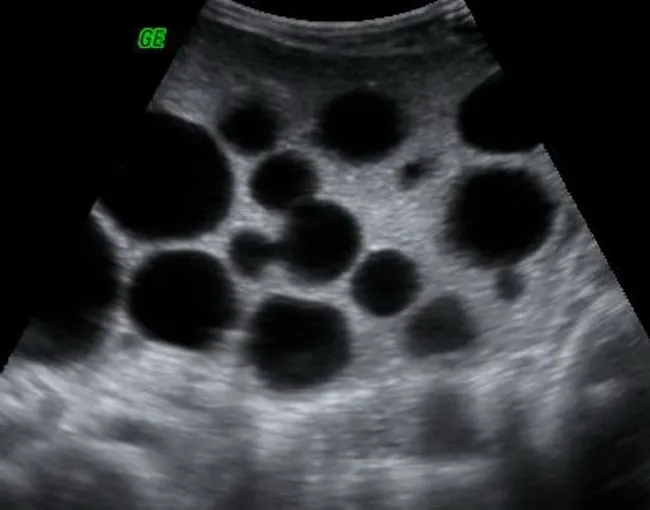
Ultrasound imaging of the peritoneal cavity demonstrates multifocal cystic lesions with well defined circular margins. The predominant appearance is multiple anechoic to hypoechoic spheres, varying in size from a few millimeters to several centimeters, with posterior acoustic enhancement and minimal internal echoes. The lesions are scattered across the peritoneal surfaces, consistent with peritoneal hydatidosis, most likely secondary to rupture or spillage from hepatic hydatid cysts; primary peritoneal hydatidosis is rare. In this case, the pattern supports secondary dissemination due to hepatic disease; the appearance may include daughter cysts and serpiginous internal septations in some cysts, creating a "cluster of grapes" or "wheel spoke" configuration. The sonographic findings correlate with echinococcosis: cysts may remain nonviable or contain viable protoscoleces, depending on host response. There is no confirmed solid nodularity or ascites; vascularity on Doppler is typically minimal. Clinically, this imaging finding suggests parasitic infection by Echinococcus species; the diagnostic significance lies in risk stratification for anaphylaxis on antigen release and in planning antihelminthic therapy (albendazole, mebendazole) and possible surgical intervention. Differential considerations include peritoneal carcinomatosis, mucinous peritoneal implants, and other cystic peritoneal diseases. Correlation with liver imaging and serology is recommended for comprehensive management. Further imaging and serology will guide therapy.
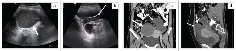
This diagnostic imaging composite displays grey-scale ultrasound and Contrast-Enhanced Computed Tomography (CECT) images of the pelvis in a 38-year-old female, demonstrating features of benign cystic mesothelioma. Panels (a) and (b) are transverse ultrasounds showing two distinct lesions: a well-defined cystic mass with low-level internal echoes ('ground glass' appearance) in the right adnexa, and an ill-defined, thin-walled multiloculated cystic lesion in the midline and left pelvis. Panels (c) and (d) provide coronal and sagittal CECT views, respectively. The right adnexal cyst is hyperdense (~33 HU), while the larger, multilobulated pelvic lesion shows fluid attenuation (~18 HU) with thin, imperceptible walls. The sagittal view (d) highlights the multiloculated lesion's relationship to pelvic anatomy, situated anterior and superior to the uterus and encasing the left ovary posteriorly. Key educational findings include the absence of internal calcifications or mural nodules, which helps differentiate this benign entity from malignant mesothelioma or peritoneal carcinomatosis. The presentation is also characteristic of peritoneal inclusion cysts often associated with endometriosis.
This diagnostic ultrasound image depicts a large, complex cystic mass located within the pelvic and lower abdominal cavity of a 38-year-old female patient. The mass demonstrates characteristic features of a multi-loculated ovarian cyst, including internal septations that divide the structure into multiple compartments. Key echogenic findings include a heterogenous internal environment with dense punctate or spot echoes dispersed throughout the fluid-filled regions, indicating the presence of debris, hemorrhage, or high-viscosity content. Additionally, certain areas of the mass exhibit vesicle-like aggregations. The image also captures evidence of peritoneal effusion, characterized by poor sound transmission and dense echoes within the free fluid. This clinical imaging is used for the preoperative assessment of ovarian pathology, illustrating complex internal morphology that aids in differentiating between simple and complex gynecological cysts.
Differential Diagnosis
1. Hydatid Disease (Echinococcosis) - TOP DIFFERENTIAL
- Mechanism: Rupture or spillage of a hepatic hydatid cyst seeds the peritoneum. Primary peritoneal hydatidosis is rare.
- Ultrasound features: Multiple cysts in varying WHO classes - internal echoes from scolices ("hydatid sand"), floating membranes ("water-lily sign"), daughter cysts creating a "cluster of grapes" or "wheel spoke" pattern, and coarse internal debris.
- Serology (IgG ELISA) and liver imaging confirm the diagnosis.
- Risk of anaphylaxis on cyst rupture.
2. Pseudomyxoma Peritonei
- Origin: Ruptured appendiceal mucinous neoplasm (low-grade appendiceal mucinous neoplasm - LAMN) or ovarian mucinous tumor.
- Ultrasound features: Loculated cystic deposits on peritoneal surfaces, internal echoes from thick mucin ("jelly belly"), scalloping of visceral surfaces (liver, spleen). Calcifications along borders may be seen.
- As described in [Yamada's Gastroenterology], implants are "often large, low in attenuation, cystic, and with calcifications along their borders."
- Key clue: Look for a dilated appendix or appendiceal mass as the primary.
3. Peritoneal Inclusion Cysts (Benign Cystic Mesothelioma)
- Occur almost exclusively in premenopausal women with prior pelvic surgery, PID, or endometriosis.
- Ultrasound features: Thin-walled multiloculated cystic mass in the pelvis; may show low-level internal echoes ("ground glass" appearance) from proteinaceous fluid.
- The ovary is often seen embedded within or adjacent to the cyst wall - a distinguishing feature.
- As per Grainger & Allison's Diagnostic Radiology: "a rare non-neoplastic mesothelial proliferation, most frequently found in the pelvis... unilocular or complex cystic mass comprising cysts of 1mm to 6cm."
4. Peritoneal Abscess / Loculated Infected Ascites
- Post-surgical, post-perforation (appendix, bowel, peptic ulcer), or from PID.
- Ultrasound features: Complex fluid collection with coarse internal echoes (pus, debris, fibrin strands), thick irregular walls, septa, possible gas bubbles (hyperechoic foci with dirty shadowing).
- Clinical context: fever, elevated WBC, peritonitis signs.
5. Peritoneal Carcinomatosis with Mucinous Deposits
- From ovarian, colorectal, gastric, or appendiceal primary tumors.
- Ultrasound features: Multiple irregular cystic deposits at omentum/peritoneal surfaces, coarse echoes from tumor necrosis or mucin, associated solid nodules, omental cake, ascites.
- As noted in Rosen's Emergency Medicine: findings suggestive of malignancy include "complex or cystic lesion with thick, irregular walls, and the presence of ascites, peritoneal metastases."
6. Hematoma (Peritoneal / Mesenteric)
- Post-trauma, anticoagulant therapy, or spontaneous (coagulopathy).
- Ultrasound features: Acute: hyperechoic. Subacute/resolving: mixed echogenicity with internal clot echoes and fluid-fluid levels. Chronic: liquefies to complex cystic.
- Key clue: History of trauma or anticoagulation; no internal vascularity on Doppler.
7. Lymphangioma (Mesenteric / Omental Cyst)
- Congenital malformation of lymphatic channels; more common in children/young adults.
- Ultrasound: Multiloculated thin-walled cystic mass in small bowel mesentery; usually anechoic but chylous content can produce low-level diffuse internal echoes.
- Distinguished from peritoneal inclusion cysts by location (mesentery vs. pelvis) and demographics (more common in males, pediatric).
8. Ovarian Cyst with Peritoneal Rupture
- Ruptured endometrioma, hemorrhagic cyst, or dermoid (teratoma) spilling into the peritoneum.
- Ultrasound features: Complex free fluid with echogenic debris, ground-glass echoes (endometrioma), or hyperechoic sebaceous material (dermoid).
Summary Table
| Diagnosis | Key Echo Feature | Key Clinical Clue |
|---|---|---|
| Hydatid disease | Daughter cysts, sand, membranes | Endemic area, eosinophilia |
| Pseudomyxoma peritonei | Thick mucin, scalloping | Appendiceal mass |
| Peritoneal inclusion cyst | Low-level echoes, ovary embedded | Prior surgery, female |
| Peritoneal abscess | Debris, gas, thick wall | Fever, post-op |
| Carcinomatosis | Nodules, omental cake, ascites | Known primary |
| Hematoma | Clot, fluid-fluid level | Trauma, anticoagulation |
| Lymphangioma | Chylous echoes, mesenteric | Pediatric, male |
| Ruptured ovarian cyst | Echogenic free fluid | Female, acute pain |
Peritoneal cavity containing cystic structures with coarse internal echoes. What would be diffrentials
peritoneal cystic structures internal echoes ultrasound differential
Ultrasound imaging of the peritoneal cavity demonstrates multifocal cystic lesions with well defined circular margins. The predominant appearance is multiple anechoic to hypoechoic spheres, varying in size from a few millimeters to several centimeters, with posterior acoustic enhancement and minimal internal echoes. The lesions are scattered across the peritoneal surfaces, consistent with peritoneal hydatidosis, most likely secondary to rupture or spillage from hepatic hydatid cysts; primary peritoneal hydatidosis is rare. In this case, the pattern supports secondary dissemination due to hepatic disease; the appearance may include daughter cysts and serpiginous internal septations in some cysts, creating a "cluster of grapes" or "wheel spoke" configuration. The sonographic findings correlate with echinococcosis: cysts may remain nonviable or contain viable protoscoleces, depending on host response. There is no confirmed solid nodularity or ascites; vascularity on Doppler is typically minimal. Clinically, this imaging finding suggests parasitic infection by Echinococcus species; the diagnostic significance lies in risk stratification for anaphylaxis on antigen release and in planning antihelminthic therapy (albendazole, mebendazole) and possible surgical intervention. Differential considerations include peritoneal carcinomatosis, mucinous peritoneal implants, and other cystic peritoneal diseases. Correlation with liver imaging and serology is recommended for comprehensive management. Further imaging and serology will guide therapy.
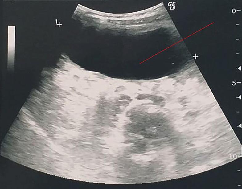
This abdominal ultrasonography (B-mode) demonstrates a large, well-defined cystic lesion within the peritoneal cavity, likely representing a primary peritoneal hydatid cyst (echinococcosis). The mass is predominantly anechoic, indicating a fluid-filled interior, with a smooth, regular wall. Significant posterior acoustic enhancement is visible distal to the cyst, which is a classic diagnostic feature of fluid-containing structures. The internal architecture reveals subtle, low-level echoes (hydatid sand or debris) settling in the dependent posterior portion of the cyst. The lesion exerts a mass effect, evidenced by the compression and displacement of adjacent heterogeneous soft tissues in the mesenteric or pelvic region. No internal septations or daughter cysts are clearly visible in this specific plane, characterizing it as a WHO type CE1 cyst. This image is an educational example of parasitic peritoneal disease and the use of diagnostic ultrasound for characterizing cystic abdominal masses.
This diagnostic ultrasound image depicts a large, complex cystic mass located within the pelvic and lower abdominal cavity of a 38-year-old female patient. The mass demonstrates characteristic features of a multi-loculated ovarian cyst, including internal septations that divide the structure into multiple compartments. Key echogenic findings include a heterogenous internal environment with dense punctate or spot echoes dispersed throughout the fluid-filled regions, indicating the presence of debris, hemorrhage, or high-viscosity content. Additionally, certain areas of the mass exhibit vesicle-like aggregations. The image also captures evidence of peritoneal effusion, characterized by poor sound transmission and dense echoes within the free fluid. This clinical imaging is used for the preoperative assessment of ovarian pathology, illustrating complex internal morphology that aids in differentiating between simple and complex gynecological cysts.
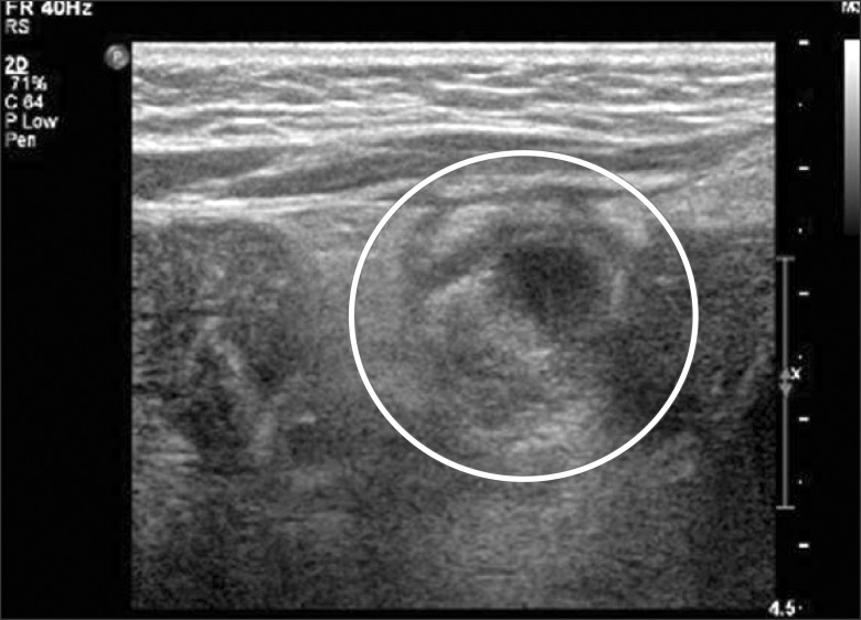
**Imaging Modality:** Abdominal Ultrasonography (USG), grayscale B-mode. **Anatomical Region:** Infraumbilical abdominal wall and underlying peritoneal cavity. **Observed Pathology:** A well-defined cystic mass is localized in the subumbilical region. The lesion demonstrates a complex internal echo pattern characterized by heterogeneous echogenic material within a predominantly hypoechoic fluid collection. **Characteristic Visual Features:** * **Morphology:** Ovoid to tubular cystic structure (highlighted by a white circular annotation). * **Internal Echoes:** Presence of low-level internal echoes and debris, indicating a non-simple fluid composition (consistent with proteinaceous material, hemorrhage, or infection). * **Relationship to Adjacent Structures:** The mass is situated deep to the rectus sheath and appears to extend toward or communicate with adjacent intestinal loops. * **Wall Characteristics:** The borders of the cyst are moderately thickened and irregular. **Clinical Context & Differential Diagnosis:** These sonographic findings are highly suggestive of an urachal anomaly, specifically an infected urachal cyst or a patent urachal remnant communicating with the bowel or bladder. The presence of internal heterogenicity suggests secondary inflammation or abscess formation.
Why "Coarse Internal Echoes" Matter
Differential Diagnoses
1. Hydatid Disease (Echinococcosis) - MOST CLASSIC
- Pathophysiology: Secondary peritoneal hydatidosis from rupture/spillage of a hepatic Echinococcus cyst. Primary peritoneal hydatidosis is rare.
- Ultrasound features:
- "Hydatid sand" - mobile echogenic foci (scolices) that shift with patient movement
- Floating membranes - "water-lily sign" (detached endocyst)
- Daughter cysts - "cluster of grapes" or "wheel-spoke" pattern
- WHO CE classification: CE1 (simple + sand) to CE5 (calcified)
- Key clues: Endemic area (Middle East, South America, Central Asia), eosinophilia, positive serology (IgG ELISA), liver cysts
2. Pseudomyxoma Peritonei
- Origin: Ruptured appendiceal mucinous neoplasm (LAMN/HAMN) - rarely from ovarian mucinous tumor. Seeding of thick gelatinous mucin across peritoneal surfaces.
- Ultrasound features:
- Multiple loculated cystic deposits with coarse internal echoes from thick mucin
- "Jelly belly" - septate, gelatinous ascites with low-level echoes
- Scalloping of liver/spleen margins - characteristic
- Calcifications along cyst borders
- Omental cake
- Key clues: Prior appendiceal or ovarian surgery, right lower quadrant mass, CT showing "scalloped viscera" and mucinous implants
3. Peritoneal Abscess / Loculated Infected Fluid
- Causes: Post-appendectomy, perforated peptic ulcer, Crohn's disease, PID, post-operative.
- Ultrasound features:
- Complex fluid collection with coarse/dirty internal echoes (pus, fibrin, debris)
- Thick irregular walls
- Internal septa
- Hyperechoic foci with dirty shadowing if gas-forming organisms
- Key clues: Fever, leukocytosis, recent surgery or perforation, point tenderness
4. Peritoneal Carcinomatosis (Mucinous Type)
- Primary tumors: Ovarian, colorectal, gastric, appendiceal, pancreatic.
- Ultrasound features:
- Irregular cystic deposits on peritoneal surfaces with internal echoes from tumor necrosis or mucin
- Solid nodules mixed with cystic components
- Omental thickening/cake
- Free or loculated ascites with echogenic debris
- Key clues: Known primary malignancy, weight loss, CA-125/CEA elevated. As noted in Grainger & Allison's Diagnostic Radiology: malignant mesothelioma and carcinomatosis appear similar - peritoneal nodularity with ascites is shared.
5. Peritoneal Inclusion Cysts (Benign Cystic Mesothelioma)
- Who: "Almost exclusively in premenopausal women with history of pelvic/abdominal surgery, pelvic inflammatory disease, or endometriosis" (Yamada's Gastroenterology)
- Mechanism: Non-neoplastic reactive mesothelial proliferation - ovarian fluid trapped within peritoneal adhesions
- Ultrasound features:
- Unilocular or complex multiloculated cystic mass, pelvis predominant
- May show low-level internal echoes (proteinaceous fluid)
- Ovary embedded within or adjacent to the cyst - pathognomonic sign
- Cysts range from 1mm to 6cm (Grainger & Allison)
- Key clues: Premenopausal woman, prior abdominopelvic surgery, chronic pelvic pain
6. Tuberculous Peritonitis (Wet/Loculated Type)
- Ultrasound features:
- Loculated ascites with fine internal echoes ("dirty" fluid)
- Fibrin strands, septa, and thickened peritoneum
- Omental thickening - "omental cake" may appear cystic with echogenic nodules
- Mesenteric lymphadenopathy
- Key clues: TB endemic region, HIV, constitutional symptoms (fever, night sweats, weight loss), high ADA in ascitic fluid. Grainger & Allison note: "a smooth peritoneum with minimal thickening and marked enhancement suggests tuberculous peritonitis."
7. Hematoma (Peritoneal / Mesenteric)
- Causes: Trauma, anticoagulation, ruptured ectopic pregnancy, ruptured hemorrhagic ovarian cyst, spontaneous mesenteric bleeding.
- Ultrasound features:
- Acute: hyperechoic solid-appearing clot
- Subacute (days-weeks): complex cystic with coarse internal echoes, fluid-fluid levels
- Chronic: liquefied complex cyst with debris
- Key clues: Trauma, anticoagulants, sudden onset pain, falling hemoglobin
8. Endometrioma with Peritoneal Spillage / Chocolate Cyst
- As per Berek & Novak's Gynecology: "typical ultrasound features of an endometriotic ovarian cyst... described as ground-glass echogenicity" - diffuse homogeneous low-level internal echoes from degraded blood products.
- When ruptured, this content spills into the peritoneum producing complex free fluid and cystic deposits with coarse echoes.
- Key clues: Dysmenorrhea, dyspareunia, known endometriosis, premenopausal woman, sudden pelvic pain (rupture)
9. Mesenteric / Omental Cysts (Lymphangioma)
- Congenital lymphatic malformations, more common in children.
- Usually anechoic, but chylous content or hemorrhage into the cyst produces diffuse low-level internal echoes.
- Located in small bowel mesentery (vs. peritoneal inclusion cysts which are pelvic).
Summary Table
| Diagnosis | Echo Character | Key Distinguishing Feature |
|---|---|---|
| Hydatid disease | Sand, membranes, daughter cysts | Endemic area, liver cysts, serology |
| Pseudomyxoma peritonei | Thick mucin, low-level diffuse | Appendiceal mass, scalloped liver |
| Peritoneal abscess | Debris, gas, dirty echoes | Fever, post-op, leukocytosis |
| Carcinomatosis | Tumor necrosis, solid+cystic | Known primary malignancy |
| Peritoneal inclusion cyst | Low-level, ground-glass | Premenopausal, prior surgery, ovary inside cyst |
| TB peritonitis | Fine echoes, fibrin strands | TB risk, ADA elevated, lymphadenopathy |
| Hematoma | Fluid-fluid level, clot | Trauma, anticoagulation, falling Hb |
| Endometrioma/spillage | Homogeneous ground-glass | Endometriosis history, acute pain |
| Lymphangioma | Chylous echoes | Pediatric, mesenteric location |
- Hydatid
- Abscess
- Pseudomyxoma peritonei
- TB peritonitis
- Endometrioma / hematoma
- Carcinomatosis
In male with history of ca esophagus and pyonephrosis and grade 3 medical renal changes what are differentials
Clinical Context: What Is Being Asked
- Carcinoma of the esophagus (primary malignancy + treatment history)
- Pyonephrosis (infected, obstructed kidney - a urological emergency)
- Grade 3 medical renal disease (on ultrasound = increased cortical echogenicity beyond liver/spleen, with poor corticomedullary differentiation)
Understanding Grade 3 Medical Renal Disease (Ultrasound)
- Grade 0: Normal - cortex less echogenic than liver/spleen
- Grade 1: Cortex = liver echogenicity
- Grade 2: Cortex > liver but < sinus fat
- Grade 3: Cortex = sinus fat echogenicity (most echogenic, severe parenchymal disease)
Differential Diagnoses
A. OBSTRUCTIVE / POSTRENAL CAUSES
1. Chronic Obstructive Nephropathy from Esophageal Cancer
- Esophageal cancer - particularly squamous cell carcinoma - can metastasize to retroperitoneal/para-aortic lymph nodes which encircle and compress the ureters.
- As Harrison's notes: "early in the course of obstruction or if the ureters are unable to dilate (e.g., encasement by pelvic or periureteral tumors or by retroperitoneal fibrosis), the ultrasound examination may be negative."
- Chronic bilateral obstruction → progressive parenchymal fibrosis → grade 3 echogenicity.
2. Pyonephrosis-Related Parenchymal Destruction
- Pyonephrosis (infected hydronephrosis with pus in the collecting system) causes direct renal parenchymal damage through:
- Suppurative destruction of renal tubules and interstitium
- Elevated intrarenal pressure → ischemic atrophy
- Xanthogranulomatous pyelonephritis (XGP) - chronic severe form
- The contralateral kidney undergoes compensatory hypertrophy, while the affected kidney develops progressive echogenic fibrosis.
B. NEPHROTOXIC / TREATMENT-RELATED CAUSES
3. Cisplatin Nephrotoxicity
- Esophageal cancer is commonly treated with cisplatin + 5-FU or cisplatin + immunotherapy.
- As per Brenner & Rector's The Kidney: "The principal site of renal damage with cisplatin is the proximal tubule. The nephrotoxicity of cisplatin is dose-dependent, yet AKI can result from a single exposure."
- Cumulative cisplatin causes chronic tubulointerstitial nephritis → parenchymal fibrosis → echogenic kidneys.
- Electrolyte disturbances: hypomagnesemia, hypokalemia are hallmarks.
4. Checkpoint Inhibitor Nephritis (Immunotherapy)
- Modern esophageal cancer regimens include nivolumab, pembrolizumab.
- Brenner & Rector: "Checkpoint inhibitors, such as nivolumab, may cause a variety of immune-related adverse events, including acute interstitial nephritis (AIN)."
- AIN → if untreated or recurrent → chronic interstitial nephritis → parenchymal scarring.
5. Radiation Nephropathy
- Radiation to the chest/upper abdomen for esophageal cancer can involve the left kidney (field exposure).
- Radiation nephritis: months to years post-exposure, presents as hypertension + proteinuria + renal impairment → parenchymal fibrosis.
6. Contrast Nephropathy / NSAID Use
- As per Brenner & Rector's Box 29.4 on AKI in cancer patients: "Exogenous nephrotoxins - chemotherapy, antibiotics, contrast agents" and "NSAIDs for pain management" are all common culprits in the cancer patient.
- Repeated contrast exposure during CT staging/surveillance + NSAID analgesia for dysphagia pain → cumulative tubular injury.
C. SEPSIS-RELATED CAUSES
7. Sepsis-Associated AKI / ATN
- Pyonephrosis causes urosepsis - a life-threatening complication.
- Harrison's: "Ischemic ATN is observed most frequently in patients who have undergone... overwhelming sepsis."
- Sepsis induces renal vasoconstriction, endothelial injury, and microvascular thrombi → ischemic ATN → if recurrent or prolonged → parenchymal fibrosis.
- Grade 3 echogenicity can follow repeated septic AKI episodes.
D. INTRINSIC/PARENCHYMAL CAUSES
8. Hypertensive Nephrosclerosis
- Esophageal cancer patients are often middle-aged to elderly males with pre-existing hypertension.
- Longstanding hypertension → arterionephrosclerosis → cortical thinning and increased echogenicity (grade 3).
9. Diabetic Nephropathy
- Comorbid diabetes (common in this age/sex group) causes glomerulosclerosis → increased cortical echogenicity.
10. Obstructive Uropathy from Tumor Direct Extension
- Esophageal cancer rarely (but possibly) involves the thoracic duct, aorta, or left renal vein by direct invasion.
- More commonly, mediastinal/para-aortic metastatic nodes compress renal vasculature or ureters.
11. Paraneoplastic Glomerulonephritis
- Esophageal carcinoma (especially SCC) can rarely cause membranous nephropathy or IgA nephropathy as a paraneoplastic phenomenon.
- Presents with proteinuria, haematuria, progressive renal impairment.
12. Amyloidosis (AA type)
- Chronic infection (pyonephrosis = chronic suppuration) + chronic malignancy → secondary (AA) amyloidosis.
- Amyloid deposits in glomeruli → nephrotic syndrome → progressive renal failure → echogenic kidneys.
- Brenner & Rector note amyloid as a cause of AKI in the cancer setting.
Summary: Organized Differential
| Category | Diagnosis | Key Mechanism |
|---|---|---|
| Obstructive | Retroperitoneal LN compression (metastatic) | Ureteral encasement by nodes |
| Obstructive | Pyonephrosis-related parenchymal destruction | Suppurative ATN + fibrosis |
| Nephrotoxic | Cisplatin nephrotoxicity | Proximal tubular necrosis → fibrosis |
| Nephrotoxic | Checkpoint inhibitor AIN | Immune-mediated interstitial nephritis |
| Nephrotoxic | Radiation nephropathy | Field-exposure fibrosis |
| Nephrotoxic | Contrast/NSAID injury | Cumulative tubular toxicity |
| Septic | Urosepsis-related ischemic ATN | Hemodynamic + endothelial injury |
| Vascular | Hypertensive nephrosclerosis | Arterial wall thickening + scarring |
| Metabolic | Diabetic nephropathy | Glomerulosclerosis |
| Paraneoplastic | Membranous nephropathy | Immune complex deposition |
| Infiltrative | AA Amyloidosis | Chronic infection + malignancy |
Clinical Priority
- Cisplatin nephrotoxicity - extremely common in esophageal cancer treatment
- Urosepsis/ATN from pyonephrosis - the acute-on-chronic injury driver
- Chronic obstructive nephropathy - from retroperitoneal nodal disease
- Pre-existing hypertensive/diabetic nephropathy - background parenchymal disease
- AA amyloidosis - must be considered given dual insults of chronic infection + malignancy
Peritoneal cavity containing cystic structures with coarse internal echoes with grade 3 renal parenchymal changes known case of ca esophagus with chemotherapy. What would be diffrentials
peritoneal metastasis cystic echoes ultrasound esophageal cancer
Ultrasound imaging of the peritoneal cavity demonstrates multifocal cystic lesions with well defined circular margins. The predominant appearance is multiple anechoic to hypoechoic spheres, varying in size from a few millimeters to several centimeters, with posterior acoustic enhancement and minimal internal echoes. The lesions are scattered across the peritoneal surfaces, consistent with peritoneal hydatidosis, most likely secondary to rupture or spillage from hepatic hydatid cysts; primary peritoneal hydatidosis is rare. In this case, the pattern supports secondary dissemination due to hepatic disease; the appearance may include daughter cysts and serpiginous internal septations in some cysts, creating a "cluster of grapes" or "wheel spoke" configuration. The sonographic findings correlate with echinococcosis: cysts may remain nonviable or contain viable protoscoleces, depending on host response. There is no confirmed solid nodularity or ascites; vascularity on Doppler is typically minimal. Clinically, this imaging finding suggests parasitic infection by Echinococcus species; the diagnostic significance lies in risk stratification for anaphylaxis on antigen release and in planning antihelminthic therapy (albendazole, mebendazole) and possible surgical intervention. Differential considerations include peritoneal carcinomatosis, mucinous peritoneal implants, and other cystic peritoneal diseases. Correlation with liver imaging and serology is recommended for comprehensive management. Further imaging and serology will guide therapy.
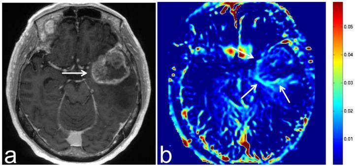
This diagnostic image pair features magnetic resonance imaging (MRI) of a brain metastasis from esophageal cancer in a 70-year-old male. Image (a) is an axial, three-dimensional, contrast-enhanced T1-weighted sequence showing a large, peripherally enhancing (ring-enhancing) lesion in the left temporal lobe. The metastasis exhibits an irregular border and a hypointense central area, suggesting central necrosis or cystic change. Image (b) shows the corresponding axial Amide Proton Transfer-weighted Chemical Exchange Saturation Transfer (APT-CEST) sequence. This molecular imaging modality displays a relatively high signal intensity (SI) at the tumor margin (highlighted by white arrows), represented by yellow-red hues on the accompanying color bar (approx. 0.03 to 0.05). The high APT signal at the periphery correlates with increased concentrations of endogenous mobile proteins and peptides, often associated with high cellular proliferation in malignant tumor margins. In contrast, the internal regions and surrounding healthy brain tissue show lower signal intensities (blue-green hues), providing metabolic differentiation of the lesion.
This diagnostic ultrasound image depicts a large, complex cystic mass located within the pelvic and lower abdominal cavity of a 38-year-old female patient. The mass demonstrates characteristic features of a multi-loculated ovarian cyst, including internal septations that divide the structure into multiple compartments. Key echogenic findings include a heterogenous internal environment with dense punctate or spot echoes dispersed throughout the fluid-filled regions, indicating the presence of debris, hemorrhage, or high-viscosity content. Additionally, certain areas of the mass exhibit vesicle-like aggregations. The image also captures evidence of peritoneal effusion, characterized by poor sound transmission and dense echoes within the free fluid. This clinical imaging is used for the preoperative assessment of ovarian pathology, illustrating complex internal morphology that aids in differentiating between simple and complex gynecological cysts.
peritoneal cystic lesions coarse echoes esophageal cancer chemotherapy renal parenchymal changes differential diagnosis
Clinical Framework
| Finding | Significance |
|---|---|
| Peritoneal cystic lesions with coarse internal echoes | Complex peritoneal disease - infection, mucin, blood, or malignant deposits |
| Grade 3 renal parenchymal changes | Severe cortical echogenicity = significant renal parenchymal disease |
| CA esophagus on chemotherapy | Known malignancy + nephrotoxic/immunosuppressive treatment |
PART 1: Differentials for the Peritoneal Cystic Lesions with Coarse Echoes
1. Peritoneal Carcinomatosis with Cystic/Necrotic Deposits - TOP DIFFERENTIAL in this setting
- Esophageal carcinoma (both SCC and adenocarcinoma) does spread to the peritoneum, though less commonly than gastric or ovarian cancer. When it occurs, it is a Stage M1 disease.
- Cystic deposits with coarse echoes = necrotic tumor implants on peritoneal surfaces, seeded via transcoelemic spread or direct extension through the gastroesophageal junction.
- Associated findings: echogenic/loculated ascites, omental cake, hepatic deposits.
- The PMC peritoneal disease review confirms: "Metastatic disease should be the initial concern in a patient with ascites and peritoneal nodularity at imaging."
2. Chemotherapy-Related Peritoneal Pseudocysts / Loculated Ascites
- Chemotherapy (cisplatin, 5-FU, taxanes) causes peritoneal irritation, mesothelial injury, and adhesion formation → loculated fluid collections.
- These collections develop internal echoes from debris, fibrin, and inflammatory exudate.
- Chemotherapy-induced thrombocytopenia can also lead to peritoneal hemorrhage → hematoma with coarse echoes.
3. Peritoneal Abscess / Opportunistic Infection
- Chemotherapy causes immunosuppression (neutropenia) → susceptibility to peritoneal infections:
- Bacterial peritonitis (gram-negatives, anaerobes)
- Fungal peritonitis (Candida, Aspergillus) - particularly in patients on prolonged antibiotics/steroids
- Tuberculous peritonitis (reactivation in immunosuppressed)
- All produce loculated complex cystic collections with coarse internal echoes.
4. Pseudomyxoma Peritonei
- Less likely given the primary is esophageal, but esophageal adenocarcinoma (especially at the GEJ) can theoretically produce mucinous peritoneal implants.
- Classic features: thick mucinous echoes, scalloping of liver/spleen, loculated "jelly-belly" ascites.
5. Hydatid Disease (if endemic region)
- Immunosuppressed patients cannot contain hydatid cysts - risk of rupture and peritoneal seeding increases.
- "Cluster of grapes," daughter cysts, hydatid sand = characteristic coarse internal echoes.
PART 2: Differentials for Grade 3 Renal Parenchymal Changes
1. Cisplatin Nephrotoxicity - MOST LIKELY in this context
- Standard esophageal cancer chemotherapy = cisplatin + 5-FU or FLOT (5-FU, leucovorin, oxaliplatin, docetaxel).
- As established in Brenner & Rector's The Kidney: "The principal site of renal damage with cisplatin is the proximal tubule. The nephrotoxicity is dose-dependent... electrolyte disturbances including hypomagnesemia and hypokalemia are common."
- Cumulative cycles → progressive tubulointerstitial fibrosis → grade 3 cortical echogenicity.
- Cumulative cisplatin causes:
- Acute proximal tubular necrosis
- Chronic interstitial fibrosis
- Reduction in GFR (irreversible beyond certain cumulative dose)
2. Checkpoint Inhibitor-Induced Nephritis
- Modern regimens for esophageal cancer now include nivolumab (CheckMate-649, CheckMate-577) and pembrolizumab (KEYNOTE-590).
- Immune checkpoint inhibitors cause acute interstitial nephritis (AIN) via T-cell-mediated renal tubular injury.
- Untreated AIN → chronic interstitial fibrosis → increased cortical echogenicity.
3. Urosepsis / Sepsis-Associated ATN
- If pyonephrosis is also present (as in the previous presentation), urosepsis-driven ATN contributes to parenchymal damage.
- Repeated ischemic-septic insults → grade 3 changes.
4. Pre-existing Hypertensive/Diabetic Nephropathy
- Background comorbidities in this age/sex group (middle-aged to elderly male) amplified by the nephrotoxic stress of chemotherapy.
5. Obstructive Nephropathy from Retroperitoneal Nodal Metastases
- Esophageal cancer metastatic to para-aortic nodes → ureteral encasement → obstructive nephropathy → parenchymal atrophy with grade 3 echogenicity.
6. Tumour Lysis Syndrome
- During induction chemotherapy, particularly with bulky disease → hyperuricemia, hyperphosphatemia → urate/calcium-phosphate nephropathy.
- Brenner & Rector: "Tumor lysis syndrome... is a well-recognized cause of AKI in patients with cancer."
7. Radiation Nephropathy
- Upper abdominal radiation for esophageal cancer (particularly left-sided or GEJ tumors) can involve the left kidney field → radiation nephritis months to years later.
8. AA Amyloidosis
- Chronic malignancy + chronic infection (if concurrent pyonephrosis/sepsis) → secondary AA amyloidosis.
- Amyloid deposition in glomeruli → nephrotic syndrome → progressive renal echogenicity.
PART 3: UNIFIED Differentials - Conditions That Explain BOTH Findings Together
| Unifying Diagnosis | Peritoneal Echoes | Renal Grade 3 | Mechanism |
|---|---|---|---|
| Advanced esophageal cancer with peritoneal + renal metastasis | Necrotic cystic implants | Parenchymal infiltration or obstructive uropathy | M1 disease, bilateral |
| Cisplatin toxicity + chemo-related peritoneal disease | Loculated ascites, hemorrhage | Proximal tubular necrosis → fibrosis | Drug toxicity |
| Opportunistic infection (immunosuppressed) | Peritoneal abscess, TB, fungal | Sepsis-ATN + tubulonephritis | Chemotherapy neutropenia |
| AA Amyloidosis | Peritoneal deposits (rare) | Glomerular amyloid deposition | Chronic malignancy + infection |
| Retroperitoneal lymphadenopathy from metastases | Obstructed ascites/loculations | Bilateral ureteral obstruction | Nodal bulk disease |
Priority Differential List (Most to Least Likely)
- Peritoneal carcinomatosis + cisplatin nephrotoxicity - the most common unified explanation; stage M1 esophageal cancer with coexisting chemotherapy-related renal damage
- Checkpoint inhibitor nephritis (AIN) + peritoneal immunotherapy-related reaction - if on nivolumab/pembrolizumab
- Opportunistic peritoneal infection (abscess/TB/fungal) + sepsis-ATN - immunosuppression-related
- Retroperitoneal nodal metastases - causing both peritoneal seeding and bilateral ureteral obstruction
- Pseudomyxoma peritonei + obstructive nephropathy
- AA Amyloidosis - late complication of combined malignancy and infection
What to Do Next
| Investigation | Purpose |
|---|---|
| CT abdomen/pelvis with contrast | Characterize peritoneal deposits, look for nodal disease, ureteral obstruction |
| Serum creatinine, eGFR, urine PCR | Quantify renal injury |
| Serum Mg, K | Cisplatin tubulopathy signature |
| Urine cytology + urinalysis | AIN (eosinophiluria), malignant cells |
| Beta-2 microglobulin, urine protein electrophoresis | Proximal tubular dysfunction |
| Ascitic fluid cytology + culture | Malignant vs infectious peritoneal disease |
| Serum ADA, fungal cultures | TB / fungal peritonitis if immunosuppressed |
| Renal biopsy | If cause of grade 3 changes remains unclear after workup |