I now have comprehensive content from multiple authoritative textbooks on all three topics. Let me compose the full, well-structured answer.
Cell Biology: Lysosomes, Cell Membrane & Transport, Cytoskeleton
1. LYSOSOMES
Structure
Lysosomes are membrane-bound vesicular organelles that bud off from the trans-Golgi network. They are typically spherical, ranging from 0.05 to 0.75 µm in diameter, and filled with ~40 different hydrolytic (acid hydrolase) enzymes - proteases, nucleases, lipases, phosphatases, glycosidases, and sulfatases. In TEM they appear electron-dense.
- The lysosomal pH is ~4.8-5.0, maintained by proton pumps in the lysosomal membrane that actively pump H⁺ into the lumen.
- At the neutral cytosolic pH (~7.2), leaked lysosomal enzymes become inactive - a key safety mechanism protecting the cell.
- Lysosomes are most numerous in cells with high phagocytic activity (macrophages, neutrophils).
Lysosomal Enzyme Targeting (M6P Pathway)
Lysosomal enzymes are synthesized in the rough ER, then transported to the Golgi apparatus. In the cis-Golgi, a phosphotransferase adds mannose-6-phosphate (M6P) residues to the N-linked oligosaccharides of hydrolases destined for lysosomes. M6P receptors in the trans-Golgi network recognize these proteins and package them into vesicles that deliver them to lysosomes - diverting them away from the secretory pathway.
Clinical pearl - I-cell disease: Phosphotransferase deficiency means M6P tags cannot be added, so lysosomal enzymes are secreted extracellularly instead of reaching lysosomes. Cells accumulate undigested material (inclusions), affecting the skeleton and nervous system.
Functions of Lysosomes
| Function | Mechanism |
|---|
| Heterophagy | Phagosomes/pinocytotic vesicles fuse with lysosomes → heterolysosome → digestion; indigestible material remains as a residual body |
| Autophagy | Defunct organelles wrapped in double-membrane autophagosome (derived from ER); fuses with lysosome for digestion and nutrient recycling |
| Autolysis | Severe cell damage → lysosomal membrane rupture → hydrolases digest entire cell contents |
| Tissue regression | Uterine involution post-partum, muscle atrophy during inactivity, mammary gland regression after lactation |
| Bactericidal | Lysosomes contain lysozyme (dissolves bacterial cell wall), lysoferrin (sequesters iron), and acid pH (~4.8) that inactivates bacterial metabolism |
| Bone resorption | Osteoclasts secrete lysosomal enzymes into a restricted extracellular space to degrade bone matrix |
- Residual bodies accumulate as lipofuscin granules in long-lived post-mitotic cells (neurons, cardiac muscle).
Lysosomal Storage Diseases
Caused by deficiency of a specific lysosomal enzyme → substrate accumulates in secondary lysosomes/residual bodies → cellular dysfunction.
| Disease | Defective Enzyme | Substrate Stored | Tissue Affected |
|---|
| Tay-Sachs | Hexosaminidase A (GM₂-gangliosidase) | Gangliosides | Nervous system |
| Gaucher | Glucocerebrosidase | Glucocerebroside | Liver, spleen |
| Hurler syndrome (MPS I) | α-L-Iduronidase | Glycosaminoglycans | Skeleton, CNS |
| I-cell disease | Phosphotransferase (M6P formation) | Multiple substrates | Skeleton, CNS |
| McArdle syndrome | Muscle phosphorylase | Glycogen | Skeletal muscle |
2. CELL MEMBRANE AND TRANSPORT (★ HIGH YIELD)
Cell Membrane Structure
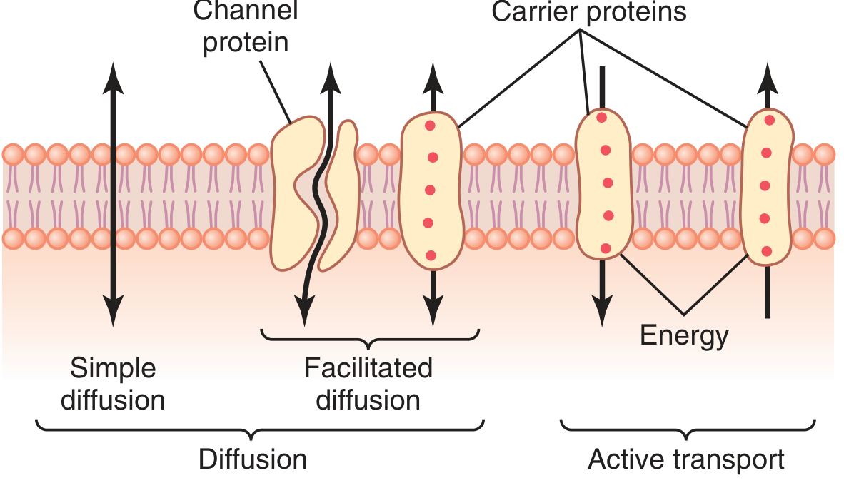
The plasma membrane is a phospholipid bilayer with two main types of proteins:
- Channel proteins - have watery pores allowing nearly free movement of water and selected ions (e.g., aquaporins, ion channels)
- Carrier proteins - bind specific molecules/ions, undergo conformational change to transport them across
The lipid bilayer itself is permeable to lipid-soluble substances (O₂, CO₂, steroid hormones, ethanol) but impermeable to most water-soluble and charged substances.
Figure: Transport pathways - simple diffusion, facilitated diffusion, and active transport (Guyton & Hall)
A. DIFFUSION (Passive Transport - no energy required)
Simple Diffusion
- Random molecular movement down a concentration gradient through lipid bilayer interspaces or protein channels.
- Rate follows Fick's Law: proportional to the concentration gradient, membrane surface area, and membrane permeability.
- Factors driving diffusion across a membrane:
- Concentration gradient (high → low)
- Electrical potential (for ions): positive charge attracts anions, repels cations
- Pressure difference: higher pressure side has more kinetic energy → net movement toward lower pressure
Nernst Potential
At equilibrium for an ion, the electrical and concentration gradients exactly balance each other. The voltage at which this occurs:
EMF (mV) = ±61 × log (C₁/C₂) (at 37°C for monovalent ions)
Facilitated Diffusion
- Uses carrier proteins - moves substances down their concentration gradient but faster than simple diffusion.
- No energy required - movement is still from high to low concentration.
- Example: glucose transport into cells (GLUT transporters); the rate shows saturation kinetics.
Osmosis
- Net diffusion of water through a selectively permeable membrane from lower solute concentration → higher solute concentration.
- The osmotic pressure that would exactly prevent net water movement is the osmotic pressure of the solution.
- Red blood cell membrane allows water diffusion equivalent to ~100× its volume per second.
B. ACTIVE TRANSPORT (Energy required, moves against gradient)
Primary Active Transport
- Uses ATP directly. Key example: Na⁺-K⁺ ATPase pump
Na⁺-K⁺ ATPase pump mechanism:
- 3 Na⁺ bind on the inside of the pump
- 2 K⁺ bind on the outside
- ATPase activity is activated → ATP is cleaved to ADP + Pᵢ
- Conformational change extrudes 3 Na⁺ out and brings 2 K⁺ in
- Net result: one positive charge moved outward per cycle (electrogenic)
Functions of the Na⁺-K⁺ pump:
- Maintains low intracellular Na⁺ and high intracellular K⁺
- Controls cell volume - without it, osmotic entry of water would cause cells to swell and burst (intracellular anions attract cations → osmosis)
- Generates the resting membrane potential (electrogenic - produces inside-negative)
- In electrically active neurons: 60-70% of total energy expenditure goes to this pump
The Na⁺-K⁺ pump can run in reverse - if electrochemical gradients are experimentally large enough, it synthesizes ATP from ADP + Pi.
Secondary Active Transport (Co-transport / Counter-transport)
- Uses the Na⁺ gradient created by Na⁺-K⁺ ATPase as the energy source (indirect).
- Co-transport (symport): Na⁺ and another solute move in the same direction (e.g., Na⁺-glucose co-transporter in intestinal epithelium)
- Counter-transport (antiport): Na⁺ and another solute move in opposite directions (e.g., Na⁺-Ca²⁺ exchanger - Ca²⁺ pumped out while Na⁺ enters)
Summary Table: Types of Transport
| Type | Energy | Direction | Example |
|---|
| Simple diffusion | No | Down gradient | O₂, CO₂, lipid-soluble drugs |
| Facilitated diffusion | No | Down gradient | Glucose (GLUT), fructose |
| Osmosis | No | Water down osmotic gradient | Water via aquaporins |
| Primary active | ATP | Against gradient | Na⁺-K⁺ pump, Ca²⁺-ATPase |
| Secondary active (symport) | Na⁺ gradient | Against gradient | Na⁺-glucose, Na⁺-amino acid |
| Secondary active (antiport) | Na⁺ gradient | Against gradient | Na⁺-Ca²⁺ exchanger, Na⁺-H⁺ |
C. Vesicular Transport (Bulk Transport)
- Endocytosis: cell membrane invaginates to engulf extracellular material
- Phagocytosis: large particles (bacteria, cell debris) - mainly in macrophages/neutrophils
- Pinocytosis: fluid droplets with dissolved substances
- Receptor-mediated endocytosis: specific molecules bind cell surface receptors → clathrin-coated pit → vesicle (e.g., LDL receptor pathway)
- Exocytosis: vesicle fuses with plasma membrane to release contents extracellularly (opposite of endocytosis)
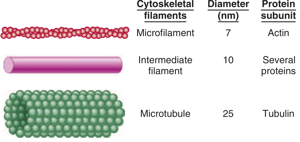
3. CYTOSKELETON
The cytoskeleton is an intracellular scaffold of structural proteins that maintains cell shape, polarity, organelle organization, and enables cell migration and division. It is composed of three major filament types, constantly undergoing assembly and disassembly.
Figure: The three cytoskeletal elements with their diameters and protein subunits (Ganong's)
A. Microtubules
- Diameter: 25 nm (hollow tubes); walls ~5 nm thick, inner cavity 15 nm
- Composition: α- and β-tubulin heterodimers polymerize into 13-protofilament hollow tubes
- γ-tubulin: associated with centrosomes (MTOCs) and initiates microtubule nucleation
- Polarity: "+" end (plus end) elongates rapidly; "-" end (minus end) typically embedded in the MTOC/centrosome near the nucleus
- Dynamics: temperature-sensitive; assembly at (+) end, disassembly at (-) end; GTP binding facilitates formation
- Functions:
- Tracks for molecular motor proteins (kinesins, dyneins) to transport vesicles, organelles, secretory granules, mitochondria
- Form the mitotic spindle - segregates sister chromatids during cell division
- Core of primary cilia (9+0 arrangement) - regulate proliferation/differentiation; defects cause polycystic kidney disease
- Core of motile cilia (9+2 arrangement) and flagella
Kinesins (anterograde motors): move cargo toward the "+" end (away from nucleus)
Dyneins (retrograde motors): move cargo toward the "-" end (toward nucleus/centrosome)
Drugs affecting microtubules:
- Colchicine / Vinca alkaloids (vincristine, vinblastine): inhibit microtubule polymerization → disrupt mitotic spindle → arrest cell division
- Taxol (paclitaxel): stabilizes microtubules (prevents depolymerization) → also arrests mitosis
B. Intermediate Filaments
- Diameter: 8-14 nm (between micro and macro - hence "intermediate")
- Composition: large, heterogeneous family of ropelike polymers
- Key property: do not actively reorganize like actin/microtubules; provide tensile strength and resist mechanical stress
- Tissue-specific expression (used as tumor markers):
| Protein | Cell Type | Clinical Use |
|---|
| Vimentin | Mesenchymal cells (fibroblasts, endothelium) | Marker for sarcomas |
| Desmin | Muscle cells | Scaffold for actin-myosin contraction |
| Cytokeratins (30+ types) | Epithelial cells | Markers for carcinomas; lung vs. GI distinction |
| Neurofilaments | Neurons | Structural strength and rigidity of axons |
| GFAP (Glial fibrillary acidic protein) | Glial cells | Marker for astrocytomas |
| Nuclear lamins | All nucleated cells (nuclear lamina) | Define nuclear shape, gene regulation |
- Connect desmosomes and hemidesmosomes → link neighboring cells and ECM mechanically
- Absent/abnormal IF → skin blistering (epidermolysis bullosa)
C. Microfilaments (Actin Filaments)
- Diameter: 5-9 nm (thinnest)
- Composition: G-actin (globular monomers) polymerize into F-actin (filamentous, double-stranded helix)
- Most abundant protein in mammalian cells - up to 15% of total cellular protein; sequence highly conserved (88% identical between yeast and rabbit)
- Polarity: new subunits added at "+" end, removed from "-" end (actin treadmilling)
- Functions:
- Cell shape changes and migration (lamellipodia formation, focal adhesion complexes)
- Reach tips of microvilli (intestinal brush border)
- Interaction with myosin → muscle contraction (sarcomeres) and non-muscle motility
- Vesicular transport and epithelial barrier regulation
- Actin nucleating proteins (e.g., Arp2/3 complex) regulate branching/polymerization
Molecular Motors
Three superfamilies of ATPase motor proteins (100-500 kDa) transport cargo along cytoskeletal tracks:
| Motor | Track | Direction | Functions |
|---|
| Kinesin | Microtubules | Anterograde (- to + end) | Vesicle/organelle transport away from nucleus; mitosis |
| Dynein (cytoplasmic) | Microtubules | Retrograde (+ to - end) | Transport toward nucleus/centrosome |
| Myosin II | Actin | - | Muscle contraction; cytokinesis |
| Myosin V | Actin | - | Vesicle transport; organelle positioning |
Clinical Correlations - Cytoskeleton
- Polycystic kidney disease: mutations in proteins of the primary cilia complex (microtubule-based)
- Kartagener syndrome (primary ciliary dyskinesia): dynein arm defects → immotile cilia → bronchiectasis, situs inversus, male infertility
- E-cadherin loss (linked to actin via adherens junctions): explains discohesive invasion in lobular breast carcinoma and some gastric cancers
- Epidermolysis bullosa: defective keratin or desmosomal proteins → skin blistering under mechanical stress
Sources:
- Guyton and Hall Textbook of Medical Physiology - Chapters 2 & 4
- Robbins, Cotran & Kumar Pathologic Basis of Disease - Chapter 1
- Ganong's Review of Medical Physiology - Chapter 2
- Junqueira's Basic Histology - Text and Atlas, 17e - Chapter 2