
Course of blood flow from the heart to the uterus and vagina
I'll look up the detailed vascular pathway from the heart to the uterus and vagina in the medical library.
Reading File
Now let me search for the relevant anatomy in parallel:
Finding Sources
Finding Sources
Finding Sources
Finding Sources
Excellent results. Now let me read the most relevant sections from Gray's Anatomy, Campbell-Walsh Urology, and the Color Atlas simultaneously:
Reading File
Reading File
Reading File
I now have detailed information from multiple authoritative sources. Let me also fetch the key anatomical diagram to include it:
Loading Image
Reading File
I now have everything needed for a thorough, well-sourced answer. Here is the complete course of blood flow from the heart to the uterus and vagina:
Blood Flow from the Heart to the Uterus and Vagina
Step-by-Step Arterial Pathway
1. Heart - Left Ventricle
Oxygenated blood is pumped from the left ventricle into the aorta.
2. Abdominal Aorta
The aorta descends through the thorax and enters the abdomen. At the level of L4, it bifurcates into the two common iliac arteries.
Note: The ovarian arteries branch directly off the abdominal aorta (below the renal arteries) and provide an additional, anastomosing blood supply to the uterus and ovaries.
3. Common Iliac Artery
Each common iliac artery divides into:
- External iliac artery (continues to lower limb)
- Internal iliac artery (hypogastric artery) -- supplies the pelvic viscera
4. Internal Iliac Artery (Hypogastric Artery)
The internal iliac artery descends into the pelvic cavity along the posterior wall and divides into:
- Anterior trunk -- source of visceral branches to bladder, uterus, vagina, rectum
- Posterior trunk -- parietal branches to the gluteal region and sacrum
The uterine artery arises from the anterior trunk of the internal iliac artery. (Campbell-Walsh Urology)
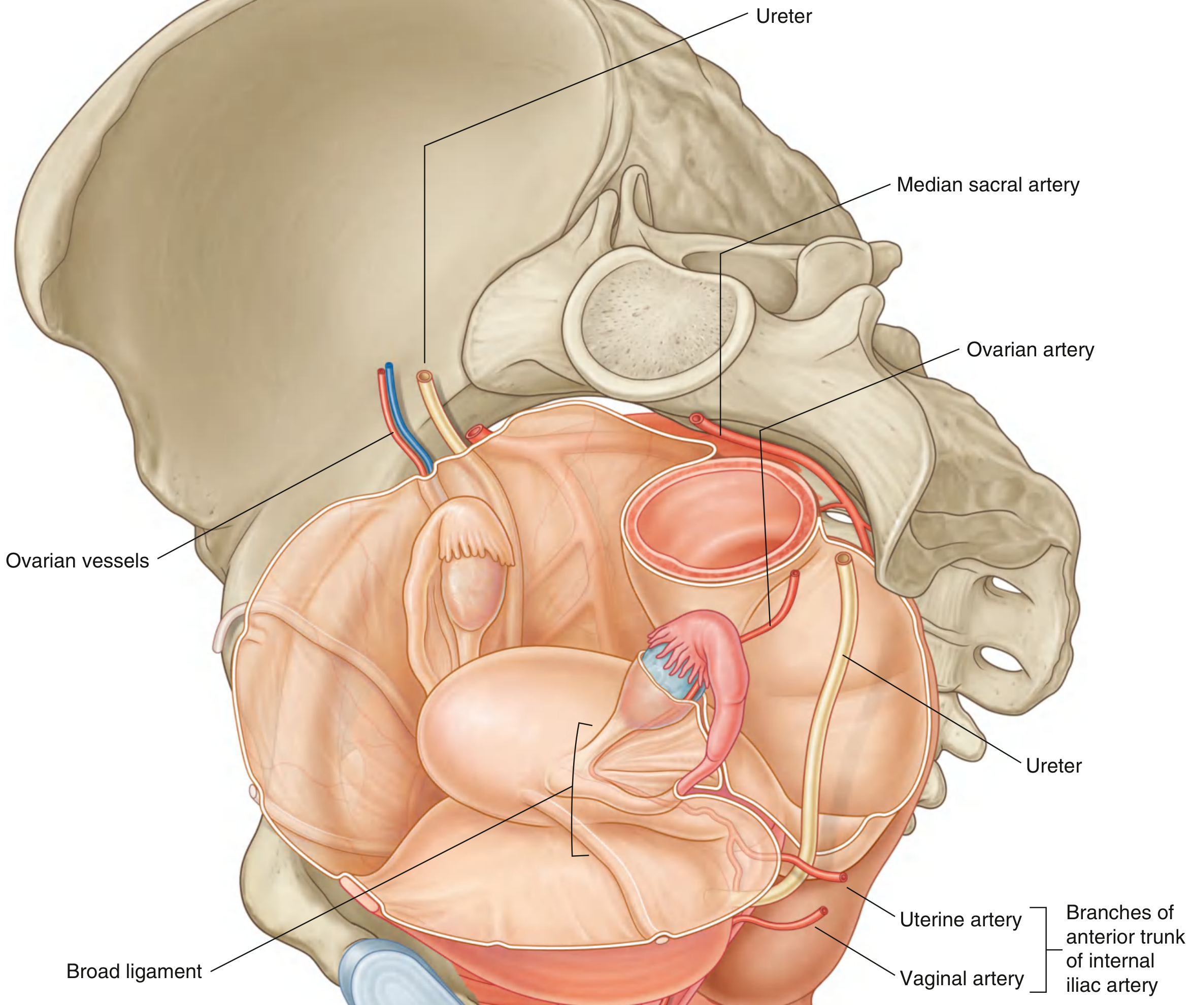
5. Uterine Artery
This is the principal artery of the uterus. Its course:
- Arises from the anterior division of the internal iliac artery
- Travels medially and anteriorly in the base of the broad ligament (in subperitoneal connective tissue)
- Crosses over the ureter - an important clinical landmark: "water under the bridge" (ureter is vulnerable to ligation here during hysterectomy)
- Passes superiorly to the lateral vaginal fornix
- Reaches the cervix and then ascends tortuously along the lateral wall of the uterus
- At the uterine fundus, it curves laterally toward the uterine tube
- Anastomoses with the ovarian artery at the superior end
From the uterine artery, a descending branch (vaginal artery) runs downward to supply the cervix and upper vagina. - Color Atlas of Human Anatomy, Vol. 2
6. Vaginal Artery
The vagina receives blood from two main sources:
| Source | Part of Vagina Supplied |
|---|---|
| Uterine artery (descending branch) | Upper vagina and cervix |
| Vaginal artery (branch of internal iliac / sometimes directly off uterine artery) | Middle and lower vagina |
| Internal pudendal artery (anterior trunk of internal iliac) | Lower vagina and vulva/perineum |
"The blood supply to the upper part of the vagina comes from the uterine and vaginal arteries, which are branches of the internal iliac artery." - Roberts and Hedges' Clinical Procedures in Emergency Medicine
Summary Flowchart
Left Ventricle
      ↓
   Aorta
      ↓
Abdominal Aorta (L4)
      ↓
Common Iliac Artery
      ↓
Internal Iliac Artery (Hypogastric Artery)
      ↓ (Anterior Trunk)
Uterine Artery
      ├──→ Ascending branch → lateral wall of uterus → anastomoses with ovarian artery (fundus)
      └──→ Descending branch (vaginal artery) → cervix → upper vagina
                                         ↓
                           Internal Pudendal Artery → lower vagina / perineum
Anatomical Diagram
Here is a diagram from Gray's Anatomy for Students showing the uterine and vaginal arteries as branches of the anterior trunk of the internal iliac artery:
Venous Drainage (Return Flow)
- The uterine venous plexus (valveless veins around the uterine body and cervix) drains via uterine veins into the internal iliac veins.
- The vaginal venous plexus joins the uterine plexus to form the uterovaginal venous plexus, also draining into the internal iliac veins.
- The internal iliac veins drain into the common iliac veins, then the inferior vena cava, and return to the right atrium.
Key Clinical Points
- The ureter crosses under the uterine artery ("water under the bridge") - at risk during hysterectomy
- The uterine artery enlarges significantly during pregnancy to meet increased demand
- Uterine artery embolization (UAE) exploits this vascular pathway to treat fibroids by cutting off blood supply via catheterization through the femoral artery
- The ovarian arteries (direct aortic branches) provide important collateral supply and anastomose with the uterine artery at the fundus
Sources: Gray's Anatomy for Students, p. 575-576 | Color Atlas of Human Anatomy Vol. 2, p. 452 | Campbell-Walsh Urology, p. 3216 | Roberts and Hedges' Clinical Procedures in Emergency Medicine, p. 1382
This is a shared conversation. Sign in to Orris to start your own chat.