
• Identify the correct anatomical landmarks for placing a stethoscope on the anterior, posterior, and lateral thorax. • Describe the physiologic mechanisms that produce normal breath sounds (vesicular, bronchovesicular, and bronchial). • Demonstrate proper technique during auscultation, including a side-to-side comparison pattern and instructing the patient on effective deep breathing. • Distinguish adventitious sounds (such as crackles, wheezes, rhonchi, stridor, and pleural friction rubs) during a patient examination with AV aids. • Accurately document auscultation findings using correct clinical terminology and recognize data that requires immediate provider notification.
lung auscultation anatomical landmarks anterior posterior thorax stethoscope placement
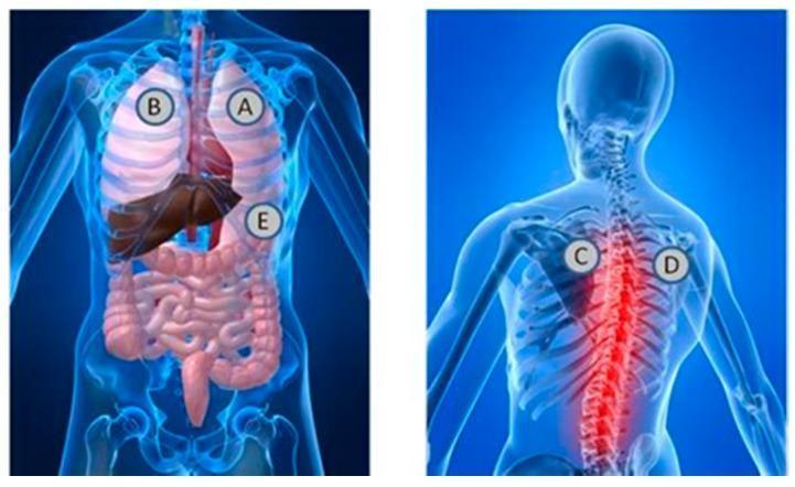
This medical anatomical diagram illustrates the standard stethoscope placement locations for auscultation of heart and lung sounds. The image consists of two panels: an anterior (front) view and a posterior (back) view of a semi-transparent human torso. In the anterior view, labels A and B are positioned over the apical regions of the right and left lungs, respectively, while label E is located at the lower left costal margin, corresponding to the cardiac apex and mitral valve area. Visible internal structures include the lungs, heart, liver, and intestinal tract. In the posterior view, labels C and D are placed in the interscapular region at the level of the upper thoracic spine, typically used for assessing posterior bronchial and vesicular breath sounds. This view highlights the vertebral column, ribs, and scapulae, with a red glow emphasizing the spinal region. This illustration serves as an educational guide for clinical physical examination techniques, demonstrating the relationship between surface landmarks and underlying thoracic and abdominal viscera.
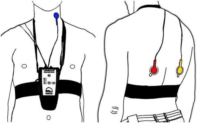
An anatomical diagram illustrating the placement of the LEOSound lung-sound monitor and its bio-acoustical sensors on a patient. The illustration includes two views: an anterior (front) view and a posterior (back) view. In the anterior view, a recording device is secured to the mid-thorax by a black chest belt and a neck strap. A blue tracheal microphone is positioned on the neck near the trachea. In the posterior view, two additional microphones are attached using adhesive pads to the inferior region of the left (red microphone) and right (yellow microphone) scapula. The system is designed for mobile, long-term recording and automated analysis of respiratory sounds such as coughing and wheezing. The diagrams emphasize the standardized anatomical landmarks required for optimal auscultation and data collection in clinical or home settings.
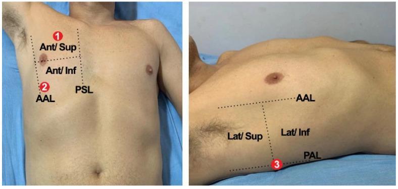
Two clinical photographs demonstrate a standardized lung auscultation protocol for ICU patients, specifically targeting the right lung. The images show a male torso with anatomical landmarks and auscultation zones delineated by dotted grid lines and numeric labels. The anterior view (left image) identifies Point 1 in the 'Ant/Sup' (Anterior/Superior) and 'Ant/Inf' (Anterior/Inferior) regions, bounded medially by the Parasternal Line (PSL) and laterally by the Anterior Axillary Line (AAL). Point 2 is situated more inferiorly along the AAL. The lateral view (right image) shows Point 3, located posteriorly to the AAL and near the Posterior Axillary Line (PAL). This region is divided into 'Lat/Sup' (Lateral/Superior) and 'Lat/Inf' (Lateral/Inferior) zones. These anatomical markers serve as a clinical guide for healthcare providers to ensure consistent positioning of the stethoscope during physical examinations, particularly for monitoring respiratory conditions such as COVID-19 in a critical care setting. The visual aid emphasizes the importance of using specific axillary and parasternal landmarks to capture sounds from the upper and lower lobes of the right lung.
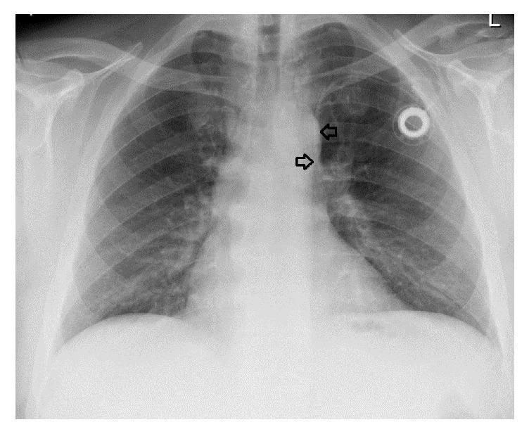
This anterior-posterior (AP) chest X-ray illustrates the placement of a subcutaneous port catheter system in a human thorax. The image displays standard anatomical landmarks, including the ribs, clavicles, lung fields, and mediastinal silhouette. A port reservoir is visible in the left upper chest wall. The radiopaque catheter tracks from the reservoir toward the mediastinum; black arrows highlight the distal segment of the catheter, which descends along the left paramediastinal border. This finding is characteristic of a catheter residing within a persistent left-sided superior vena cava (PLSSVC), a congenital venous anomaly. The lung parenchyma appears clear, and the cardiomediastinal contour is otherwise unremarkable. This diagnostic image is significant for demonstrating atypical medical device positioning necessitated by vascular variation, serving as an educational example of how congenital anomalies can influence clinical procedures such as central venous access.
adventitious breath sounds crackles wheezes rhonchi diagram
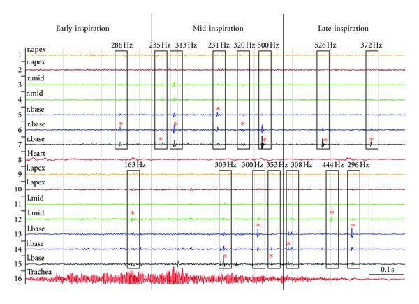
This diagnostic visualization is a multi-channel time-amplitude stack plot of inspiratory lung sounds from a patient with pneumonia, demonstrating multichannel lung sound analysis. The plot consists of 16 vertically arranged channels: channels 1–7 represent the right lung (apex, mid, and base), channels 9–15 represent the left lung (apex, mid, and base), with additional channels for the heart (8) and trachea (16). The horizontal axis denotes time, partitioned by thick vertical lines into early-inspiration, mid-inspiration, and late-inspiration phases. Adventitious lung sounds, specifically crackles, are visualized as discrete high-amplitude spikes. These are grouped into 'crackle families' indicated by black rectangular borders. Key diagnostic annotations include the crackle pitch frequency (Hz) labeled at the top of each border and a red star (*) identifying the 'mother crackle' (the primary or originating deflection). The data illustrates a clinical trend of increasing crackle frequency and density as inspiration progresses from the mid to late phases, particularly noticeable in the basal segments of both lungs.
This medical anatomical diagram illustrates the standard stethoscope placement locations for auscultation of heart and lung sounds. The image consists of two panels: an anterior (front) view and a posterior (back) view of a semi-transparent human torso. In the anterior view, labels A and B are positioned over the apical regions of the right and left lungs, respectively, while label E is located at the lower left costal margin, corresponding to the cardiac apex and mitral valve area. Visible internal structures include the lungs, heart, liver, and intestinal tract. In the posterior view, labels C and D are placed in the interscapular region at the level of the upper thoracic spine, typically used for assessing posterior bronchial and vesicular breath sounds. This view highlights the vertebral column, ribs, and scapulae, with a red glow emphasizing the spinal region. This illustration serves as an educational guide for clinical physical examination techniques, demonstrating the relationship between surface landmarks and underlying thoracic and abdominal viscera.
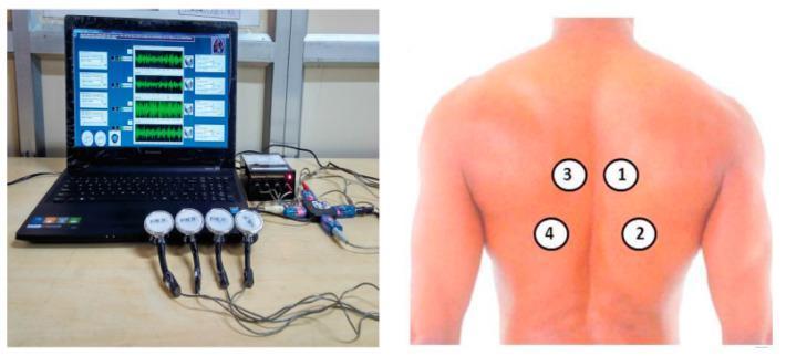
This procedural image illustrates a multi-channel data acquisition setup for collecting and analyzing respiratory sound signals. The left panel shows the physical components: four circular electronic acoustic sensors (transducers) with integrated handles, connected via cables to a central signal processing unit, which is interfaced with a laptop. The laptop display exhibits four synchronized real-time audio waveforms, corresponding to the four input channels. The right panel is a clinical photograph of a human back with numbered markers (1-4) indicating the anatomical sensor placement for auscultation. Markers 1 and 3 are positioned in the upper interscapular region, lateral to the thoracic spine, while markers 2 and 4 are situated in the lower infrascapular area. This setup is designed for computerized pulmonary examination, allowing for the detection and classification of normal and adventitious lung sounds (e.g., wheezing, crackles) across different anatomical zones of the lungs simultaneously.
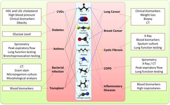
A medical infographic and conceptual flowchart illustrating the integration of exhaled breath (EB) analysis into the diagnostic workflow for various clinical conditions. The diagram is organized into three vertical panels. The left and right green panels list traditional diagnostic parameters and tests, including blood pressure, glucose levels, spirometry, CT imaging, and sputum culture. The central pink panel lists major disease categories, such as CVDs, Diabetes, Asthma, Lung Cancer, and COPD, flanking a central column of molecular ball-and-stick models. These models represent volatile organic compounds (VOCs) found in breath, including Acetone, Nitric Oxide, Carbon Disulphide, Formaldehyde, Pentane, Ammonia, Ethane, Isoprene, and Hydrogen Cyanide. Connecting lines visually map specific clinical biomarkers and diagnostic tests to their corresponding diseases and associated volatile metabolites. This conceptual diagram demonstrates how breathomics and the identification of metabolic biomarkers can augment existing diagnostic modalities to accelerate disease detection and personalized monitoring.
breath sounds auscultation pattern side to side comparison technique
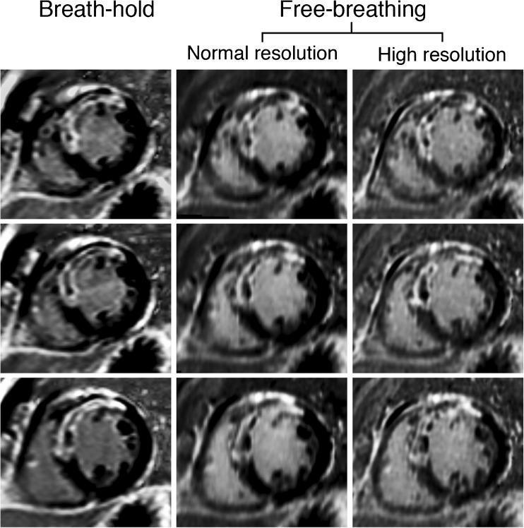
This diagnostic image set displays Late Gadolinium Enhancement Cardiac Magnetic Resonance (LGE-CMR) short-axis views of the left ventricle in a patient with ischemic cardiomyopathy. The 3x3 grid provides a side-by-side comparison of three acquisition techniques: breath-hold (left column), free-breathing normal resolution (middle column), and free-breathing high resolution (right column) across three different slices. The images demonstrate transmural LGE in the anteroseptal wall, indicative of a full-thickness myocardial infarction. A key clinical feature visualized is the presence of central dark areas within the hyperenhanced scar tissue, representing 'no-reflow' zones or microvascular obstruction. The comparison highlights the superior diagnostic clarity of the high-resolution free-breathing technique, which shows significantly sharper scar-to-myocardium borders and more defined internal morphology of the no-reflow zones compared to the blurrier margins seen in the breath-hold and normal-resolution acquisitions. This visual evidence supports the educational concept of improved scar edge sharpness (SES) through high spatial resolution imaging in cardiovascular radiology.
This medical anatomical diagram illustrates the standard stethoscope placement locations for auscultation of heart and lung sounds. The image consists of two panels: an anterior (front) view and a posterior (back) view of a semi-transparent human torso. In the anterior view, labels A and B are positioned over the apical regions of the right and left lungs, respectively, while label E is located at the lower left costal margin, corresponding to the cardiac apex and mitral valve area. Visible internal structures include the lungs, heart, liver, and intestinal tract. In the posterior view, labels C and D are placed in the interscapular region at the level of the upper thoracic spine, typically used for assessing posterior bronchial and vesicular breath sounds. This view highlights the vertebral column, ribs, and scapulae, with a red glow emphasizing the spinal region. This illustration serves as an educational guide for clinical physical examination techniques, demonstrating the relationship between surface landmarks and underlying thoracic and abdominal viscera.
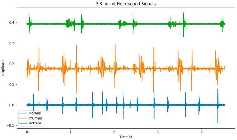
This Comparison Chart displays three distinct phonocardiogram (PCG) waveforms, representing heart sound signals over a 5-second interval. The chart illustrates three categories: Normal (blue), Murmur (orange), and Extrahs (green), plotted as amplitude over time. The 'Normal' waveform shows a rhythmic pattern of relatively low-amplitude spikes corresponding to the standard S1 and S2 heart sounds with consistent intervals. The 'Murmur' signal exhibits a more complex and erratic morphology characterized by higher amplitude spikes and high-frequency oscillations between the primary heart sounds, indicating turbulent blood flow. The 'Extrahs' (extra heart sound) signal features the highest amplitude spikes and additional sound components that appear rhythmically but at a lower frequency than the normal baseline. This diagnostic visualization is used in cardiology to demonstrate the temporal and acoustic differences between physiological and pathological heart sounds, aiding in the identification of cardiovascular diseases through digital auscultation analysis.
pleural friction rub stridor lung examination clinical findings
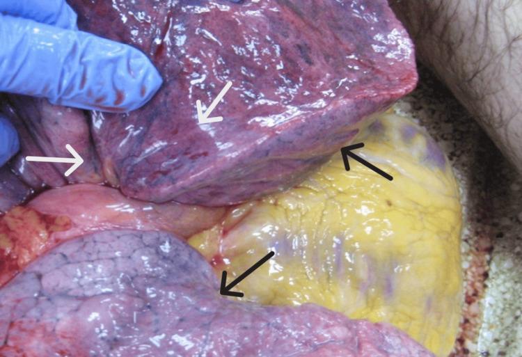
This forensic clinical photograph depicts the gross pathological findings of the lungs during an internal examination (autopsy). The lung tissue demonstrates severe pulmonary congestion, characterized by a diffuse, dark reddish-purple discoloration and a heavy, engorged appearance. Labeled with black arrows are areas of pulmonary edema, manifesting as localized swelling and a glistening, fluid-filled texture on the pleural surface. Indicated by white arrows are multiple petechial hemorrhages (Tardieu spots), appearing as small, pinpoint red subpleural spots. These findings are classic markers of mechanical asphyxia. The image also shows adjacent mediastinal structures, including epicardial fat on the heart surface. The photograph is taken in situ with a gloved hand providing retraction to expose the diaphragmatic and costal surfaces of the lung lobes. This visual is intended for forensic pathology and medical education to illustrate the non-specific signs of rapid death due to hypoxia or asphyxiation.
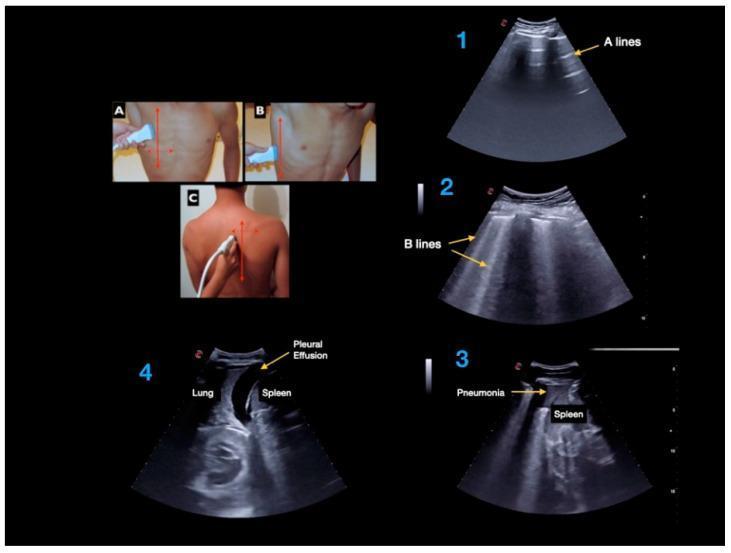
A composite clinical educational image detailing Point-of-Care Ultrasound (PoCUS) techniques and findings for lung examination. The left panels (A, B, C) demonstrate transducer placement for anterior, lateral, and posterior thoracic scans. The right panels display four diagnostic ultrasound frames illustrating key pulmonary artifacts and pathologies. Frame 1 shows 'A-lines', which are horizontal, repetitive echogenic artifacts parallel to the pleural line, indicative of normal aerated lung. Frame 2 demonstrates 'B-lines', vertical laser-like hyperechoic artifacts extending from the pleural line to the bottom of the screen, suggesting interstitial syndrome or pulmonary edema. Frame 3 shows lung consolidation labeled as 'Pneumonia' adjacent to the spleen, characterized by a tissue-like echotexture (hepatization). Frame 4 depicts a 'Pleural Effusion', visible as an anechoic (black) fluid collection superior to the diaphragm, separating the lung from the spleen. This visual resource is intended for medical training in respiratory diagnostics and bedside sonography.
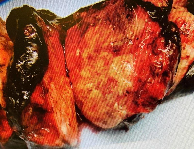
This clinical photograph shows a gross pathological examination of a resected human lung lobectomy specimen. The specimen features a large, well-circumscribed mass lesion that is predominantly grey-white in color with visible focal areas of hemorrhage and congestion. The tumor displays a firm consistency and occupies a significant portion of the parenchymal volume. Key anatomical findings include the mass protruding into the bronchial lumen and causing significant retraction of the overlying pleura, indicative of aggressive growth. The surrounding lung tissue shows secondary changes related to the mass effect. Clinically, this image demonstrates the macroscopic appearance of a primary lung malignancy, such as pulmonary carcinosarcoma, illustrating its expansive growth pattern and interaction with pleural and bronchial structures. This visual is suitable for pathology education regarding surgical staging and gross tumor morphology in thoracic oncology.
lung auscultation anatomical landmarks anterior posterior lateral thorax stethoscope placement technique clinical
https://pressbooks.library.torontomu.ca/assessmentnursing/ch…
Lung Auscultation: A Complete Clinical Guide
1. Anatomical Landmarks for Stethoscope Placement
Anterior Thorax
| Location | Landmark | Lobe Sampled |
|---|---|---|
| Apices | Just below the clavicle, lateral to the mid-clavicular line | Upper lobes (R & L) |
| 1st-2nd ICS, lateral to sternum | Parasternal line | Upper lobe (R), upper/middle lobe transition |
| 3rd-4th ICS, anterior axillary region | At or medial to the anterior axillary line (AAL) | Right middle lobe / left upper lobe lingula |
| 5th-6th ICS, lateral to mid-clavicular line | Near the costal margin | Lower lobe bases |
Key point: The right lung has 3 lobes; the left has 2. The anterior thorax predominantly overlies the upper and middle lobes.
Posterior Thorax
| Location | Landmark | Lobe Sampled |
|---|---|---|
| Apices | Above the scapular spine (T1-T3 level) | Upper lobes |
| Interscapular region | Between the medial scapular border and spine (T4-T7) | Upper/lower lobe transition (bronchial sounds heard here normally) |
| Infrascapular region | Below the inferior scapular angle (T8-T10) | Lower lobes |
| Bases | Costophrenic angle / lower ribs | Lower lobe bases |
Key point: The posterior thorax is primarily the lower lobes. This is where vesicular sounds are loudest and where pathology (pneumonia, effusion, atelectasis) most commonly hides.
Lateral Thorax
- Right lateral: 3 locations (upper, mid, lower) between the AAL and posterior axillary line (PAL) - samples the right middle lobe and right lower lobe
- Left lateral: 2 locations between AAL and PAL - samples the left lower lobe
- Ask the patient to raise their arm or place their hand behind their head to open the intercostal spaces
2. Physiologic Mechanisms of Normal Breath Sounds
Vesicular Sounds
- Where heard: Over most of the peripheral lung fields - posterior and lateral thorax
- Character: Soft, low-pitched, rustling (like wind through leaves)
- I:E ratio: Inspiration is longer than expiration (approx. 3:1); the expiratory phase is quiet and short
- Mechanism: Air flowing into the small bronchioles and alveolar ducts creates low-frequency turbulence. The chest wall filters out higher frequencies. During expiration, airways narrow and airflow diminishes quickly, so the sound drops off fast
- Clinical note: These are the sounds you expect in healthy peripheral lung
Bronchovesicular Sounds
- Where heard: 1st and 2nd ICS beside the sternum (anteriorly); between the scapulae (posteriorly) - the interscapular area overlies the mainstem bronchi
- Character: Medium-pitched, moderate intensity
- I:E ratio: Inspiration and expiration are roughly equal in duration
- Mechanism: Produced by air moving through the larger, segmental and lobar bronchi. The proximity of these airways to the chest wall allows a higher-frequency component to come through
Bronchial (Tracheal) Sounds
- Where heard: Directly over the trachea and manubrium anteriorly; at the posterior base of the neck / upper thoracic spine posteriorly
- Character: Loud, high-pitched, hollow or tubular quality
- I:E ratio: Expiration is longer than inspiration (roughly 1:2); there is a brief gap between the two phases
- Mechanism: High-velocity airflow through the trachea and large central airways generates intense turbulence. Heard over peripheral lung, bronchial sounds are always abnormal (indicating consolidation, which conducts sound more efficiently)
| Sound Type | Pitch | Intensity | Inspiration vs. Expiration | Normal Location |
|---|---|---|---|---|
| Vesicular | Low | Soft | Insp > Exp | Peripheral lung fields |
| Bronchovesicular | Medium | Moderate | Insp = Exp | 1st-2nd ICS / interscapular |
| Bronchial/Tracheal | High | Loud | Exp > Insp (with gap) | Trachea / manubrium |
3. Proper Auscultation Technique
Equipment and Environment
- Use the diaphragm of the stethoscope for breath sounds (higher-pitched sounds)
- Warm the diaphragm in your hands before placing it on the patient - cold metal causes discomfort and muscle tension
- Perform in a quiet room - ambient noise masks subtle sounds
- Never auscultate through clothing; always place on bare skin with full seal contact
Patient Positioning
- Preferred: Seated upright, arms relaxed at sides or crossed in front (this moves the scapulae laterally, opening the posterior fields)
- If the patient cannot sit, roll them side-to-side and auscultate each posterior field in turn
- For patients with breast tissue, ask them to reposition the breast to allow flat diaphragm contact - you may demonstrate the motion over your own uniform
Instructing Effective Deep Breathing
- Breathing through the open mouth reduces upper airway noise
- Deep, slow breaths (not rapid or shallow) maximize airflow and make sounds more audible
- For children: "Pretend you're blowing out birthday candles" or "blow the stethoscope away"
- Patients with dyspnea may fatigue - watch for lightheadedness (hyperventilation) and allow rest between sites
The Side-to-Side Comparison Pattern (Zigzag / Stepladder)
Posterior pattern (top view from behind):

  Left    Right
   L   R     ← Apices
    L   R    ← Interscapular upper
     L   R   ← Interscapular mid
      L   R  ← Infrascapular
       L  R  ← Bases
- Listen to one complete respiratory cycle (full inspiration + expiration) at each site before moving
- Compare: equal air entry? Equal pitch and intensity? Any added sounds?
- Finish posterior fields before moving to lateral, then anterior - this follows a systematic top-to-bottom sequence
4. Adventitious (Abnormal) Breath Sounds
- Discontinuous (interrupted): Crackles
- Continuous: Wheezes, rhonchi
- Extra-pulmonary: Stridor, pleural friction rub
Crackles (formerly Rales)
| Feature | Fine Crackles | Coarse Crackles |
|---|---|---|
| Sound | Brief, high-pitched "popping" or "Velcro-tearing" | Louder, lower-pitched, bubbly or gurgling |
| Timing | Late inspiratory | Early inspiratory and/or expiratory |
| Mechanism | Sudden reopening of collapsed small airways and alveoli that were held shut by surface tension or fluid | Movement of secretions in larger airways |
| Cleared by coughing? | No | Sometimes yes |
| Clinical association | Pulmonary fibrosis, pulmonary edema, early pneumonia | COPD, bronchitis, pneumonia with secretions |
| Location | Bilateral posterior bases (fibrosis, edema); focal (pneumonia) | Central/bilateral |
Wheezes
- Sound: High-pitched, musical, continuous (>250 ms), "squeaky"
- Timing: Predominantly expiratory; can be both phases in severe obstruction
- Mechanism: Partial obstruction of a medium-to-small airway causes wall oscillation as high-velocity air forces through a narrowed lumen (like air through a reed instrument)
- Clinical associations: Asthma (diffuse bilateral expiratory wheezes), COPD exacerbation, bronchospasm, foreign body (unilateral fixed wheeze)
- Note: Wheezing implies airway narrowing - does not mean the patient is "moving air well"
Rhonchi
- Sound: Low-pitched, continuous, snoring or rumbling
- Timing: More prominent on expiration but can be biphasic
- Mechanism: Secretions in the larger central airways (trachea, mainstem bronchi) create turbulence and partial obstruction. Lower pitch compared to wheezes because of the larger airway diameter
- Cleared by coughing? Yes - a hallmark feature that distinguishes rhonchi from other sounds
- Clinical associations: Bronchitis, COPD, mucus plugging, post-operative retained secretions
Stridor
- Sound: High-pitched, harsh, loud, monophonic (single pitch), inspiratory (or biphasic)
- Mechanism: Severe narrowing of the upper airway (larynx/trachea) - the sound is generated at the site of critical obstruction
- Heard: Often audible without a stethoscope; best auscultated over the trachea/throat
- Clinical associations: Croup, epiglottitis, anaphylaxis with laryngeal edema, foreign body aspiration, post-extubation laryngospasm, tracheal tumor
- URGENT: Stridor always signals a potentially life-threatening airway emergency
Pleural Friction Rub
- Sound: Creaking, leathery, grating - like two pieces of leather rubbing together
- Timing: Biphasic - heard on both inspiration AND expiration (moves with breathing)
- Mechanism: Inflamed pleural surfaces (parietal and visceral pleura) lose their normal fluid lubrication and rub against each other during respiratory excursion
- Location: Usually localized to the area of pleural inflammation
- Distinguish from pericardial rub: A pleural rub disappears when the patient holds their breath; a pericardial rub persists (it moves with the heartbeat, not respiration)
- Clinical associations: Pleuritis, pulmonary embolism with infarction, pleurodesis
5. Documentation and Immediate Notification Criteria
Standard Documentation Template
Respiratory Assessment: Patient seated, breathing comfortably. Respiratory rate 16/min, unlabored. Chest expansion symmetric. Auscultation performed in a systematic side-to-side pattern, posterior then lateral then anterior. [Sound type] heard [timing: inspiratory/expiratory/biphasic] at [location: e.g., bilateral posterior bases, right mid-lung field, left apex], [quality: fine/coarse, wheeze/rhonchi, etc.]. Cleared with cough: [yes/no]. Air entry equal bilaterally in upper and mid fields. No adventitious sounds at remaining fields.
Examples of Correct Terminology
| Finding | Correct Documentation |
|---|---|
| Normal | "Clear breath sounds bilaterally, all fields" |
| Crackles at bases | "Fine inspiratory crackles bilateral posterior bases, not cleared by cough" |
| Wheeze | "Expiratory wheeze audible throughout bilateral lung fields, greater at mid-chest" |
| Rhonchi | "Low-pitched rhonchi bilateral mid-lung anteriorly, partially cleared with cough" |
| Absent sounds | "Breath sounds absent right lower lobe; dull to percussion same region" |
| Bronchial over periphery | "Bronchial breath sounds right lower lobe posterior - consolidation suspected" |
| Friction rub | "Pleural friction rub audible left lateral thorax, biphasic, localized" |
Findings Requiring Immediate Provider Notification
- Stridor - upper airway obstruction; potential life-threatening emergency
- Absent breath sounds unilaterally - may indicate pneumothorax, massive pleural effusion, or complete atelectasis
- New-onset diffuse crackles + respiratory distress - pulmonary edema, ARDS
- New-onset wheeze with signs of anaphylaxis (hives, hypotension, facial swelling) - anaphylaxis
- Bronchial sounds over peripheral fields in an acutely unwell patient - consolidation/lobar pneumonia with hemodynamic compromise
- Sudden deterioration in air entry from a prior baseline in any patient
Notify the provider promptly (non-urgent) for: new crackles in a stable patient, new rhonchi, new focal wheeze, or any sound that represents a change from the documented baseline.
Quick Reference Summary Card
| Sound | Continuous? | Pitch | Phase | Cleared by Cough | Key Association |
|---|---|---|---|---|---|
| Vesicular (normal) | No | Low | Insp > Exp | N/A | Healthy lung periphery |
| Bronchovesicular (normal) | No | Medium | Insp = Exp | N/A | Mainstem bronchus region |
| Bronchial (normal at trachea) | No | High | Exp > Insp | N/A | Trachea only |
| Fine crackles | No | High | Late insp | No | Fibrosis, pulmonary edema |
| Coarse crackles | No | Low | Early insp/exp | Sometimes | COPD, secretions |
| Wheeze | Yes | High | Expiratory | No | Asthma, obstruction |
| Rhonchi | Yes | Low | Expiratory | Yes | Bronchitis, mucus |
| Stridor | Yes | High | Inspiratory | No | AIRWAY EMERGENCY |
| Pleural friction rub | No | Medium | Biphasic | No | Pleuritis, PE |
- Auscultation of the Respiratory System (PMC) - peer-reviewed technique review
- Anterior Thorax Auscultation - Nursing Assessment - step-by-step with audio clips
- Posterior and Lateral Thorax Auscultation - Nursing Assessment - includes Audio 3.1-3.4 (normal breath sounds, wheezes, stridor, crackles)
- Lung Auscultation Points - Complete Anatomy/3D4Medical - interactive 3D anatomy