
Rotational vertebral artery syndrome
"rotational vertebral artery syndrome" OR "bow hunter syndrome"
rotational vertebral artery syndrome bow hunter syndrome vertebral artery compression head rotation
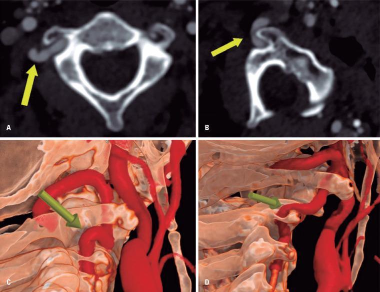
This composite diagnostic image illustrates dynamic vertebral artery compression, also known as Bow Hunter's Syndrome. Panels A and B are axial Computed Tomography Angiography (CTA) images. Panel A shows a dominant right vertebral artery in a neutral position with tortuosity at the V2 to V3 segment transition but maintained vessel caliber. Panel B, captured during contralateral head rotation, demonstrates severe focal stenosis of the same artery caused by mechanical compression between the superolateral aspect of the C2 transverse process and the adjacent bony structures. Panels C and D provide corresponding 3D volume-rendered reconstructions. Panel C shows the normal spatial relationship and vessel patency in a neutral state. Panel D highlights the dynamic narrowing and pinch effect on the vertebral artery during cervical rotation. The educational focus is on the radiological presentation of rotational vertebral artery occlusion and the utility of dynamic CTA in diagnosing vascular syndromes related to cervical spine anatomy.
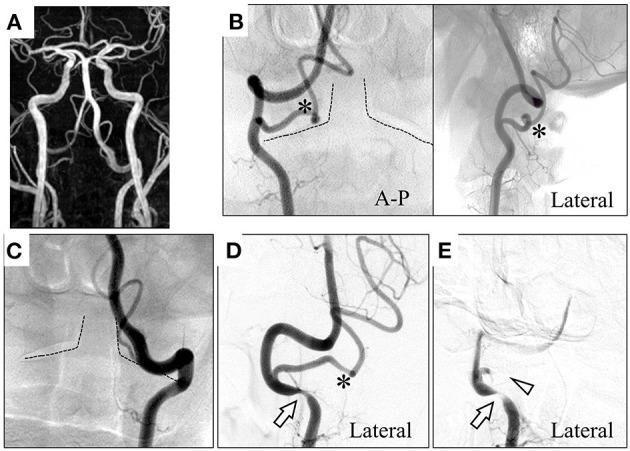
Educational neuroimaging series demonstrating dynamic vertebral artery compression in Bow Hunter's Syndrome. Image (A) is a maximum intensity projection MRA showing bilateral vertebral arteries (VAs) merging into the basilar artery, with left-sided dominance. Images (B) and (C) are Digital Subtraction Angiography (DSA) frames in anterior-posterior and lateral views identifying anatomical variations: the right posterior inferior cerebellar artery (PICA, marked with an asterisk) originates at the C1-C2 level and enters the spinal canal. Images (D) and (E) show dynamic DSA during head rotation. At 45° leftward rotation (D), there is focal narrowing/compression of the right VA at the V3 segment (arrow). At 60° rotation (E), the compression of the right VA worsens (arrow) and the right PICA becomes completely occluded (arrowhead). This sequence illustrates rotational occlusion of the posterior circulation, a hallmark of Bow Hunter's Syndrome caused by mechanical compression during cervical rotation at the atlantoaxial level.
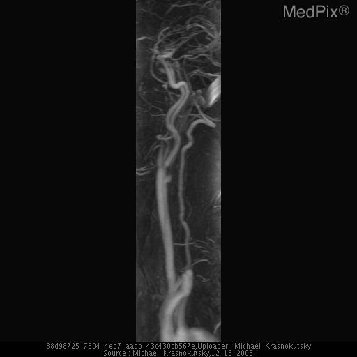
**Imaging Modality:** Magnetic Resonance Angiography (MRA) maximum intensity projection (MIP) image. **Anatomical Region:** Cervical region focusing on the posterior cerebral circulation. **Observed Pathology:** The image demonstrates the course of the vertebral artery. Clinical context suggests rotational vertebral artery occlusion, also known as Bow Hunter’s syndrome. This condition is characterized by mechanical compression of the vertebral artery during head rotation. **Characteristic Visual Features:** - A longitudinal view of the vertebral artery ascending through the cervical spine. - The vessel exhibits its characteristic tortuous course as it approaches the atlanto-axial (C1-C2) segment. - Potential narrowing or signal dropout is typically localized at the level of the C1-C2 joint, where the artery exits the foramen transversarium and travels over the posterior arch of the atlas. **Key Diagnostic Features:** The image highlights the V2 (foraminal) and V3 (extraspinal) segments of the vertebral artery. In the context of Bow Hunter’s syndrome, the primary finding is dynamic stenosis or occlusion triggered by physiological neck rotation, frequently secondary to osteophytic compression or ligamentous hypertrophy at the C1-C2 level.
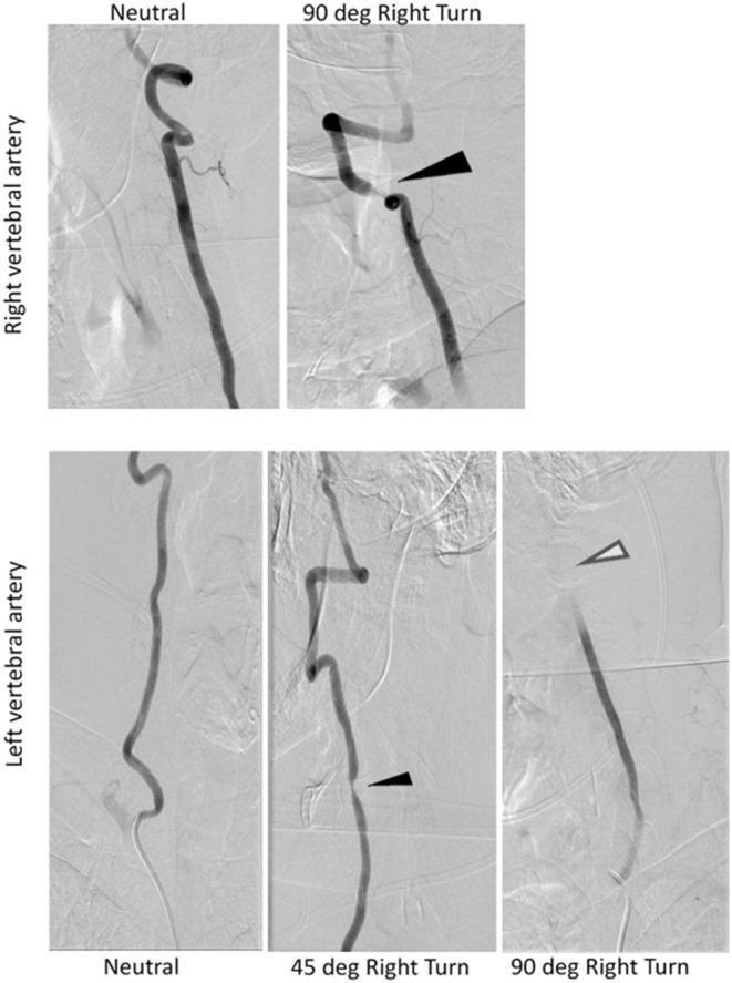
This diagnostic image set consists of dynamic digital subtraction angiography (DSA) frames of the vertebral arteries, illustrating rotational vertebral artery occlusion, also known as Bow Hunter's syndrome. The top row focuses on the right vertebral artery (RVA), comparing a neutral head position to a 90-degree right turn. In the neutral position, the RVA shows normal, patent flow. Upon a 90-degree right turn, a black arrow indicates significant focal narrowing/stenosis at the mid-cervical level (C4-C5), suggesting mechanical compression. The bottom row demonstrates the left vertebral artery (LVA) in three positions: neutral, 45-degree right turn, and 90-degree right turn. In neutral, the LVA is patent. At 45 degrees, a small black arrow highlights emerging partial stenosis. At 90 degrees, an open white arrow points to complete cessation of contrast flow, indicating total dynamic occlusion at the C1-C2 level. These images serve as a classic educational example of how head rotation can cause transient cerebrovascular insufficiency through mechanical obstruction by osteophytes or ligamentous stretching, correlating with clinical symptoms like vertigo or nystagmus.
PMID: 39640315
Rotational Vertebral Artery Syndrome (RVAS)
Historical Note
Anatomy and Pathophysiology
- One hypoplastic, absent, or occluded contralateral VA (so collateral flow is inadequate)
- Dynamic compression of the dominant (only functional) VA on head rotation
- Osteophytes from uncovertebral joint degeneration (most common)
- Ligamentous hypertrophy (especially at the atlantoaxial joint)
- Fibrous bands
- Cervical disk herniation
- Bony anomalies (os odontoideum, atlantoaxial instability)
- Rarely, tumors or abnormal muscle insertions
Clinical Features
| Symptom | Notes |
|---|---|
| Vertigo | Most common; paroxysmal, triggered by head rotation |
| Nystagmus | Initially downbeat, with horizontal and torsional components beating toward the compressed VA side |
| Ataxia / drop attacks | From cerebellar or brainstem hypoperfusion |
| Dysarthria | Brainstem involvement |
| Visual disturbance | Diplopia, blurred vision, oscillopsia |
| Syncope / presyncope | Severe flow reduction |
| TIA or stroke | Posterior circulation territory; may be the presenting event |
Diagnosis
1. Dynamic CTA (CT Angiography)
- Patent vessel in neutral
- Focal stenosis or occlusion at the compression point during rotation
2. Dynamic DSA (Digital Subtraction Angiography)
- Complete occlusion of the dominant VA during ipsi- or contralateral rotation
- Compensation (or lack thereof) via the contralateral VA
- Degree of posterior fossa perfusion compromise
3. MRA (MR Angiography)
4. Transcranial Doppler (TCD)
Management
Conservative (Non-surgical)
- Cervical collar / neck brace to restrict head rotation
- Antiplatelet therapy (aspirin, clopidogrel) - to reduce thromboembolic risk from dynamic stenosis
- Anticoagulation - if there is evidence of in situ thrombus or embolism
- Physical therapy with movement restriction education
- Suitable for mild/infrequent symptoms or surgically unfit patients
Surgical
- Anterior approach (40% of cases) - had the best outcomes (84% good recovery). Includes anterior cervical decompression with or without fusion, uncovertebral joint osteophyte removal.
- Posterior approach (30% of cases) - more cases with only partial recovery (19% vs 16%). Includes C1-C2 fusion, posterior decompression, or resection of a compressive structure.
- Endovascular options (angioplasty/stenting) - used in selected cases, particularly where anatomy precludes open surgery.
Important caveat: Evidence remains inconclusive on the optimal surgical approach. No RCTs exist; the literature consists of case series and reviews. Management must be individualised based on the level of compression, VA anatomy, and patient fitness.
Chiropractic neck manipulation
Differential Diagnosis
| Condition | Key Differentiating Feature |
|---|---|
| BPPV | Triggered by head positioning (Dix-Hallpike), not rotation per se; no posterior circulation ischemia |
| Vestibular neuritis | Sustained (not paroxysmal), not positional |
| Posterior fossa tumour | Persistent symptoms; imaging shows mass lesion |
| VA dissection | Neck pain, Horner's syndrome, often post-trauma |
| Cervicogenic vertigo | Disputed entity; neck pain predominant; no vascular compression on imaging |
| Subclavian steal | Exercise-related, subclavian stenosis proximal to VA origin |
Key Points Summary
- RVAS (= Bow Hunter's Syndrome) = dynamic VA compression during head rotation, most often at C1-C2
- Requires a hypoplastic/absent contralateral VA for symptoms to manifest
- Presents with paroxysmal vertigo, nystagmus (initially downbeat), ataxia, and drop attacks triggered by head turning
- Diagnosis requires dynamic CTA or DSA during the provocative head position
- Initial nystagmus: downbeat, with horizontal-torsional component beating toward the compressed VA side
- Treatment: cervical collar + antiplatelets first; surgery (anterior approach preferred) if conservative measures fail
- Avoid chiropractic neck manipulation in at-risk patients
- Bradley & Daroff's Neurology in Clinical Practice (9780323642613)
- Localization in Clinical Neurology, 8e (9781975160241)
- Firestein & Kelley's Textbook of Rheumatology (9780323935364)
- Elizondo-Ramirez et al. (2024). Bow Hunter's syndrome surgical approach and outcome. Surg Neurol Int. PMID 39640315.