

Hey. So this is a CBL, uh, case-based learning in, uh, our med school. So basically, uh, what this is, is basically a patient's history and the procedures that were performed on him. So, um, you know, in medical, everything is important from the age, uh, to his, to the gender, to, you know, like, every single thing. So I want you to take out every single line. And for example, a 42-year-old, uh, woman presents with a painless swelling on the left side of her neck that she first noticed two months earlier. So everything that has a significance, like her age, her gender, her, uh, painless swelling, and, uh, especially on the left side of her neck, and, uh, the time duration, and, uh, the examination that was done, uh, done on her, um, the, you know, the qualities of the, uh, lymph node, um, and, uh, the, uh, node, uh, the location of the lymph node. I hope you get me. Like, you know, just take out a single line from this case and, you know, define it. And, you know, you also have to give me differential diagn osys for every single thing. Like, for example, a painless swelling on the left side of her neck. Apart from this diagnosed case, what other, you know, like, diseases or diagnoses could have been made? You know, uh, you should get me. So basically, uh, what you have to do is divide the CBL into lines and, uh, just break down those lines and, uh, give me s- every single thing, like, you know, differential diagnosis. For example, uh, types of, uh, lymph node, uh, masses, tender, non-tender, firm, mobile, uh, non-mobile, um, you know, or every single thing. And, uh, types of, uh, biopsies, why are they done, and, uh, you know, the durations of time. Why is she drooping? What other, you know, uh, cause, uh, diseases cause drooping? You know, uh, uh, I think you get me. Yeah.
posterior triangle of the neck boundaries subdivisions diagram
spinal accessory nerve course posterior triangle trapezius
cervical plexus cutaneous branches sensory distribution neck
CBL-3 Full Line-by-Line Breakdown
"Shoulder Droop Following a Neck Biopsy: Applied Anatomy of Neck Triangles"
THE CASE TEXT (Full)
"A 42-year-old woman presents with a painless swelling on the left side of her neck that she first noticed two months earlier. Examination reveals a firm, mobile, non-tender lymph node in the upper part of the posterior triangle, between the posterior border of sternocleidomastoid and the anterior border of trapezius. An excisional biopsy is performed through the lateral neck. The surgical dissection is carried out close to the midpoint of the posterior border of sternocleidomastoid. Ten days later, the patient reports drooping of the left shoulder, weakness while shrugging, and difficulty raising her left upper limb above shoulder level. The left shoulder lies lower than the right, producing asymmetry of the neck-shoulder line. She also reports reduced sensation over the angle of the mandible, anterior aspect of the neck and the skin over the clavicle and shoulder."
LINE 1: "A 42-year-old woman"
- Age (42 years): Middle-aged adult. This is the peak age range for lymphoma (both Hodgkin's - peaks 15-35 and 55+, and non-Hodgkin's), thyroid carcinoma, and metastatic cervical lymphadenopathy from head/neck primaries. It is also the age group where reactive lymphadenopathy from infections remains common.
- Sex (Female): Women have higher incidence of thyroid carcinoma (3:1 female-to-male), autoimmune lymphadenopathy (e.g., Sjögren's, SLE), and breast cancer (which can metastasize to axillary or supraclavicular nodes).
| Cause | Notes |
|---|---|
| Reactive lymphadenopathy | Most common overall; viral (EBV, CMV), bacterial (TB) |
| Lymphoma (Hodgkin's / Non-Hodgkin's) | NHL more common at this age |
| Thyroid cancer metastasis | Papillary carcinoma - very common, often presents as neck node first |
| Squamous cell carcinoma metastasis | From oropharynx, larynx, nasopharynx |
| Breast cancer metastasis | Spreads to supraclavicular nodes |
| Salivary gland tumor | Parotid, submandibular |
| Branchial cyst | Lateral neck cyst, can present in adults |
| Lipoma | Soft, lobulated, non-tender |
LINE 2: "Painless swelling on the left side of her neck"
- Painless: This is a red flag. Painful swellings tend to be inflammatory/infective (tender lymphadenitis, abscess). A painless neck mass strongly suggests malignancy (lymphoma, metastatic carcinoma) or a non-inflammatory process.
- Left-sided: Clinically important. The left supraclavicular fossa contains Virchow's node (also called Troisier's node), which when enlarged signals distant metastasis from abdominal/thoracic malignancy (gastric cancer, lung cancer, pancreatic cancer). Although this node is in the upper posterior triangle rather than the left supraclavicular fossa, left-sided nodes in the neck can also drain abdominal organs via the thoracic duct.
- Two months duration: Subacute to chronic course. Acute (<2 weeks) favors infection; chronic (>6 weeks) favors malignancy. Two months without resolution is a major red flag for malignancy.
| Condition | Pain? | Duration hint |
|---|---|---|
| Lymphoma | Painless | Weeks-months |
| TB lymphadenitis | Can be painless | Weeks-months |
| Metastatic carcinoma | Painless | Weeks-months |
| Branchial cyst | Painless | Can be years |
| Thyroglossal cyst | Usually painless | Congenital/years |
| Reactive lymph node | Usually tender | Days-weeks |
| Abscess | Painful + hot | Days |
| Lipoma | Painless | Months-years |
LINE 3: "Examination reveals a firm, mobile, non-tender lymph node"
Firm
- Firmness (rubbery) = Lymphoma (classically described as "rubbery-firm")
- Hard, rock-like = Metastatic carcinoma (the node has been replaced by tumor)
- Soft = Reactive/infective (edematous node), abscess, lipoma, cyst
Mobile
- Mobile = early-stage disease or benign; the node has not infiltrated surrounding tissue
- Fixed/immobile = malignancy with extracapsular spread and infiltration of adjacent structures (advanced cancer, can indicate unresectability)
- Mobile cystic swellings in this region = branchial cyst, thyroglossal cyst
Non-tender
- Non-tender = malignancy (lymphoma, carcinoma) or chronic infection (TB)
- Tender = acute infection, abscess, reactive lymphadenitis
Summary: Firm + Mobile + Non-tender = Classic for Lymphoma (especially early), but also consistent with early metastatic carcinoma before extracapsular spread
| Quality | Suggests |
|---|---|
| Soft, fluctuant | Abscess, suppurative lymphadenitis |
| Firm, rubbery | Lymphoma |
| Hard, stony | Metastatic carcinoma |
| Mobile | Benign or early malignancy |
| Fixed | Advanced malignancy (extracapsular spread) |
| Tender | Infection, inflammation |
| Non-tender | Malignancy, TB, sarcoid |
| Matted (nodes stuck together) | TB, lymphoma, metastasis |
| Single node | Could be anything |
| Multiple nodes | Lymphoma, widespread infection, metastasis |
LINE 4: "In the upper part of the posterior triangle, between the posterior border of sternocleidomastoid and the anterior border of trapezius"
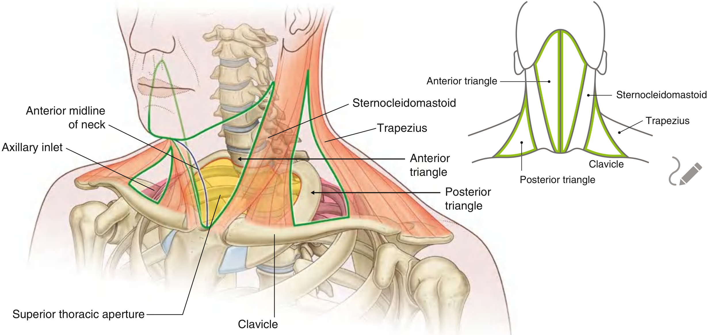
The Posterior Triangle of the Neck
-
Boundaries:
- Anterior: posterior border of sternocleidomastoid (SCM)
- Posterior: anterior border of trapezius
- Inferior: middle one-third of the clavicle
- Roof: investing layer of deep cervical fascia
- Floor: prevertebral fascia covering scalene muscles, levator scapulae, splenius capitis
-
Subdivisions - the inferior belly of the omohyoid divides it into:
- Occipital triangle (upper, larger part) - where this lymph node is located
- Supraclavicular (subclavian) triangle (lower, smaller part)
Contents of the Posterior Triangle (clinically important at-risk structures):
- Spinal accessory nerve (CN XI) - crosses the triangle superficially - THIS IS THE KEY NERVE IN THIS CASE
- Cutaneous branches of cervical plexus (emerge at Erb's point at the midpoint of the posterior border of SCM)
- Brachial plexus roots (lower part)
- Subclavian artery (lower part, supraclavicular triangle)
- External jugular vein
- Occipital, supraclavicular, transverse cervical lymph nodes
- Transverse cervical and suprascapular vessels
Why the upper posterior triangle matters:
Differential Diagnosis for masses specifically in the posterior triangle:
| Mass | Features |
|---|---|
| Lymphoma | Firm, rubbery, non-tender |
| Metastatic node (nasopharyngeal, thyroid) | Firm to hard |
| TB lymphadenitis | Matted nodes, may have sinus tract |
| Branchial cyst (Type II) | Smooth, cystic, transilluminates |
| Cervical rib | Bony, inferior to clavicle level |
| Lipoma | Soft, lobulated |
| Cystic hygroma (in children) | Brilliantly transilluminates |
| Subclavian artery aneurysm | Pulsatile (lower posterior triangle) |
LINE 5: "An excisional biopsy is performed through the lateral neck. The surgical dissection is carried out close to the midpoint of the posterior border of sternocleidomastoid."
Types of Biopsy and When They Are Used
| Biopsy Type | Description | Used When |
|---|---|---|
| Fine Needle Aspiration Cytology (FNAC) | Thin needle extracts cells (not core tissue) | First-line investigation for neck masses; quick, cheap, minimal risk. Gives cytology but not architecture. |
| Core needle biopsy (Tru-cut) | Larger needle, extracts a tissue core | When FNAC is inconclusive; gives architecture (e.g., needed to distinguish lymphoma subtypes) |
| Excisional biopsy | Entire lymph node is surgically removed | When needle techniques are inconclusive, when lymphoma is suspected (need whole-node architecture for classification), or when an excisional specimen is needed for diagnosis |
| Incisional biopsy | Only part of the mass is removed | Large masses; rarely done for lymph nodes |
| Sentinel lymph node biopsy | The "first draining" node is identified by dye/radiotracer and removed | Staging of melanoma and breast cancer |
| Bone marrow biopsy | Posterior iliac crest; trephine needle | Staging lymphoma, leukemia workup |
Why excisional biopsy here?
Why is "midpoint of the posterior border of SCM" critical?
LINE 6: "Ten days later, the patient reports drooping of the left shoulder, weakness while shrugging, and difficulty raising her left upper limb above shoulder level. The left shoulder lies lower than the right, producing asymmetry of the neck-shoulder line."
Anatomy of CN XI (Spinal Accessory Nerve)
- Origin: Spinal cord (C1-C5/6), exits via foramen magnum, joins the cranial root briefly, then exits the skull via the jugular foramen
- Course in the neck: Exits the jugular foramen, crosses the jugular vein, enters and pierces the SCM (supplying it), then crosses the posterior triangle superficially (at risk!)
- Terminal supply: Enters the trapezius muscle
- Action of trapezius:
- Upper fibers: elevate the shoulder (shrugging)
- Middle fibers: retract scapula
- Lower fibers: depress scapula
- All together: stabilize and rotate the scapula during arm elevation above shoulder level
Why does CN XI injury cause each symptom?
| Symptom | Explanation |
|---|---|
| Shoulder droop (left lower than right) | Loss of trapezius tone; scapula droops and rotates |
| Weakness while shrugging | Upper trapezius - CN XI |
| Cannot raise arm above shoulder | Without trapezius stabilizing/rotating the scapula (upward rotation), the deltoid+rotator cuff cannot complete shoulder abduction above 90 degrees |
| Neck-shoulder line asymmetry | Loss of trapezius bulk and tone on left |
| Winging of scapula (may be present) | Scapula not held against chest wall |
Why was the nerve injured in this case?
Differential Diagnoses for Shoulder Droop (other causes)
| Cause | Mechanism | Distinguishing Feature |
|---|---|---|
| CN XI palsy (this case) | Direct injury to nerve | Isolated trapezius weakness; SCM may be intact (if distal injury) |
| Brachial plexus injury (upper trunk, C5-C6) | Erb's palsy | "Waiter's tip" posture; also involves deltoid, biceps |
| Rotator cuff tear | Tendon rupture | Normal nerve conduction; positive drop arm test; MRI confirms |
| Suprascapular nerve injury | Compression or traction | Supraspinatus + infraspinatus weakness; pain in shoulder |
| Long thoracic nerve injury | Serratus anterior denervation | Medial winging of scapula (different from CN XI winging) |
| Shoulder dislocation | Posterior/anterior | X-ray confirms; different muscle pattern |
| Stroke (cortical) | UMN lesion | Hemiplegia, facial droop, speech change |
| Cervical myelopathy (C3-C4) | Cord compression | Bilateral symptoms, hyperreflexia, Lhermitte's sign |
| Myopathy (e.g., Duchenne, limb-girdle) | Muscle disease | Bilateral, proximal weakness; elevated CK |
| Neuralgic amyotrophy (Parsonage-Turner) | Brachial neuritis | Sudden severe pain then weakness; no surgical history |
| Adhesive capsulitis (frozen shoulder) | Capsular contracture | Global restriction of movement; not true droop |
LINE 7: "She also reports reduced sensation over the angle of the mandible, anterior aspect of the neck and the skin over the clavicle and shoulder."
Cervical Plexus Cutaneous Branches
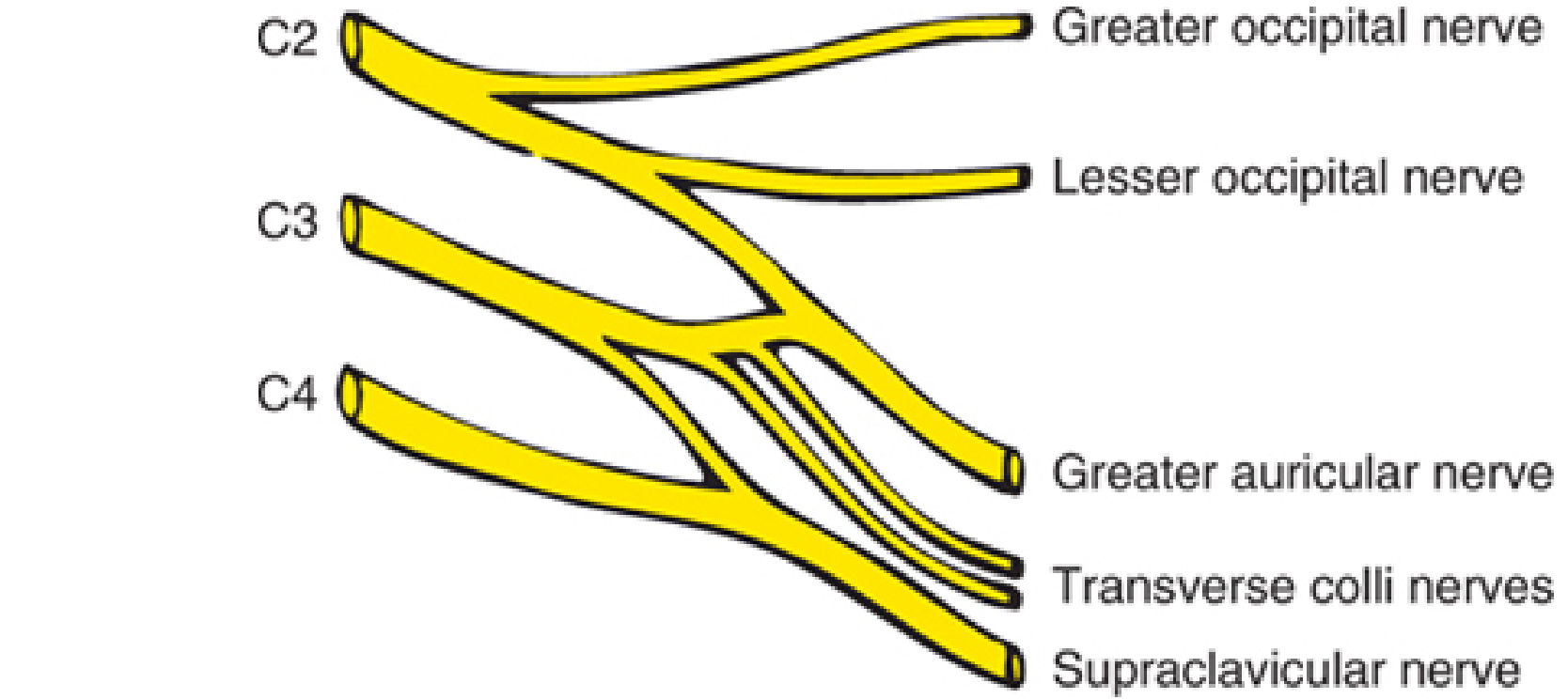
| Branch | Spinal Level | Area Supplied |
|---|---|---|
| Lesser occipital nerve | C2 | Skin behind the ear, lateral scalp |
| Great auricular nerve | C2-C3 | Skin over angle of the mandible, lower cheek, lower ear, upper neck - AFFECTED IN THIS CASE |
| Transverse cervical nerve | C2-C3 | Anterior aspect of the neck - AFFECTED IN THIS CASE |
| Supraclavicular nerves | C3-C4 | Skin over clavicle, upper shoulder, upper chest - AFFECTED IN THIS CASE |
Important: This is a separate injury from CN XI. The combination tells us the dissection simultaneously damaged:
- CN XI (motor to trapezius)
- The cutaneous cervical plexus branches
Differential Diagnoses for Reduced Sensation at the Angle of the Mandible / Anterior Neck / Shoulder
| Cause | Features |
|---|---|
| Cervical plexus injury (this case) | Follows exact dermatomal territory; post-surgical |
| Cervical radiculopathy (C3-C4) | Neck pain, dermatomal numbness; MRI shows disc prolapse |
| Herpes zoster (shingles) of C3-C4 | Vesicular rash in the same dermatomal distribution |
| Diabetic neuropathy | Diffuse, glove-stocking pattern; not localized |
| Brachial neuritis | Pain precedes numbness; affects whole plexus |
| Post-radiation neuropathy | History of radiation; progressive |
| Lymphoma compressing cervical plexus | The underlying disease itself |
| Cervical rib / thoracic outlet syndrome | Usually C8-T1 (little finger, medial forearm) |
SUMMARY TABLE: The Full CBL Decoded
| Clinical Finding | Anatomical Structure | Diagnosis/Mechanism | Key Differentials |
|---|---|---|---|
| 42-year-old woman | - | Middle age, female - thyroid/lymphoma risk | Lymphoma, thyroid Ca, metastasis, TB |
| Painless swelling | Lymph node | Red flag for malignancy | Lymphoma, carcinoma, branchial cyst |
| 2 months duration | - | Chronic = malignancy likely | Any chronic lymphadenopathy |
| Left neck | Left posterior triangle | Left-sided drainage territory | Virchow's (supraclavicular) node for abdominal mets |
| Firm, rubbery | Node consistency | Lymphoma | Metastasis (hard), abscess (soft) |
| Mobile | No extracapsular spread | Early disease | Fixed = advanced malignancy |
| Non-tender | No inflammation | Malignancy / TB | Reactive (tender) |
| Upper posterior triangle | Occipital subdivision | CN XI and cervical plexus territory | Branchial cyst, lipoma, TB |
| Excisional biopsy | Surgical technique | Needed for lymphoma classification | FNAC, core biopsy alternatives |
| Dissection near midpoint of SCM (Erb's point) | CN XI + cervical plexus emerge here | Simultaneous injury to nerve and plexus | Preventable with nerve identification |
| Shoulder droop | Trapezius denervation | CN XI (spinal accessory) palsy | Rotator cuff tear, C5-C6 palsy, neuralgic amyotrophy |
| Can't shrug | Upper trapezius | CN XI | Brachial plexus (Erb's) |
| Can't raise arm above 90° | Scapular rotation (trapezius) | CN XI - no upward scapular rotation | Suprascapular nerve, rotator cuff |
| Asymmetric neck-shoulder line | Loss of trapezius tone | CN XI palsy | Scoliosis, hemi-atlanto-axial sublux |
| Numbness at angle of mandible | Great auricular nerve (C2-C3) | Cervical plexus injury | Zoster, radiculopathy |
| Numbness anterior neck | Transverse cervical nerve (C2-C3) | Cervical plexus injury | Cervical radiculopathy |
| Numbness over clavicle/shoulder | Supraclavicular nerves (C3-C4) | Cervical plexus injury | C4 radiculopathy, thoracic outlet |
ANSWERING THE CBL QUESTIONS
- Prevention: Pre-operative nerve mapping; intraoperative nerve monitoring (electromyography); careful surgical technique with direct nerve visualization before cutting any structure in the posterior triangle
- Recognition: Intraoperatively - stimulate the nerve before cutting. Postoperatively - any shoulder droop or inability to shrug after posterior triangle surgery should prompt nerve function testing and EMG/nerve conduction studies
- Management: Early recognition within days to weeks - surgical re-exploration and nerve repair (end-to-end anastomosis, nerve grafting). Late presentation - physiotherapy to strengthen remaining shoulder muscles; if nerve intact but in continuity - watchful waiting (up to 12-18 months for recovery)
- Angle of mandible = Great auricular nerve (C2-C3)
- Anterior neck = Transverse cervical nerve (C2-C3)
- Clavicle and shoulder skin = Supraclavicular nerves (C3-C4) All emerge at Erb's point (midpoint of posterior border of SCM)
- Internal jugular vein runs deep to SCM, inside the carotid sheath - not directly in the field but at risk if dissection goes deeper and medially
- External jugular vein crosses the posterior triangle superficially and is visible; can be inadvertently cut
- Common carotid artery + internal carotid = deep, within carotid sheath, anteromedial to the operative field - risk if dissection goes deeper toward carotid sheath
- Subclavian artery = passes through the lower posterior triangle (supraclavicular triangle, beneath the clavicle) - risk if dissection extends inferiorly
- Inferiorly into lower posterior triangle: Brachial plexus trunks (especially C5, C6 - upper trunk), subclavian artery and vein, phrenic nerve (on anterior surface of scalenus anterior, in lower part)
- Deeper anteromedially beneath SCM toward carotid sheath: Common carotid artery, internal jugular vein, vagus nerve (CN X), and sympathetic trunk (Horner syndrome if injured)
...А-а-а, Uh, so, you know, uh, you didn't give me differential diagnosis for this case. Like, you know, we don't know what the patient initially had, uh, so we cannot, you know, just point out that this patient had a, you know, infection or the metastasis of cancer to his supraclavicular lymph nodes, and, uh, that, uh, led us to do a biopsy or, you know, excision of the lymph nodes that led to the damage of the spinal accessory nerve 9. Okay. So, uh, I want you to give me the differential diagnosis, you know, that we would initially suspect and, uh, ultimately funnel it down to, you know, our diagnosis that we made. And, uh, so the preoperative symptoms and the postoperative symptoms this patient presented with, I want differential diagnoses for that.
Differential Diagnosis: The Full Clinical Funnel
The Two Clinical Phases
- Pre-op phase: A 42-year-old woman with a painless, firm, mobile, non-tender lymph node in the upper posterior triangle lasting 2 months
- Post-op phase (10 days later): Shoulder droop + can't shrug + can't raise arm above shoulder + sensory loss over the angle of mandible, anterior neck, clavicle, and shoulder
PHASE 1 - PRE-OPERATIVE DIFFERENTIALS
"What did this patient actually have in her neck?"
STEP 1 - Broadest Category: What causes a neck mass?
| Category | Examples |
|---|---|
| Inflammatory / Infective | Reactive lymphadenopathy, bacterial lymphadenitis, TB, cat-scratch disease, EBV/CMV mononucleosis |
| Congenital / Developmental | Branchial cleft cyst, thyroglossal duct cyst, cystic hygroma |
| Neoplastic | Lymphoma, metastatic carcinoma, primary neck tumors |
From Harrison's Principles of Internal Medicine 22e: "In primary care practice, more than two-thirds of patients with lymphadenopathy have nonspecific causes or upper respiratory illnesses and <1% have a malignancy."
STEP 2 - Apply the First Clinical Filter: Duration
- Acute (<2 weeks), bilateral = viral infection
- Acute (<2 weeks), unilateral = bacterial infection
- Subacute/chronic (>4-6 weeks) = granulomatous infection (TB, sarcoid) or malignancy
- Lymphoma
- Metastatic carcinoma
- Tuberculosis / atypical mycobacteria
- Sarcoidosis
- Toxoplasmosis (rare, but can cause persistent adenopathy)
STEP 3 - Apply the Second Filter: Node Characteristics
| Finding | What it Eliminates | What it Favors |
|---|---|---|
| Painless | Acute bacterial adenitis (tender), abscess | Lymphoma, metastatic carcinoma, TB |
| Firm (rubbery) | Soft cystic lesions (branchial cyst, lipoma), abscess | Lymphoma (classically "rubbery-firm") |
| Mobile | Fixed = advanced malignancy with extracapsular spread | Early malignancy OR benign process |
| Non-tender | Suppurative nodes, reactive adenitis | Malignancy, TB, sarcoid |
| Single node | Generalized lymphadenopathy conditions (EBV, HIV, SLE, leukemia) | Local pathology or early systemic disease |
STEP 4 - Apply the Third Filter: Location (Upper Posterior Triangle)
- Scalp (occipital and posterior scalp)
- Nasopharynx (important - nasopharyngeal carcinoma commonly presents as a posterior triangle node)
- Thyroid gland (papillary thyroid carcinoma metastasizes here early)
- Posterior oral cavity
- The posterior triangle also receives lymph from the upper limb
| Possible Primary Source | Key Features |
|---|---|
| Nasopharyngeal carcinoma (NPC) | Very common cause of posterior triangle nodes; associated with EBV; Southeast Asian/Chinese populations; may have no other symptoms for a long time |
| Papillary thyroid carcinoma | Spreads to lateral cervical nodes early; check thyroid on examination |
| Squamous cell carcinoma of oropharynx / hypopharynx | ENT exam would show primary lesion |
| Scalp / skin melanoma or SCC | Check scalp and posterior ear |
| Unknown primary (occult head/neck cancer) | Node is the first sign; primary never found in 10-20% of cases |
THE NARROWED DIFFERENTIAL (most likely, ranked)
| Rank | Diagnosis | Why it fits |
|---|---|---|
| 1. Lymphoma (Hodgkin's or Non-Hodgkin's) | Firm, rubbery, mobile, non-tender, 2-month duration, classic age group. Hodgkin's lymphoma characteristically presents as an isolated cervical node in young-to-middle-aged adults. The posterior triangle (along the internal jugular chain) is a very common first site. | |
| 2. Metastatic carcinoma | Firm, non-tender, 2-month duration. Most likely NPC or thyroid carcinoma given the location. "Occult primary" must also be considered. | |
| 3. Tuberculosis lymphadenitis (scrofula) | Can produce painless, firm nodes in this region. Nodes can be matted. May show central necrosis on biopsy. More common in endemic areas or immunocompromised patients. The node is typically described as matted/fixed in later stages, but early TB nodes can be mobile. | |
| 4. Sarcoidosis | Bilateral hilar lymphadenopathy is classic, but isolated cervical nodes occur. Diagnosis requires non-caseating granulomas on biopsy. | |
| 5. Cat-scratch disease (Bartonella henselae) | Usually tender, but occasionally painless. 2-month duration is possible. History of cat exposure would be sought. | |
| 6. Toxoplasmosis | Painless cervical adenopathy, subacute course. Usually self-limiting. Serology would confirm. |
What is effectively ruled out at this point:
- Reactive/viral lymphadenopathy (too long a duration, single painless node)
- Abscess (not fluctuant, non-tender)
- Branchial cyst (typically cystic/soft, not firm; usually fluctuant; younger patients)
- Thyroglossal cyst (midline, moves with swallowing - not posterior triangle)
- Lipoma (soft, lobulated, not lymph node territory)
Why an Excisional Biopsy Was Done
- Lymphoma subtyping requires whole-node architecture (follicular pattern vs. diffuse, Reed-Sternberg cells in Hodgkin's, etc.) - you cannot classify lymphoma on cytology alone
- Excisional biopsy gives the definitive diagnosis that determines whether it is Hodgkin's lymphoma (highly curable with chemo-radiation), Non-Hodgkin's lymphoma, or a carcinoma (requiring a search for the primary)
PHASE 2 - POST-OPERATIVE DIFFERENTIALS
"What caused the shoulder droop and sensory loss 10 days later?"
- Left shoulder droop
- Weakness while shrugging (left)
- Cannot raise left arm above shoulder level
- Left shoulder lower than right (asymmetric neck-shoulder line)
- Reduced sensation: angle of mandible + anterior neck + clavicle + shoulder skin
PART A: Differentials for the Motor Deficit (Shoulder Droop + Can't Shrug + Can't Raise Arm)
Funnel Approach
- UMN (brain/spinal cord): Would give spasticity, hyperreflexia, Babinski sign, facial involvement, speech change (stroke, myelopathy)
- LMN (nerve/muscle): Flaccidity, wasting, hyporeflexia, localized to a muscle group
| Structure | Muscles Affected | Clinical Picture |
|---|---|---|
| CN XI (spinal accessory nerve) | Trapezius (± SCM) | Shoulder droop, can't shrug, can't abduct >90°, winging of scapula |
| C5-C6 nerve roots (upper brachial plexus - Erb's) | Deltoid, biceps, supraspinatus, infraspinatus, brachioradialis | "Waiter's tip" posture; also affects elbow flexion, forearm rotation |
| Suprascapular nerve | Supraspinatus + infraspinatus only | Shoulder external rotation + abduction weakness; no drooping |
| Long thoracic nerve (C5-C7) | Serratus anterior | Medial winging of scapula (different pattern); no shrug weakness |
| Axillary nerve | Deltoid + teres minor | Cannot abduct arm; no shrug weakness; patch of numbness over deltoid |
| Dorsal scapular nerve | Rhomboids | Mild lateral winging; no shoulder drop |
- Shoulder droop = loss of trapezius tone (upper fibers no longer holding scapula up)
- Can't shrug = trapezius upper fibers perform shoulder elevation (shrugging)
- Can't raise arm above 90° = trapezius rotates the scapula upward during full arm elevation; without this, the glenoid cannot face upward for the deltoid/rotator cuff to complete the motion
Complete Motor Differential for Shoulder Droop
| Diagnosis | How to Distinguish | Fits this Case? |
|---|---|---|
| CN XI palsy (spinal accessory nerve injury) | Isolated trapezius weakness; SCM may be spared (distal injury); history of posterior triangle surgery | YES - primary diagnosis |
| Upper brachial plexus (Erb's palsy, C5-C6) | Deltoid + biceps also weak; "waiter's tip" posture; elbow flexion impaired | No - elbow/biceps not mentioned |
| Rotator cuff tear | No neural deficit; tenderness at shoulder; MRI confirms; normal nerve conduction | No - acute nerve injury pattern fits better |
| Neuralgic amyotrophy (Parsonage-Turner syndrome) | Sudden severe shoulder pain THEN weakness; can affect multiple nerves including CN XI; no surgical cause needed | Possible co-diagnosis but surgical context makes CN XI injury far more likely |
| Suprascapular nerve injury | Supraspinatus + infraspinatus only; external rotation impaired; no shoulder droop or shrug weakness | No - shrug is impaired |
| Long thoracic nerve injury | Serratus anterior paralysis; medial (vertebral border) winging of scapula; no shoulder drop or shrug weakness | No - lateral displacement of scapula, different winging pattern |
| Cervical myelopathy (C3-C4 level) | Bilateral symptoms; hyperreflexia; bowel/bladder changes; Lhermitte's sign | No - unilateral, post-surgical |
| Myopathy (limb-girdle, inflammatory) | Bilateral; proximal symmetrical; elevated CK; no sensory loss | No - unilateral, acute, post-surgical |
| Stroke (middle cerebral artery territory) | UMN signs; arm + face + leg; contralateral; speech or language change | No - no central signs |
| Shoulder dislocation | X-ray confirms; global restriction; trauma history | No |
PART B: Differentials for the Sensory Deficit
- Angle of the mandible
- Anterior aspect of the neck
- Skin over the clavicle and shoulder
| Area of Sensory Loss | Nerve | Level |
|---|---|---|
| Angle of mandible + lower cheek | Great auricular nerve | C2-C3 |
| Anterior neck skin | Transverse cervical nerve | C2-C3 |
| Skin over clavicle | Supraclavicular nerves (medial, intermediate, lateral) | C3-C4 |
| Skin over shoulder | Supraclavicular nerves (lateral branch) | C3-C4 |
| Diagnosis | Features | Fits? |
|---|---|---|
| Cervical plexus injury at Erb's point (this case) | Post-surgical; exact dermatomal territory of C2-C4 branches; onset 10 days (neuropraxia/axonotmesis takes days to present) | YES - primary diagnosis |
| Cervical radiculopathy C3-C4 | Neck pain; disc prolapse on MRI; gradual onset; no surgery | No - acute onset post-surgery |
| Herpes zoster (shingles) C2-C4 | Burning pain precedes rash; vesicular dermatomal rash visible; can occur post-surgery as immune suppression trigger | Possible but vesicles would be expected |
| Brachial neuritis (Parsonage-Turner) | Severe pain then sensory loss + motor deficit; can affect cervical plexus branches | Possible co-occurrence but less likely |
| Hematoma compressing nerves | Post-surgical; gradual onset over hours; usually motor > sensory | Less clean anatomical fit |
| Diabetic neuropathy | Diffuse, bilateral, distal, glove-stocking; unrelated to surgery | No |
| Thoracic outlet syndrome | C8-T1 distribution (medial forearm, little finger); not this territory | No - wrong territory |
THE FINAL DIAGNOSIS (What this CBL is actually concluding)
- CN XI (spinal accessory nerve) palsy → surgical injury during dissection close to the midpoint of the posterior border of SCM, where the nerve crosses superficially
- Cervical plexus cutaneous branch injury (great auricular + transverse cervical + supraclavicular nerves) → also injured at Erb's point during the same dissection

Explain the learning objectives Use bd churasia human anatomy and Snells clinical anatomy and klm anatomy books as references
Learning Objectives - Explained
Anatomy CBL-3: Shoulder Droop Following a Neck Biopsy
LO 1 - Identify the Boundaries and Subdivisions of the Anterior and Posterior Triangles of the Neck
THE POSTERIOR TRIANGLE
| Wall | Structure |
|---|---|
| Anterior | Posterior border of SCM |
| Posterior | Anterior border of trapezius |
| Base (inferior) | Middle one-third of the clavicle |
| Apex | Meeting point of SCM and trapezius at the superior nuchal line |
| Roof | Investing layer of deep cervical fascia (skin + platysma over it) |
| Floor (from above down) | Semispinalis capitis, splenius capitis, levator scapulae, scalenus medius, scalenus posterior - all covered by prevertebral fascia |
| Subdivision | Also Called | Position |
|---|---|---|
| Occipital triangle | Superior/larger part | Above the omohyoid |
| Supraclavicular (subclavian) triangle | Inferior/smaller part | Below the omohyoid |
BD Chaurasia: "The posterior triangle of the neck is covered by the investing layer of the deep cervical fascia and contains the spinal accessory nerve, the cutaneous branches of the cervical plexus, the lower part of the brachial plexus, the third part of the subclavian artery, the transverse cervical and suprascapular vessels, and the external jugular vein."
Snell's: "The omohyoid muscle subdivides the posterior triangle into the larger occipital triangle above and the smaller supraclavicular triangle below."
KLM: "The posterior triangle is clinically important because the spinal accessory nerve (CN XI) passes through it superficially, making it vulnerable during surgical procedures."
THE ANTERIOR TRIANGLE
| Wall | Structure |
|---|---|
| Anterior (medial) | Midline of the neck (from chin to jugular notch) |
| Posterior (lateral) | Anterior border of SCM |
| Superior (base) | Lower border of the mandible |
| Apex | Jugular notch (inferiorly) |
| Triangle | Boundaries | Key Contents |
|---|---|---|
| Submental triangle (unpaired, midline) | Anterior bellies of digastric (both sides) + hyoid bone | Submental lymph nodes, small veins |
| Digastric (submandibular) triangle | Lower border of mandible + both bellies of digastric | Submandibular gland, submandibular lymph nodes, facial artery & vein, hypoglossal nerve |
| Carotid triangle | Posterior belly of digastric + superior belly of omohyoid + anterior border of SCM | Common carotid artery (bifurcation), internal + external carotid arteries, internal jugular vein, vagus nerve (CN X), hypoglossal nerve - clinically most important |
| Muscular (omotracheal) triangle | Superior belly of omohyoid + anterior border of SCM + midline | Infrahyoid (strap) muscles, thyroid gland, trachea, esophagus |
BD Chaurasia: "The carotid triangle is most important clinically as it contains the carotid sheath and its contents - the common carotid artery, internal jugular vein, and vagus nerve."
LO 2 - Describe the Clinically Relevant Contents of the Posterior and Carotid Triangles
Contents of the Posterior Triangle
| Structure | Significance |
|---|---|
| Spinal accessory nerve (CN XI) | Enters the triangle by piercing the SCM (approximately at the junction of its upper 1/3 and lower 2/3), crosses the triangle diagonally from anterosuperior to posteroinferior, and exits by deep to the trapezius. It is entirely superficial here - only covered by skin, platysma, and the investing fascia. This makes it the most vulnerable structure in any posterior triangle surgery. |
| Cutaneous branches of cervical plexus | Emerge at Erb's point (midpoint of posterior border of SCM). Fan out as: lesser occipital (C2), great auricular (C2-C3), transverse cervical (C2-C3), supraclavicular (C3-C4) |
| Brachial plexus (lower part) | The trunks of the brachial plexus emerge between scalenus anterior and medius and appear in the lower part of the posterior triangle (supraclavicular triangle). C5+C6 = upper trunk; C7 = middle trunk; C8+T1 = lower trunk |
| Third part of subclavian artery | Lies in the supraclavicular triangle, arching over the first rib; can be palpated here |
| Transverse cervical artery | From thyrocervical trunk; crosses the posterior triangle |
| Suprascapular artery | Also from thyrocervical trunk |
| External jugular vein | Forms behind the ear, crosses the SCM, then pierces the investing fascia at the posterior triangle to drain into the subclavian vein |
| Cervical lymph nodes | Spinal accessory chain of nodes runs along CN XI in the posterior triangle |
Snell's: "The spinal accessory nerve is the key structure in the posterior triangle. Its superficial position makes it prone to injury during lymph node biopsy in this region."
KLM: "The subclavian artery (third part) can be compressed against the first rib in the supraclavicular triangle to control haemorrhage from the upper limb."
Contents of the Carotid Triangle
| Structure | Detail |
|---|---|
| Common carotid artery | Bifurcates at the level of the upper border of thyroid cartilage (C4) into internal and external carotid arteries |
| Carotid sinus | Dilation at the bifurcation; baroreceptor (IX nerve); clinically - carotid sinus massage can slow the heart rate in SVT |
| Carotid body | Chemoreceptor at the bifurcation; detects changes in blood O2, CO2, pH |
| Internal carotid artery | Enters the carotid canal; no branches in the neck |
| External carotid artery | 8 branches in the neck (superior thyroid, ascending pharyngeal, lingual, facial, occipital, posterior auricular, superficial temporal, maxillary) |
| Internal jugular vein | Medial to the carotid sheath; drains the brain, face, neck |
| Vagus nerve (CN X) | Lies posteriorly between carotid artery and jugular vein inside the carotid sheath |
| Hypoglossal nerve (CN XII) | Loops through the triangle; gives off the superior root of ansa cervicalis |
| Ansa cervicalis | Loop of nerves (C1-C3); supplies all infrahyoid (strap) muscles except thyrohyoid |
BD Chaurasia: "The carotid triangle is bounded by the SCM posteriorly, the posterior belly of digastric superiorly, and the superior belly of omohyoid inferiorly. It contains the bifurcation of the common carotid artery - one of the most surgically important landmarks in the neck."
LO 3 - Trace the Accessory Nerve and Explain Its Vulnerability During Lymph Node Biopsy
Origin
- Cranial root: From the nucleus ambiguus in the medulla oblongata - joins the vagus nerve and is distributed with it. Not the functionally significant part.
- Spinal root (the true CN XI): Arises from the anterior horn cells of the spinal cord C1-C5/6. The rootlets emerge from the lateral side of the spinal cord, join together, ascend through the foramen magnum, and exit the skull through the jugular foramen.
Course in the Neck
- Exits jugular foramen in the posterior cranial fossa (together with CN IX and CN X)
- Passes anterior to the internal jugular vein (in most people) in the upper neck
- Enters and pierces the SCM at the junction of its upper 1/3 and lower 2/3 - giving a branch to it (motor)
- Emerges from the posterior border of SCM approximately at the junction of its upper 1/3 and lower 2/3 - at approximately the level of the midpoint of the posterior border - this is the most vulnerable point
- Crosses the posterior triangle diagonally (from the SCM, passing supero-anteriorly to infero-posteriorly), lying on the levator scapulae muscle, covered only by the investing fascia
- Passes deep to the anterior border of the trapezius and enters it - providing its motor supply
Why Is It So Vulnerable?
| Factor | Explanation |
|---|---|
| Superficial position | In the posterior triangle, the nerve lies between the investing fascia and the prevertebral fascia - only 1-2 cm deep to the skin. There is no deep structure protecting it. |
| No fat or fascia covering | Unlike most major nerves, CN XI has minimal connective tissue protection in the posterior triangle. |
| Crosses at the exact site of biopsy | Lymph nodes of the spinal accessory chain lie alongside the nerve. Removing them without identifying the nerve first almost guarantees nerve contact. |
| Erb's point proximity | The nerve exits SCM near the same point where the cutaneous cervical plexus emerges, so a single incision can damage both. |
| Anatomical variation | In up to 20-30% of people, the nerve's position within the triangle varies - some nerve fibers are more superficial, some more medial. |
KLM: "The spinal accessory nerve is the 'nerve at risk' during operations in the posterior triangle of the neck. Surgeons must identify it before clamping or cutting any structure in this region."
Snell's: "The nerve traverses the posterior triangle of the neck in a relatively superficial position and is therefore vulnerable to damage during surgical procedures such as lymph node biopsy or radical neck dissection."
BD Chaurasia: "The nerve crosses the posterior triangle of the neck on the levator scapulae and supplies the trapezius muscle. Injury results in inability to shrug the shoulder and difficulty in raising the arm above the head due to loss of scapular rotation."
LO 4 - Describe the Formation, Emergence, and Cutaneous Distribution of the Cervical Plexus
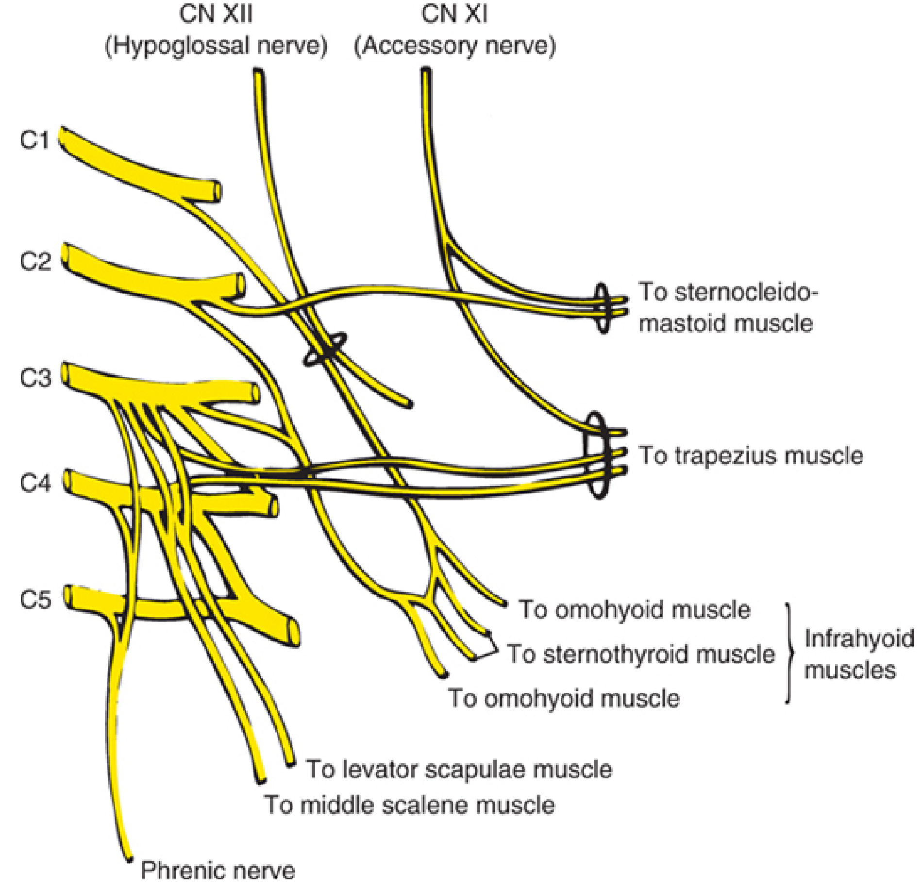
Formation
- These rami emerge from the intervertebral foramina and form a series of anastomotic loops
- The plexus lies deep to the SCM, on the surface of the middle scalene and levator scapulae muscles, covered by the prevertebral fascia
Emergence - Erb's Point
- Location: Midpoint of the posterior border of the SCM
- All four groups of cutaneous branches fan out from this point like the four quadrants of a compass
BD Chaurasia: "The cutaneous branches of the cervical plexus emerge from behind the middle of the posterior border of the sternocleidomastoid muscle - this point is called Erb's point."
Cutaneous (Sensory) Branches and Their Distribution
| Branch | Spinal Level | Direction from Erb's Point | Area Supplied | Relation to This Case |
|---|---|---|---|---|
| Lesser occipital nerve | C2 | Upward, along posterior border of SCM | Skin of lateral scalp + behind the ear | Not affected in this case |
| Great auricular nerve | C2-C3 | Upward and forward | Lower cheek, skin over angle of the mandible, lower part of the ear, upper neck | AFFECTED - explains numbness at angle of mandible |
| Transverse cervical nerve (transverse colli) | C2-C3 | Forward, horizontally across SCM | Anterior and lateral aspects of the neck | AFFECTED - explains numbness over anterior neck |
| Supraclavicular nerves (medial, intermediate, lateral) | C3-C4 | Downward, fanning out | Skin over the clavicle, upper chest, upper shoulder, skin of the shoulder | AFFECTED - explains numbness over clavicle and shoulder |
Motor Branches of the Cervical Plexus
| Branch | Levels | Muscle(s) Supplied |
|---|---|---|
| Ansa cervicalis (superior + inferior roots) | C1-C3 | All infrahyoid muscles (sternohyoid, sternothyroid, omohyoid, thyrohyoid) |
| Phrenic nerve | C3-C5 (mainly C4) | Diaphragm (sole motor supply) - "C3, 4, 5 keep the diaphragm alive" |
| Branches to SCM and trapezius | C2-C4 | Proprioceptive fibers to SCM and trapezius (CN XI is the main motor supply; cervical plexus provides proprioception) |
| Branches to scalene and levator scapulae | C3-C5 | Scalenes, levator scapulae |
KLM: "The cervical plexus is formed by the ventral rami of the first four cervical nerves. Its cutaneous branches supply the skin of the back of the head, the lateral face and neck, and the upper chest and shoulder."
Snell's: "All four cutaneous branches emerge from the posterior border of the SCM at approximately its midpoint. This point is important surgically as damage here can affect all the cutaneous branches simultaneously."
LO 5 - Enumerate the Superficial and Deep Cervical Lymph Nodes and Their Drainage Territories
Superficial Cervical Lymph Nodes
| Group | Location | Drains |
|---|---|---|
| Submental nodes | Below the chin, in the submental triangle | Midline structures: tip of tongue, lower central incisors, central lower lip, floor of mouth |
| Submandibular nodes | In the submandibular triangle, around the submandibular gland | Cheeks, lateral lip, lateral tongue, upper and lower teeth (except central incisors), part of nasal cavity |
| Parotid (preauricular) nodes | On and in the parotid gland | Scalp (frontotemporal), external ear, skin around the eye, parotid gland |
| Mastoid (postauricular) nodes | Behind the ear | Posterior scalp, external ear, mastoid region |
| Occipital nodes | Base of skull posteriorly | Posterior scalp |
| Superficial cervical nodes (along EJV) | Along the external jugular vein, superficial to SCM | Lower face, parotid region, ear |
| Anterior cervical nodes | Along the anterior jugular vein | Larynx, thyroid, trachea |
Deep Cervical Lymph Nodes
Major Groups:
- Located around the upper part of the internal jugular vein, near the carotid bifurcation
- Key node: Jugulodigastric node - the largest and most accessible; lies below the posterior belly of digastric at the angle of the jaw
- Drains: Tonsil, posterior tongue, oropharynx - first palpable node in tonsillar/throat infections
- Located around the lower internal jugular vein, near the subclavian vein
- Key node: Jugulo-omohyoid node - where the omohyoid crosses the internal jugular vein
- Drains: Tongue (anterior 2/3), submental nodes, submandibular nodes
- Run along the course of CN XI in the posterior triangle
- Drain: Scalp, ear, posterior neck
- This is the group in our CBL patient
- Along the transverse cervical vessels
- Virchow's node (Troisier's sign): Left supraclavicular node - when enlarged, signals metastasis from abdomen (gastric cancer, left testicular cancer) or thorax (lung cancer), because the thoracic duct drains here on the left
Lymphatic Drainage Summary:
| Region | First-echelon Nodes |
|---|---|
| Scalp (anterior) | Parotid + submandibular |
| Scalp (posterior) | Occipital + mastoid |
| Face | Parotid + submandibular |
| Nose + nasal sinuses | Submandibular + upper deep cervical |
| Tonsil + soft palate | Jugulodigastric node |
| Tongue (anterior 2/3) | Submental + submandibular + jugulo-omohyoid |
| Tongue (posterior 1/3) | Upper deep cervical directly |
| Larynx (above cords) | Upper deep cervical |
| Larynx (below cords) | Pretracheal + lower deep cervical |
| Thyroid | Pretracheal + upper deep cervical |
| All nodes eventually | Drain into the jugular trunk → right lymphatic duct (right side) or thoracic duct (left side) → subclavian vein |
BD Chaurasia: "The deep cervical lymph nodes form a chain along the internal jugular vein. They receive lymph directly from head and neck structures as well as indirectly from all the superficial groups. The jugulodigastric node is the 'sentinel' node of the tonsil."
KLM: "The lymph from the entire head and neck ultimately drains to the deep cervical nodes. On the right, the jugular trunk joins the right lymphatic duct; on the left, it joins the thoracic duct."
LO 6 - Trace the Carotid and Subclavian Arteries and Major Venous Drainage Pathways
THE CAROTID SYSTEM
Common Carotid Artery
- Right CCA: Arises from the brachiocephalic (innominate) trunk behind the right sternoclavicular joint
- Left CCA: Arises directly from the arch of the aorta in the thorax
- Both ascend in the carotid sheath alongside the internal jugular vein and vagus nerve
- Bifurcation: At the level of the upper border of thyroid cartilage (C4) - in the carotid triangle
- The CCA has no branches in the neck
Internal Carotid Artery (ICA)
- Ascends medially, with no branches in the neck
- Enters the skull through the carotid canal in the petrous temporal bone
- Supplies: Brain (80%), eye (ophthalmic artery), forehead
External Carotid Artery (ECA)
- Has 8 branches in the neck - supplies face, scalp, thyroid, pharynx, meninges
- Mnemonic: "Some Anatomists Like Freaking Out Poor Medical Students"
- Superior thyroid, Ascending pharyngeal, Lingual, Facial, Occipital, Posterior auricular, Maxillary, Superficial temporal
| Branch | Area Supplied |
|---|---|
| Superior thyroid | Upper thyroid, larynx |
| Ascending pharyngeal | Pharynx, meninges |
| Lingual | Tongue, floor of mouth |
| Facial | Face (lower 2/3) |
| Occipital | Posterior scalp |
| Posterior auricular | Behind the ear |
| Superficial temporal | Frontotemporal scalp - palpable at the temple |
| Maxillary | Deep face, teeth, dura mater (middle meningeal artery) |
THE SUBCLAVIAN ARTERY
- Right: Branch of the brachiocephalic trunk
- Left: Direct branch of the aortic arch
- Both arch upward, crossing the first rib, and enter the axilla to become the axillary artery
| Part | Position | Branches |
|---|---|---|
| 1st part | Medial to scalenus anterior | Vertebral artery, thyrocervical trunk, internal thoracic artery |
| 2nd part | Behind scalenus anterior | Costocervical trunk |
| 3rd part | Lateral to scalenus anterior - in the supraclavicular triangle (posterior triangle) | Dorsal scapular artery (sometimes) |
The 3rd part of the subclavian in the supraclavicular triangle can be compressed against the first rib to control upper limb haemorrhage. This is clinically important in the posterior triangle.
- Inferior thyroid artery
- Transverse cervical artery - crosses the posterior triangle
- Suprascapular artery - crosses the posterior triangle
MAJOR VENOUS DRAINAGE PATHWAYS
Internal Jugular Vein (IJV)
- Begins at the jugular foramen as a continuation of the sigmoid sinus
- Descends in the carotid sheath, lateral to the carotid artery
- Receives: facial vein, lingual vein, pharyngeal veins, superior and middle thyroid veins
- Joins the subclavian vein behind the sternoclavicular joint to form the brachiocephalic (innominate) vein
- The right brachiocephalic + left brachiocephalic veins join to form the superior vena cava
External Jugular Vein (EJV)
- Formed behind the angle of the jaw (parotid gland area) by the union of the posterior auricular vein and the posterior division of the retromandibular vein
- Crosses the SCM superficially (visible through the skin)
- Crosses the posterior triangle and pierces the investing fascia
- Drains into the subclavian vein
- Clinical use: Visible when venous pressure is raised (e.g., right heart failure, cardiac tamponade) - JVP assessment
Anterior Jugular Vein
- Forms near the chin, descends near the midline
- Joins the EJV or subclavian vein at the base of the neck
- Variable - occasionally large (important during thyroid surgery)
Subclavian Vein
- Continuation of the axillary vein at the lateral border of the first rib
- Joins the IJV to form the brachiocephalic vein
- Common site for central venous catheter (CVC) insertion - accessed in the subclavian triangle
- The thoracic duct drains into the angle between the left subclavian and left IJV
Brain/Face/Neck → Internal Jugular Vein
↘
Brachiocephalic Vein → Superior Vena Cava
↗
Upper limb → Subclavian Vein
Scalp/Face superficial → External Jugular Vein → Subclavian Vein
BD Chaurasia: "The internal jugular vein is the chief venous channel of the head and neck. It begins at the jugular foramen, runs in the carotid sheath, and ends by joining the subclavian vein to form the brachiocephalic vein."
Snell's: "The external jugular vein runs across the superficial surface of the SCM and is visible through the skin, particularly when the venous pressure is raised. It is a useful surface landmark and can be used for venous access."
KLM: "The subclavian vein lies anterior to the scalenus anterior muscle and posterior to the clavicle. It is commonly used for central venous access and lies anterior to the subclavian artery and the phrenic nerve."
How the Learning Objectives Connect Back to This Case
| LO | Relevance to the Patient |
|---|---|
| LO 1 - Triangles | The node was in the upper posterior triangle (occipital subdivision). Understanding its boundaries tells you what structures are nearby and at risk. |
| LO 2 - Contents | The spinal accessory nerve and cervical plexus cutaneous branches are the two contents injured in this case. The carotid triangle contents would be at risk if dissection went deeper/anteromedially. |
| LO 3 - Accessory nerve | The superficial course of CN XI across the posterior triangle explains exactly why it was severed - it lay right in the path of the lymph node excision. |
| LO 4 - Cervical plexus | All three of the patient's areas of numbness (mandible angle, anterior neck, shoulder/clavicle) map exactly onto the great auricular, transverse cervical, and supraclavicular branches - all emerging from Erb's point where the dissection occurred. |
| LO 5 - Lymph nodes | The node being biopsied was from the spinal accessory chain (posterior triangle group). Understanding lymph node levels helps determine what pathology was suspected and how far it may have spread. |
| LO 6 - Vessels | The carotid sheath (IJV + carotid) and subclavian artery would have been endangered had the surgeon dissected deeper or extended inferiorly - making knowledge of their position essential for safe surgery in this region. |