
explain meningitis in detail way its pathophysiology mechanism its types its symptoms initial and sympoms criteria dianostic approach physical test to diagnose it clinically and its treatment approach and give clinical exmple to explain it
Reading File
Now I'll run parallel searches across the medical library for all aspects of meningitis simultaneously.
Finding Sources
Finding Sources
Finding Sources
Excellent findings. Now let me read the key textbook sections in parallel - Harrison's (pathophysiology diagram), Robbins (pathology), Rosen's Emergency Medicine (clinical), and Bradley/Daroff's Neurology.
Reading File
Reading File
Reading File
Reading File
Excellent content. Now let me read more of Harrison's for pathophysiology details, and also get treatment section and CSF findings.
Reading File
Searching PubMed
"bacterial meningitis" management treatment guidelines
Excellent - I have rich content from Harrison's. Now let me get the Harrison's pathophysiology image and the treatment section, plus CSF analysis from Henry's:
Loading Image
Reading File
Reading File
Excellent! Now let me get the Harrison's treatment section and the Tietz CSF table, and also the Robbins brain image:
Reading File
Reading File
I now have comprehensive content from multiple authoritative textbooks. Let me also grab the Robbins brain pathology image and the Harrison's treatment table:
Loading Image
Reading File
I now have all the information needed for a comprehensive, well-sourced answer. Let me compile the full response.# Meningitis: A Complete Clinical Guide
1. Definition
Meningitis is an inflammatory process involving the leptomeninges (pia and arachnoid mater) within the subarachnoid space (SAS). When the infection extends into the underlying brain parenchyma, it is termed meningoencephalitis. It can be infectious or non-infectious (chemical meningitis from ruptured epidermoid cyst, carcinomatous meningitis from metastatic cancer).
- Robbins & Kumar Basic Pathology, p. 833
2. Types of Meningitis
| Type | Onset | Primary Causes | CSF Pattern |
|---|---|---|---|
| Acute Pyogenic (Bacterial) | Hours-days | S. pneumoniae, N. meningitidis, E. coli (neonates) | Neutrophil-dominant, low glucose, high protein |
| Aseptic (Viral) | Days | Enteroviruses, HSV-2, arboviruses, West Nile | Lymphocyte-dominant, normal glucose |
| Chronic (Tuberculous/Fungal) | Weeks-months | M. tuberculosis, Cryptococcus, Coccidioides | Lymphocytic, very low glucose |
| Fungal | Subacute | Cryptococcus neoformans (especially HIV+) | Lymphocytic, India ink positive |
| Chemical/Aseptic (non-infective) | Variable | Drugs, contrast media, tumor, post-LP | Variable, no organisms |
Common Pathogens by Age Group
| Age | Likely Organism |
|---|---|
| Neonates (<1 month) | E. coli, Group B Streptococcus (GBS), Listeria monocytogenes |
| Infants/children | Neisseria meningitidis, S. pneumoniae |
| Adolescents/young adults | N. meningitidis (most common) |
| Adults >50 / elderly | S. pneumoniae, Listeria monocytogenes |
| Immunocompromised | Listeria, Cryptococcus, CMV, TB |
| Post-neurosurgery | Staphylococci (coagulase-negative), gram-negatives, P. aeruginosa |
- Harrison's Principles of Internal Medicine 22E; Robbins Basic Pathology, p. 833
3. Pathophysiology Mechanism
This is the core of understanding meningitis. The neurologic damage is largely immune-mediated, not just from bacterial invasion directly.
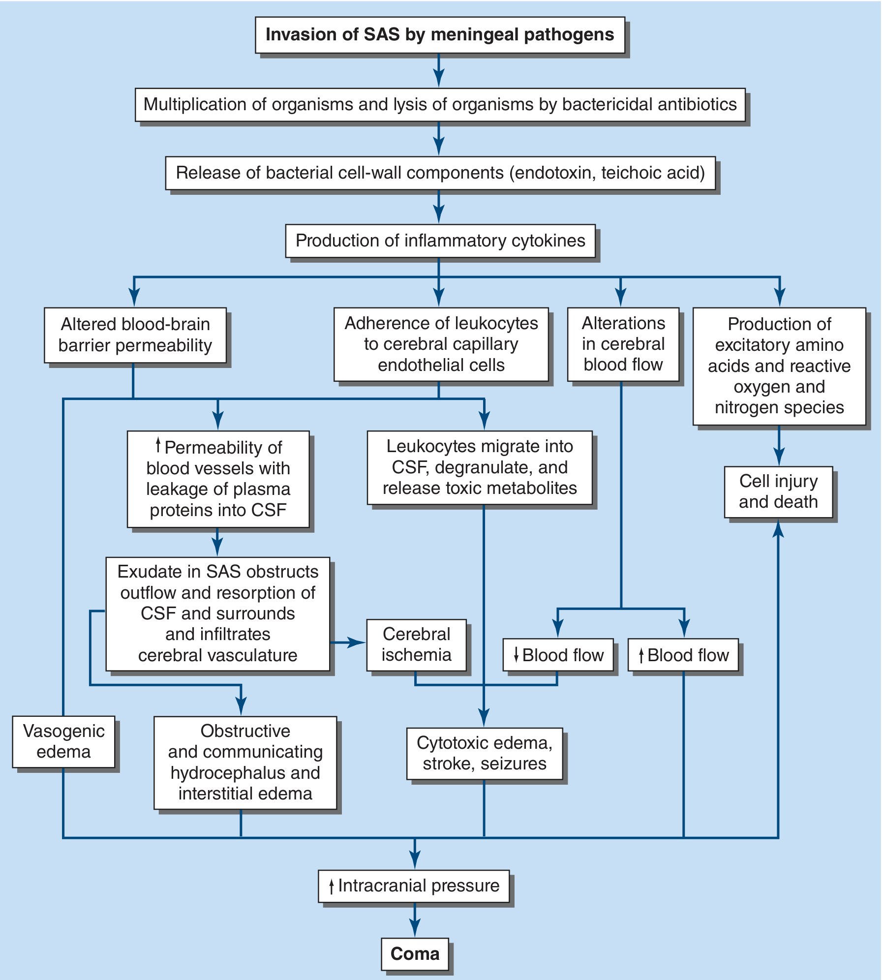
FIGURE: The pathophysiology of neurologic complications of bacterial meningitis. CSF = cerebrospinal fluid; SAS = subarachnoid space. (Harrison's Principles of Internal Medicine 22E, Fig. 143-1)
Step-by-Step Mechanism
Step 1 - Colonization and Invasion of the Bloodstream
Organisms such as S. pneumoniae and N. meningitidis first colonize the nasopharyngeal epithelium. They attach to epithelial cells and are either transported across them in membrane-bound vacuoles or invade by separating tight junctions. The bacteria's polysaccharide capsule protects them from neutrophil phagocytosis and classic complement-mediated killing once they reach the bloodstream.
Step 2 - Crossing the Blood-Brain Barrier (BBB)
Bloodborne bacteria reach the choroid plexus, directly infect choroid plexus epithelial cells, and gain access to the CSF. Some bacteria (notably S. pneumoniae) adhere to cerebral capillary endothelial cells and migrate through or between them into the CSF.
Step 3 - Multiplication in the CSF
Normal CSF has few white cells, minimal complement, and low immunoglobulin levels - this is a "safe haven" for bacteria. Poor opsonization and the fluid environment of CSF prevent effective phagocytosis, so bacteria multiply rapidly.
Step 4 - Inflammatory Cascade (the key injury mechanism)
Lysis of bacteria releases cell-wall components into the SAS:
- Gram-negative bacteria release lipopolysaccharide (LPS / endotoxin)
- S. pneumoniae releases teichoic acid and peptidoglycans
These trigger microglia, astrocytes, monocytes, and endothelial cells to produce:
- TNF-alpha and IL-1beta (within 1-2 hours of inoculation)
- Chemokines that drive leukocyte migration into the CSF
Step 5 - Four Parallel Injury Pathways (see diagram above)
- Vasogenic edema: TNF-alpha + IL-1beta increase BBB permeability, causing plasma protein leakage into the SAS.
- Obstructive hydrocephalus and interstitial edema: The proteinaceous exudate obstructs CSF flow through the ventricular system and impairs arachnoid granulation resorption, causing communicating and obstructive hydrocephalus.
- Cytotoxic edema, stroke, seizures: Leukocytes adhere to capillary endothelium, migrate into CSF, and degranulate - releasing toxic metabolites. Bacteremia and inflammatory cytokines also induce production of excitatory amino acids, reactive oxygen and nitrogen species (free radicals, nitric oxide, peroxynitrite), directly killing neurons, especially in the dentate gyrus of the hippocampus.
- Cerebrovascular injury: The purulent exudate in the SAS encroaches on large arteries at the base of the brain. Inflammatory infiltration of arterial walls causes vasculitis and intimal thickening, leading to arterial or venous thrombosis, infarction, and vasospasm.
Final Common Pathway:
All four mechanisms converge on raised intracranial pressure (ICP) → coma → cerebral herniation if untreated.
Key point: Neurologic injury can progress even after CSF sterilization by antibiotics, because the immune cascade is already in motion. This is the rationale for corticosteroid therapy.
- Harrison's 22E, pp. 1660-1669; Robbins Basic Pathology, p. 833-834
4. Histopathology
Below is a microscopic image of bacterial (pyogenic) meningitis - note the dense neutrophilic infiltrate in the subarachnoid space:
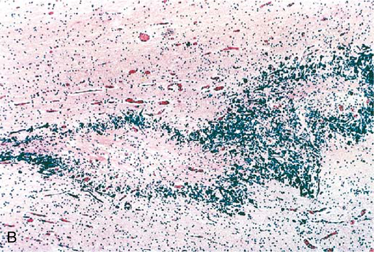
Dense neutrophilic exudate filling the subarachnoid space in acute pyogenic (bacterial) meningitis. (Robbins & Kumar Basic Pathology)
5. Symptoms and Clinical Presentation
Classic Triad (present in ~44% of patients)
- Fever
- Nuchal rigidity (neck stiffness)
- Altered consciousness (headache, confusion, lethargy)
A fourth hallmark is photophobia and phonophobia (intolerance to light and sound).
Initial / Early Symptoms (first 24 hours)
- Severe, progressively worsening headache ("worst headache of my life")
- High fever (38-40°C), rigors
- Nausea, vomiting
- Photophobia
- Malaise, myalgias
Late / Advanced Symptoms
- Neck stiffness (meningismus) - pain and resistance on neck flexion
- Altered mental status, confusion, drowsiness progressing to coma
- Seizures (20-30% of bacterial meningitis patients)
- Focal neurologic deficits
- Petechial/purpuric rash (especially with meningococcemia - typically starts on extremities)
- Papilledema (sign of raised ICP)
- Bulging fontanelle in neonates
Special Populations
-
Neonates: May only show bulging fontanelle, irritability, decreased tone, poor feeding, temperature instability - classic signs are often absent
-
Elderly/immunosuppressed: Altered mental status may be the only finding; lower threshold for LP is essential
-
Tuberculous meningitis: Subacute/chronic onset over weeks to months; fever, weight loss, night sweats, headache - may be the only manifestation of TB or accompany pulmonary TB
-
Fungal meningitis: Subtle even in healthy adults; mild headache, low-grade fever, malaise, weight loss - often not initially suspected
-
Rosen's Emergency Medicine, p. 4166-4176; Harrison's 22E
6. Diagnostic Approach
Clinical Criteria for Suspicion
Any 2 of the 4 cardinal features should prompt immediate evaluation:
- Headache
- Fever
- Neck stiffness
- Altered consciousness
Step-by-Step Diagnostic Workflow
Suspected Meningitis
        ↓
Blood cultures (STAT) + CBC, CMP, Coagulation, Procalcitonin
        ↓
Neurologic exam - any focal deficits, papilledema, seizures, LOC?
        ↓
YES → CT head FIRST             NO → Proceed directly to LP
        ↓                                    ↓
CT normal → LP              LUMBAR PUNCTURE (gold standard)
Critical rule: Do NOT delay antibiotics for CT or LP. If CT is needed, blood cultures + empiric antibiotics should start IMMEDIATELY, then LP after CT. Delay in treatment is the most critical factor determining morbidity and mortality.
7. Physical Examination Tests (Clinical Diagnosis)
A. Kernig's Sign
How to perform: Patient lies supine. Flex the hip to 90 degrees. Then attempt to extend the knee.
Positive sign: Inability to extend the knee beyond ~135 degrees due to pain and spasm of the hamstrings.
Interpretation: Positive = meningeal irritation. Sensitivity is <12% but specificity is high - if present, it strongly supports meningitis.
B. Brudzinski's Sign
How to perform: Patient lies supine. Passively flex the neck toward the chest.
Positive sign: Involuntary flexion of the hips and knees in response to neck flexion.
Interpretation: Same significance as Kernig's - low sensitivity (<12%), high specificity.
Both Kernig and Brudzinski signs have a sensitivity less than 12% but high specificity - their absence does NOT rule out meningitis, but their presence is diagnostically meaningful.
C. Jolt Accentuation of Headache
How to perform: Ask the patient to rotate their head rapidly horizontally at 2-3 rotations per second.
Positive sign: Worsening of headache with this maneuver.
Interpretation: May have higher sensitivity than Kernig/Brudzinski for meningeal irritation.
D. Fundoscopy (Papilledema)
Check for papilledema or loss of venous pulsations before LP - these are contraindications to immediate LP (mass lesion must be excluded). Bedside ocular ultrasound for optic nerve sheath diameter is an alternative to formal fundoscopy.
E. Skin Examination
Petechiae or purpura on extremities, trunk, or conjunctivae strongly suggest meningococcemia (Neisseria meningitidis). This rash can progress to ecchymoses and necrosis if Waterhouse-Friderichsen syndrome (bilateral adrenal hemorrhage) develops.
- Rosen's Emergency Medicine, p. 4168-4171
8. Lumbar Puncture - CSF Analysis (Gold Standard)
CSF Interpretation Table
| Parameter | Normal | Bacterial | Viral | TB/Fungal |
|---|---|---|---|---|
| Opening pressure | 70-180 mmH₂O | >180 mmH₂O (often >400) | Mildly elevated | Moderately elevated |
| Appearance | Clear | Turbid/purulent | Clear | Clear/slightly turbid |
| WBC count | <5/μL | 100-10,000/μL | 10-500/μL | 10-500/μL |
| Cell type | Lymphocytes | Neutrophils (PMN) | Lymphocytes | Lymphocytes |
| Glucose | 45-80 mg/dL | <40 mg/dL (<2.2 mmol/L) | Normal | Very low |
| CSF/Serum glucose ratio | >0.6 | <0.4 | >0.6 | <0.4 |
| Protein | 15-45 mg/dL | >45 mg/dL (often 100-500) | Mildly elevated | Markedly elevated |
| Gram stain | Negative | Positive >60% | Negative | Negative |
| Culture | Negative | Positive >80% | Negative | Positive (weeks) |
| PCR | - | Bacterial DNA | Viral DNA/RNA | MTB PCR |
Key discriminating rule: PMN count >1,180 cells/μL OR total WBC >2,000/μL has a 99% predictive value for bacterial meningitis. (Henry's Clinical Diagnosis and Management by Laboratory Methods)
Note: Viral meningitis may show early PMN predominance (>60%) but this shifts to lymphocytic within 2-3 days, unlike bacterial meningitis.
Additional Tests
- Blood cultures x2 (before antibiotics ideally, but do NOT delay antibiotics)
- Serum procalcitonin - elevated in bacterial vs. viral meningitis
- Multiplex PCR (meningitis/encephalitis panel) - rapid detection of bacteria, viruses, fungi
- CT head - before LP if: focal neuro deficits, papilledema, seizure, GCS < 14, HIV, immunocompromised, age >60
- MRI with gadolinium - shows meningeal enhancement (not specific for meningitis, any disease increasing BBB permeability)
- Petechial skin biopsy - in meningococcemia, may show organism on Gram stain
Organisms and Their CSF Gram Stain Patterns
| Organism | Gram Stain Pattern |
|---|---|
| S. pneumoniae | Gram-positive diplococci in pairs |
| N. meningitidis | Gram-negative diplococci |
| H. influenzae | Gram-negative coccobacillus |
| L. monocytogenes | Gram-positive rods |
| E. coli | Gram-negative rods |
- Bradley and Daroff's Neurology, Table 79.1; Harrison's 22E, Table 143-1
9. Treatment Approach
Principle: This is a MEDICAL EMERGENCY. Target: antibiotics within 60 minutes of arrival.
Step 1 - Empiric Antibiotics (Start IMMEDIATELY, before culture results)
Community-acquired meningitis (adults and children >3 months):
- Ceftriaxone 2g IV q12h (or cefotaxime) + Vancomycin 45-60 mg/kg/day IV (for penicillin-resistant S. pneumoniae) + Dexamethasone 10mg IV q6h x4 days (give 15-20 min BEFORE or with first antibiotic dose)
Add Ampicillin if: age <3 months, >55 years, pregnancy, immunocompromised, or suspected Listeria (covers L. monocytogenes, which cephalosporins do not cover)
Add Metronidazole if: otitis, sinusitis, or mastoiditis (cover gram-negative anaerobes)
Post-neurosurgery/hospital-acquired:
- Vancomycin + Meropenem (or Cefepime) to cover gram-negatives including P. aeruginosa
Step 2 - Adjunctive Corticosteroids (Dexamethasone)
Dose: 10 mg IV q6h for 4 days
Mechanism: Inhibits TNF-alpha and IL-1beta synthesis at mRNA level, decreases CSF outflow resistance, stabilizes BBB
Must be given BEFORE or concurrent with antibiotics - dexamethasone cannot suppress TNF-alpha once the cytokine cascade has been fully activated
Benefit: Reduces unfavorable outcomes (15% vs. 25%), reduces mortality (7% vs. 15%), and reduces sensorineural hearing loss - especially in pneumococcal meningitis
Caveat: Reduces vancomycin penetration into CSF - so use higher vancomycin doses (45-60 mg/kg/day) or consider intraventricular vancomycin
Step 3 - Targeted Antibiotics (Once organism identified)
| Organism | Preferred Antibiotic |
|---|---|
| S. pneumoniae (penicillin-sensitive) | Penicillin G or Amoxicillin |
| S. pneumoniae (resistant) | Ceftriaxone + Vancomycin |
| N. meningitidis (penicillin-sensitive) | Penicillin G |
| N. meningitidis (resistant) | Ceftriaxone |
| L. monocytogenes | Ampicillin + Gentamicin |
| H. influenzae | Ceftriaxone |
| Gram-negative bacilli | Cefepime or Meropenem |
Step 4 - Management of Raised ICP
- Elevate head of bed to 30-45 degrees
- Intubation and controlled hyperventilation (PaCO₂ 25-30 mmHg)
- Mannitol 0.5-1 g/kg IV for osmotic decompression
- ICU monitoring with ICP monitoring device
- Anti-seizure prophylaxis if seizures present
Viral Meningitis Treatment
Mostly supportive (hydration, analgesia, antipyretics). Exception: HSV meningitis/encephalitis requires Acyclovir 10 mg/kg IV q8h. Empiric acyclovir is often added when HSV cannot be excluded.
Prognosis (Mortality by Organism)
-
H. influenzae, N. meningitidis, Group B Strep: 3-7%
-
Listeria monocytogenes: 15-25%
-
S. pneumoniae: 20-30% (highest for bacterial meningitis)
-
Complications include sensorineural hearing loss (5-35%), cognitive impairment, hydrocephalus, brain abscess
-
Harrison's 22E, pp. 1762-1832; Rosen's Emergency Medicine
10. Clinical Example (Case Study)
Setting: Emergency Department, 3:00 AM
Patient: 19-year-old male university student, brought in by roommate
Presenting complaint: "Worst headache of my life" for 6 hours, with high fever
History: Woke up with frontal headache that rapidly worsened. Now has fever, nausea, and says light hurts his eyes. He noticed a rash on his legs about 2 hours ago.
Vitals:
- Temp: 39.4°C
- HR: 118 bpm
- BP: 98/60 mmHg (hypotensive)
- RR: 22/min
- GCS: 13 (confused)
Physical Examination:
- Multiple petechiae and purpuric lesions on bilateral lower extremities and trunk - non-blanching
- Neck stiffness: Cannot touch chin to chest
- Kernig's sign: Positive bilaterally
- Brudzinski's sign: Positive - hip/knee flexion on neck flexion
- Fundoscopy: No papilledema
- No focal neurologic deficits
Immediate Action (within 10 minutes):
- Blood cultures x2 drawn IMMEDIATELY
- Dexamethasone 10mg IV given
- Ceftriaxone 2g IV + Vancomycin 1.5g IV started within 20 minutes of arrival (no CT needed - no focal deficits, no papilledema)
- IV fluids for hypotension
LP performed 30 minutes later:
- Opening pressure: 420 mmH₂O (markedly elevated)
- Appearance: Turbid (purulent)
- WBC: 8,200/μL - 92% neutrophils
- Glucose: 18 mg/dL (serum glucose 110 mg/dL) - ratio 0.16
- Protein: 380 mg/dL
- Gram stain: Gram-negative diplococci
Diagnosis: Neisseria meningitidis meningitis (Meningococcal meningitis) with meningococcemia
Pathophysiology explained in this patient:
- N. meningitidis invaded his nasopharynx (common in crowded university dormitory settings) → bacteremia → crossed the BBB at the choroid plexus → LPS triggered TNF-alpha / IL-1beta cascade → purulent exudate in SAS → raised ICP (headache, raised opening pressure) → vasogenic edema
- Simultaneously, dermal seeding of N. meningitidis organisms caused vascular endothelial damage → non-blanching petechiae/purpura
- Hypotension signals early septic shock / possible early Waterhouse-Friderichsen syndrome
Treatment continuation:
- Ceftriaxone monotherapy (N. meningitidis is penicillin/cephalosporin-sensitive) once identified
- Dexamethasone continued for 4 days
- ICU admission; ICP management
- Public health notification and ciprofloxacin prophylaxis for close contacts
- Isolation for first 24 hours of antibiotic therapy
Outcome (with prompt treatment): Full recovery without neurologic sequelae. Without treatment, this patient would likely have progressed to coma and death within 12-24 hours.
Quick Summary Table
| Feature | Bacterial | Viral | TB/Fungal |
|---|---|---|---|
| Onset | Hours | Days | Weeks-months |
| Fever | High | Moderate | Low-grade |
| CSF glucose | Very low | Normal | Low |
| CSF cells | Neutrophils | Lymphocytes | Lymphocytes |
| CSF protein | Very high | Mildly high | High |
| Gram stain | Often + | Negative | Negative |
| Treatment | Antibiotics + Dexamethasone | Supportive (Acyclovir for HSV) | Anti-TB drugs / Antifungals |
| Mortality | 15-30% | <1% | 20-40% |
Sources:
- Harrison's Principles of Internal Medicine, 22nd Edition (2025)
- Robbins & Kumar Basic Pathology
- Rosen's Emergency Medicine: Concepts and Clinical Practice
- Bradley and Daroff's Neurology in Clinical Practice
- Henry's Clinical Diagnosis and Management by Laboratory Methods
- Tietz Textbook of Laboratory Medicine, 7th Edition
- Recent evidence: Antimicrobial resistance in meningitis (Lancet Microbe, 2026) - highlights growing resistance in S. pneumoniae, N. meningitidis, H. influenzae (PMID 41616789)That covers everything you asked for, sourced directly from Harrison's 22E, Robbins Pathology, Rosen's Emergency Medicine, Bradley-Daroff's Neurology, Henry's Lab Methods, and Tietz Laboratory Medicine. Here's a quick orientation of what was covered:
What you got:
- Definition - what meningitis is and its non-infectious forms
- Types - bacterial/viral/TB/fungal/chemical with organism-by-age table
- Pathophysiology - the complete 5-step mechanism with the Harrison's flowchart image showing how invasion leads to cytokine storm → 4 injury pathways → raised ICP → coma
- Histopathology - Robbins microscopy image of the neutrophilic subarachnoid exudate
- Symptoms - early vs. late, classic triad, special populations (neonates, elderly, TB)
- Diagnostic approach - the workflow tree including when to do CT first vs. go straight to LP
- Physical exam tests - Kernig's, Brudzinski's, jolt accentuation, fundoscopy, skin exam with sensitivity/specificity data
- CSF analysis table - normal vs. bacterial vs. viral vs. TB/fungal with the 99% predictive value cutoff
- Treatment - empiric regimen, dexamethasone rationale and timing, targeted therapy, ICP management, prognosis
- Clinical case - a 19-year-old with meningococcal meningitis tying all concepts together from presentation to outcome
This is a shared conversation. Sign in to Orris to start your own chat.