
Components of blood
Blood is made up of a liquid component (plasma, ~55%) and formed elements (~45%): red blood cells (erythrocytes), white blood cells (leukocytes), and platelets (thrombocytes). Plasma itself contains water, proteins (albumin, globulins, fibrinogen), electrolytes, nutrients, and waste products.
Searching sources...
Reading File
Finding Sources
Finding Sources
Reading File
Reading File
Reading File
Reading File
Loading Image
Loading Image
Here is a thorough breakdown of the components of blood, sourced from Histology: A Text and Atlas and the Color Atlas of Human Anatomy Vol. 2:
Components of Blood
Blood is a fluid connective tissue consisting of a liquid phase (plasma) and solid formed elements suspended within it. Total blood volume is approximately 8% of body weight.
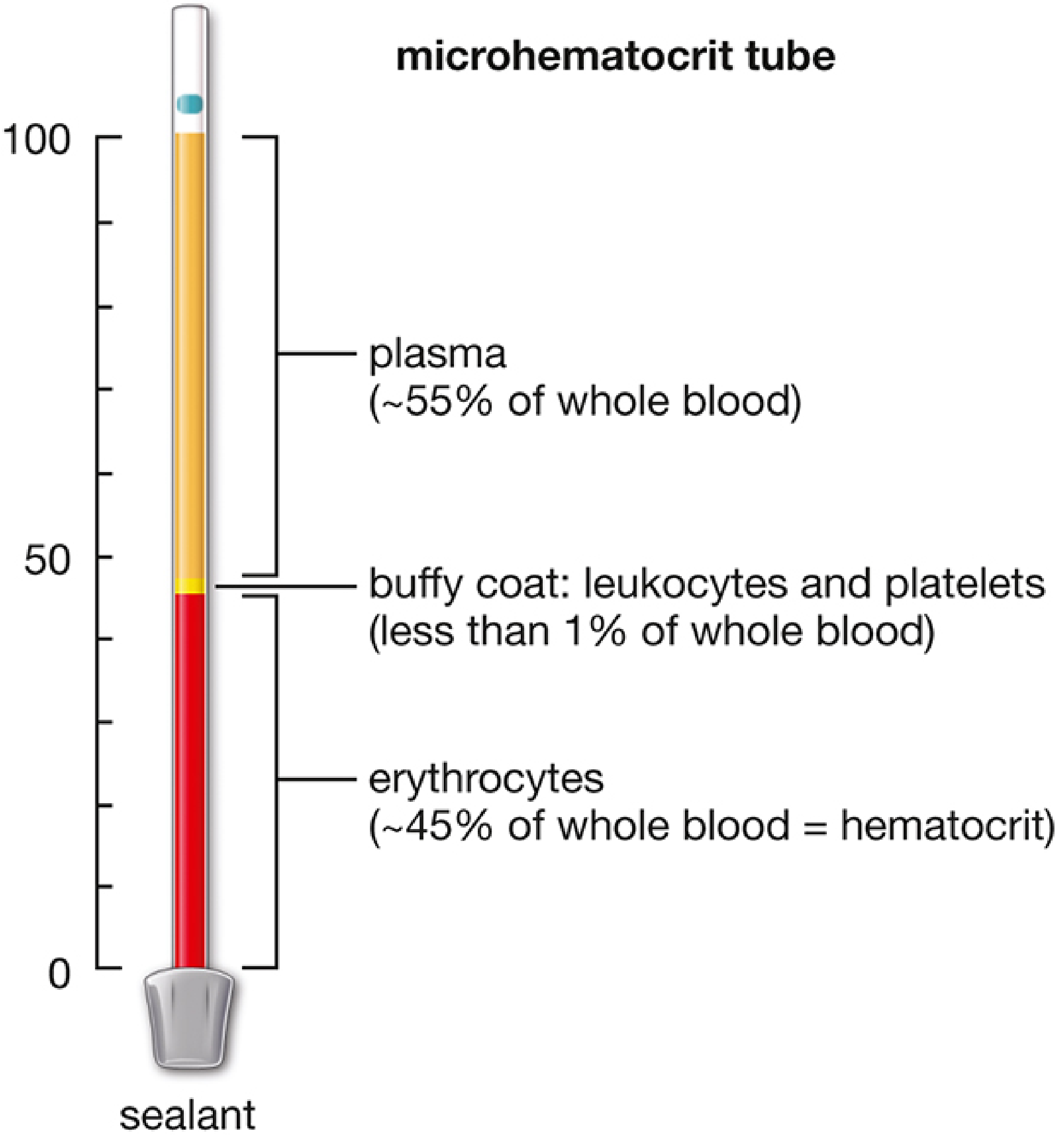
1. Plasma (~55% of whole blood)
Plasma is the protein-rich liquid extracellular matrix of blood. Its composition:
| Component | % of Plasma |
|---|---|
| Water | 91-92% |
| Proteins (albumin, globulins, fibrinogen) | 7-8% |
| Other solutes (electrolytes, nutrients, gases, waste) | 1-2% |
- Albumin: maintains osmotic pressure; transports fatty acids, hormones, bilirubin
- Globulins (alpha, beta, gamma): transport lipids/metals; gamma globulins are antibodies (immunoglobulins)
- Fibrinogen: key clotting factor; converts to fibrin during coagulation
- Serum = plasma minus clotting factors (i.e., what remains after blood clots and is centrifuged)
2. Formed Elements (~45% of whole blood)
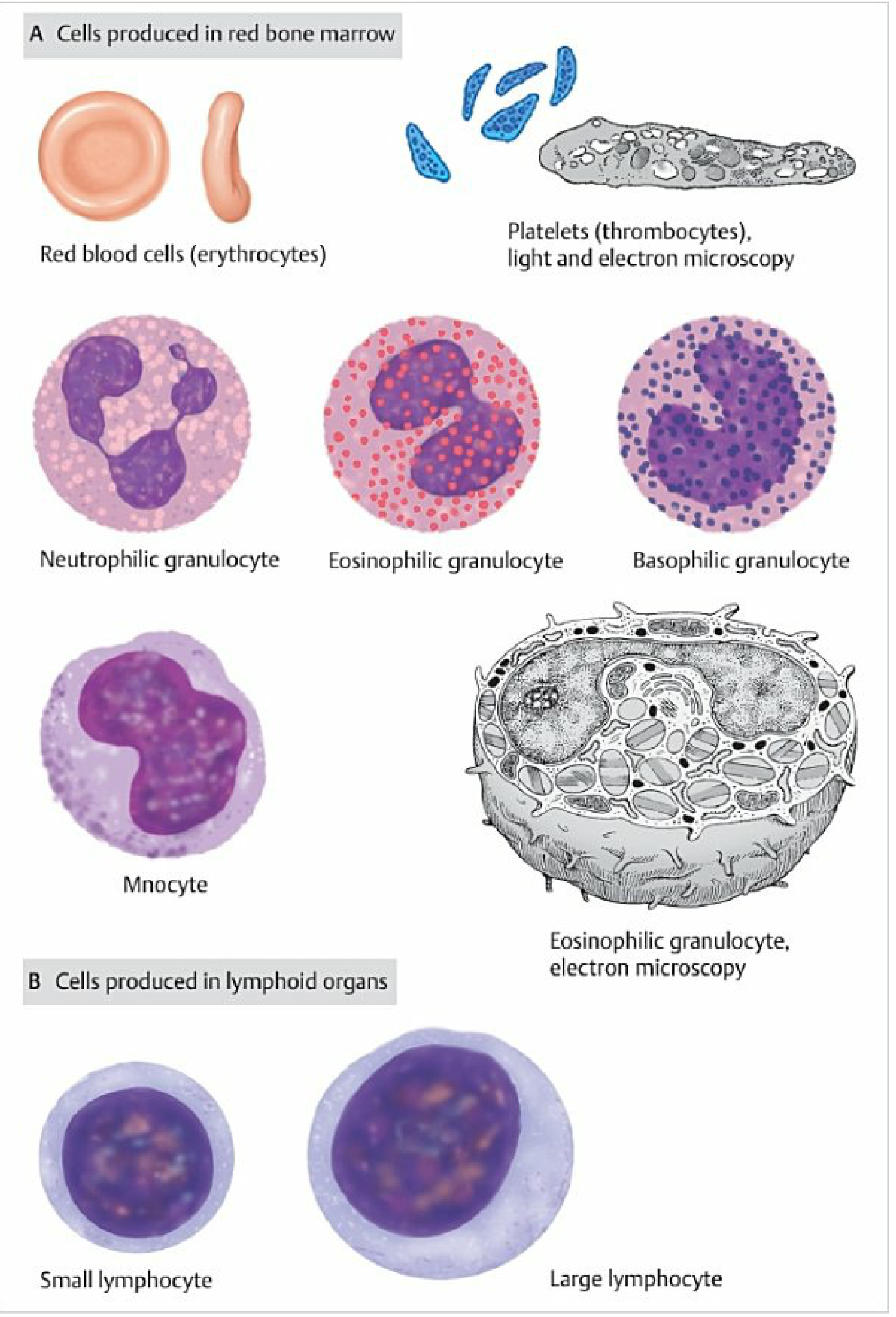
A. Erythrocytes (Red Blood Cells) - ~99% of formed elements
| Feature | Detail |
|---|---|
| Count | 4.3-5.7 × 10¹²/L (male); 3.9-5.0 × 10¹²/L (female) |
| Diameter | ~7.5 µm |
| Shape | Biconcave disc (no nucleus) |
| Lifespan | 100-120 days |
| Main content | ~90% hemoglobin (iron-containing) |
- Biconcave shape maximizes surface area for O₂/CO₂ exchange
- Oxygenated = bright red; deoxygenated = dark red
- Reticulocytes are immature erythrocytes (~1% in blood); increased after blood loss
- Destroyed mainly in the spleen and liver; iron is recycled for erythropoiesis in bone marrow
- Surface glycocalyx (glycolipids and glycoproteins) determines ABO blood group
B. Leukocytes (White Blood Cells)
Total count: 3.5-10.5 × 10⁹/L. Leukocytes function in immune defense. They are divided into two broad classes:
Granulocytes (produced in red bone marrow)
| Type | Count/µL | % of WBCs | Lifespan | Function |
|---|---|---|---|---|
| Neutrophils | 2200-6300 | 55-70% | ~6-7 hours | Phagocytosis of bacteria/viruses; first responders to infection |
| Eosinophils | 80-360 | 2-4% | ~8 hours | Defense against parasites; limits allergic reactions |
| Basophils | 0-90 | 0-1% | ~5-6 hours | Release histamine and heparin; involved in allergic/parasitic responses |
- Granulocytes have lobulated/segmented nuclei
- "Band cells" = immature granulocytes lacking nuclear segmentation (a "left shift" in infection)
Agranulocytes (produced in red bone marrow and lymphoid organs)
| Type | Count/µL | % of WBCs | Function |
|---|---|---|---|
| Lymphocytes | 900-2900 | 25-28% | Adaptive immunity (B cells = antibodies; T cells = cell-mediated) |
| Monocytes | 300-900 | ~8.6% | Phagocytosis; differentiate into macrophages in tissues |
Key WBC abnormalities:
- Leukocytosis: WBC > 10,000/µL (infection, inflammation, malignancy)
- Leukopenia: WBC < 2,000/µL (bone marrow suppression, autoimmune)
C. Thrombocytes (Platelets)
| Feature | Detail |
|---|---|
| Count | 150-450 × 10⁹/L |
| Origin | Fragments of megakaryocyte cytoplasm (not true cells) |
| Size | 2-4 µm, irregularly shaped |
- Platelets release thrombokinase, initiating the clotting cascade
- Also transport serotonin, a local vasoconstrictor
- Thrombocytopenia = platelet deficiency (bleeding risk)
- Thrombocytosis = excess platelets (thrombosis risk)
Summary Table
| Component | % of Blood | Key Role |
|---|---|---|
| Plasma | ~55% | Solvent; transport; immune proteins; clotting factors |
| Erythrocytes | ~44-45% | O₂/CO₂ transport |
| Buffy coat (WBCs + platelets) | <1% | Immune defense; hemostasis |
Sources: Histology: A Text and Atlas with Correlated Cell and Molecular Biology; Color Atlas of Human Anatomy Vol. 2, Internal Organs
Broncho pneumonia
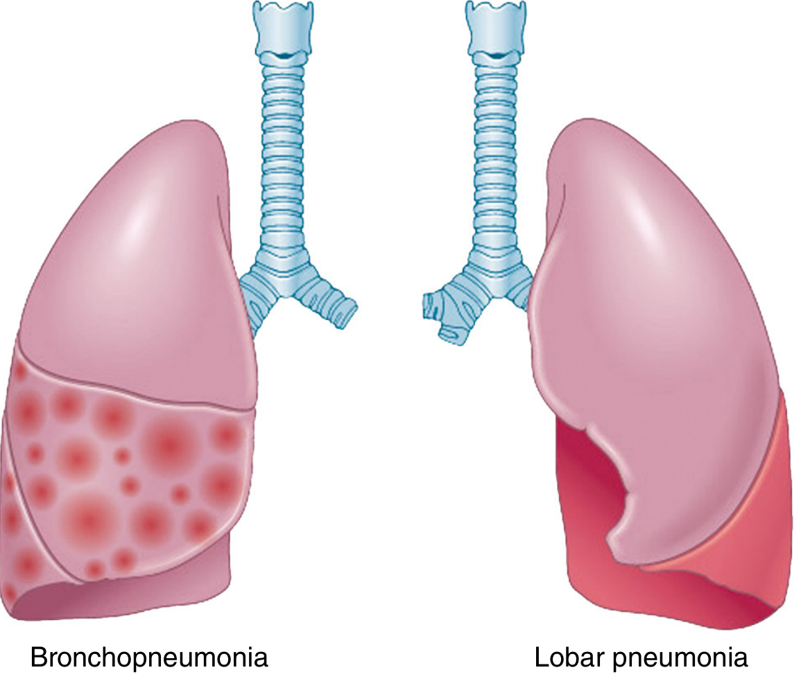
Bronchopneumonia is a pattern of acute bacterial pneumonia characterized by patchy, multifocal consolidation centered on bronchioles and surrounding alveoli, as opposed to lobar pneumonia which involves an entire lobe. It is common in the very young, elderly, and debilitated patients, and is often caused by Staphylococcus aureus, Streptococcus pneumoniae, Klebsiella, and gram-negative organisms.
Searching sources...
Finding Sources
Finding Sources
Reading File
Reading File
Reading File
Reading File
Finding Sources
Loading Image
Loading Image
Here is a comprehensive overview of bronchopneumonia from Robbins, Cotran & Kumar - Pathologic Basis of Disease, Fishman's Pulmonary Diseases and Disorders, and Grainger & Allison's Diagnostic Radiology:
Bronchopneumonia (Lobular Pneumonia)
Definition
Bronchopneumonia is a pattern of acute bacterial pneumonia characterized by patchy, multifocal consolidation of the lung centered on bronchioles and extending to surrounding peribronchiolar alveoli. It is also called lobular pneumonia because involvement is at the lobular (rather than lobar) level.
Bronchopneumonia vs. Lobar Pneumonia
| Feature | Bronchopneumonia | Lobar Pneumonia |
|---|---|---|
| Distribution | Patchy, multifocal, lobular | Entire lobe uniformly consolidated |
| Location | Often bilateral, basal | Usually unilateral |
| Onset | Insidious | Abrupt |
| Typical patients | Elderly, debilitated, infants | Previously healthy adults |
| Causative organisms | S. aureus, H. influenzae, gram-negatives | S. pneumoniae (most common) |
Pathogenesis
Infection begins in the bronchi and bronchioles, causing:
- Epithelial ulceration of bronchial/bronchiolar walls
- Fibrinopurulent exudate filling the peribronchiolar alveoli
- Patchy segmental involvement, which may become confluent producing more homogeneous consolidation
The patchy involvement tends to be bilateral and basal because respiratory secretions tend to gravitate to the lower lobes.
Etiology - Common Causative Organisms
| Organism | Notes |
|---|---|
| Staphylococcus aureus | Most typical bronchopneumonic pattern; often after viral URI; bilateral in ~40%; risk of cavitation, abscesses, pneumatoceles |
| Haemophilus influenzae (non-encapsulated) | Common bronchopneumonic pattern |
| Chlamydophila pneumoniae | Segmental bronchopneumonic pattern |
| Mycoplasma pneumoniae | Segmental pattern; common in young adults in closed communities |
| Klebsiella pneumoniae | Prone to necrosis and abscess formation |
| Escherichia coli | Multilobar, predominantly lower lobes; seen in debilitated patients |
| Pseudomonas aeruginosa | Confluent, often extensive bronchopneumonia; frequent cavitation; nosocomial |
| Viral pneumonias | Many viruses produce a bronchopneumonic consolidation pattern |
Non-infectious causes also produce a bronchopneumonic pattern: aspiration of gastric contents, sarcoidosis, hypersensitivity pneumonitis, cryptogenic organizing pneumonia (COP), and autoimmune diseases.
Morphology (Gross and Microscopic)
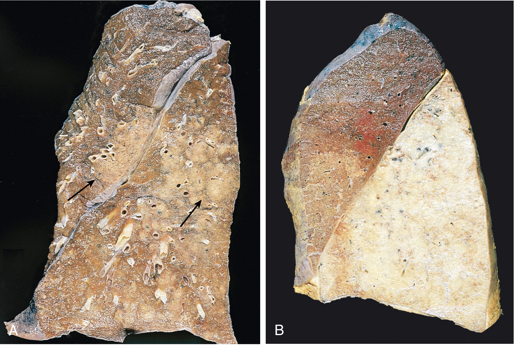
Gross appearance:
- Consolidated foci are slightly elevated, dry, granular, gray-red to yellow
- Poorly delimited margins
- Most often multilobar, bilateral, and basal
Microscopic appearance:
- Neutrophil-rich exudate filling bronchi, bronchioles, and adjacent alveolar spaces
- Epithelial ulceration of bronchial walls
- No orderly staging (unlike the four stages of lobar pneumonia)
Clinical Features
- Fever (high, often with shaking chills)
- Productive cough with mucopurulent sputum (occasionally hemoptysis)
- Dyspnea
- Pleuritic chest pain and pleural friction rub if pleuritis is present
- On imaging: focal opacities (vs. whole-lobe radiopacity in lobar pneumonia)
- Auscultation: crepitations, reduced breath sounds over consolidated areas
Radiology:
- Chest X-ray: patchy, ill-defined areas of airspace consolidation, predominantly in lower lobes
- CT: centrilobular nodules, tree-in-bud opacities, lobular/segmental consolidation
Complications
- Lung abscess - tissue destruction and necrosis (especially with Klebsiella or Staphylococcus)
- Empyema - spread of infection to pleural cavity causing intrapleural fibrinopurulent reaction
- Bacteremia and septicemia - dissemination to heart valves (endocarditis), brain (abscess), meningitis, pericarditis, suppurative arthritis
- Cavitation - especially with S. aureus and Pseudomonas
- Pneumatoceles - thin-walled air cysts, especially in staphylococcal disease
- Pleural effusion - occurs in 30-50% with staphylococcal pneumonia
Treatment
- Identify the causative organism and determine antibiotic sensitivity - the keystone of therapy
- Empirical antibiotics targeting the likely pathogen based on clinical setting (community-acquired vs. hospital-acquired)
- Appropriately treated patients typically become afebrile within 48-72 hours of starting antibiotics
- Mortality in hospitalized patients with pneumonia is less than 10% when treated effectively; deaths usually occur from complications (empyema, meningitis, endocarditis) or in debilitated patients
Sources: Robbins, Cotran & Kumar - Pathologic Basis of Disease; Fishman's Pulmonary Diseases and Disorders, 2-Volume Set; Grainger & Allison's Diagnostic Radiology
Compare clinical features of lobar vs. bronchopneumonia
Lobar pneumonia typically presents with sudden onset, high fever, single lobe involvement, and is caused mainly by S. pneumoniae in previously healthy adults. Bronchopneumonia tends to have a more insidious onset, patchy bilateral consolidation, affects the debilitated/elderly/infants, and is caused by S. aureus, gram-negatives, and others.
Searching sources...
Finding Sources
Finding Sources
Finding Sources
Finding Sources
Reading File
Reading File
Reading File
Finding Sources
Finding Sources
Finding Sources
Finding Sources
Finding Sources
Reading File
I now have rich content from all three major sources. Here is the full comparison:
Lobar Pneumonia vs. Bronchopneumonia - Clinical Comparison
1. Epidemiology & Predisposing Factors
| Feature | Lobar Pneumonia | Bronchopneumonia |
|---|---|---|
| Typical patient | Previously healthy adults | Elderly, infants, debilitated, immunocompromised |
| Predisposing conditions | Prior viral URTI, chronic heart/lung disease, alcoholism, splenectomy | COPD, chronic bronchitis, bronchiectasis, cardiac failure, post-viral infection, diabetes, renal failure |
| Seasonality | Winter/early spring (pneumococcal) | Year-round |
| Setting | Mainly community-acquired | Community-acquired and hospital-acquired |
2. Etiology
| Lobar Pneumonia | Bronchopneumonia |
|---|---|
| Streptococcus pneumoniae (most common, ~1/3 of all CAP) | Staphylococcus aureus |
| Klebsiella pneumoniae (classic upper lobe, bulging fissure) | Haemophilus influenzae (non-encapsulated) |
| Legionella pneumophila (rapidly progressive) | Chlamydophila pneumoniae, Mycoplasma pneumoniae |
| Klebsiella, E. coli, Pseudomonas (nosocomial/HAP) | |
| Viral pneumonias; aspiration; post-influenza bacterial superinfection |
3. Onset & Symptoms
| Feature | Lobar Pneumonia | Bronchopneumonia |
|---|---|---|
| Onset | Abrupt - sudden high fever, shaking chills | More insidious - gradual onset |
| Fever | High, sustained; may be absent in elderly | Present, often lower-grade |
| Chills | Prominent, rigors | Less prominent |
| Cough | Initially dry, then productive of purulent/blood-tinged ("rusty") sputum | Productive of mucopurulent sputum |
| Pleuritic chest pain | Prominent - stabbing, indicates parietal pleural involvement | Less prominent |
| Dyspnea | Present, significant | Present |
| Hemoptysis | Occasionally (blood-tinged sputum) | Occasionally |
| Myalgia/malaise | Common | Common |
| Confusion | Seen in elderly | Seen in elderly and debilitated |
In the elderly, classic features may be absent in both types - confusion or malaise without fever/cough may be the only presentation, demanding a high index of suspicion (Harrison's Principles of Internal Medicine, 22e).
4. Physical Examination Signs
| Sign | Lobar Pneumonia | Bronchopneumonia |
|---|---|---|
| Percussion | Dullness over the affected lobe (dense consolidation) | Patchy dullness (focal areas) |
| Breath sounds | Bronchial breathing (minority of cases); decreased air entry | Decreased air entry; scattered crackles |
| Adventitious sounds | Crepitations (crackles) over consolidation | Crepitations - patchy, bilateral |
| Pleural rub | Present in occasional cases | Less common |
| Chest expansion | Reduced (splinting to minimize pain) | Mildly reduced |
| Tachypnea | Present (>20 breaths/min) | Present |
| Tachycardia | Present | Present |
| Cyanosis | In severe hypoxemia | In severe cases |
| Vocal fremitus | Increased over consolidation | Patchy increase |
5. Pathological Stages (Lobar Pneumonia Only)
Lobar pneumonia has four classic stages of inflammatory response - bronchopneumonia does not follow this orderly progression:
| Stage | Gross Appearance | Microscopic Features |
|---|---|---|
| 1. Congestion | Lung heavy, boggy, red | Vascular engorgement, intra-alveolar edema, few neutrophils; bacteria numerous |
| 2. Red hepatization | Lobe red, firm, airless - liver-like | Massive exudate: neutrophils + RBCs + fibrin fill alveoli |
| 3. Gray hepatization | Lobe grayish-brown | RBCs disintegrate; fibrinopurulent exudate persists |
| 4. Resolution | Lung texture normalizes | Enzymatic digestion; debris resorbed by macrophages; normal architecture restored |
In bronchopneumonia, the lesions are consolidated areas of acute suppurative inflammation - gray-red to yellow, slightly elevated, poorly delimited, with a neutrophil-rich exudate filling bronchi, bronchioles, and adjacent alveoli.
6. Radiology
| Feature | Lobar Pneumonia | Bronchopneumonia |
|---|---|---|
| CXR pattern | Homogeneous consolidation of an entire lobe | Patchy, ill-defined multifocal opacities |
| Distribution | Usually unilateral, single lobe | Usually bilateral, predominantly lower lobes |
| Air bronchogram | Characteristic - airways outlined by surrounding alveolar fluid | Less prominent; not a defining feature |
| Segmental boundaries | Crosses segmental boundaries (nonsegmental) | Follows segmental/lobular distribution |
| Bulging fissure | Klebsiella - expanded lobe with bulging fissure | Not typical |
| Cavitation | Rare (except Klebsiella) | More common (S. aureus, Pseudomonas) |
| Pleural effusion | Up to 50% (pneumococcal) | 30-50% (staphylococcal) |
| CT findings | Dense homogeneous consolidation with air bronchograms | Centrilobular nodules, tree-in-bud opacities, lobular consolidation |
7. Complications
Both types share similar complications, but their frequency differs:
| Complication | Lobar | Bronchopneumonia |
|---|---|---|
| Lung abscess | Less common (mainly Klebsiella) | More common (S. aureus, Pseudomonas, Klebsiella) |
| Empyema | Occurs | Occurs |
| Bacteremia/septicemia | Occurs (endocarditis, meningitis, pericarditis) | Occurs |
| Cavitation/pneumatoceles | Rare | More common (S. aureus) |
| Pleural adhesions | Can follow pleuritis | Less organized pleuritis |
| Resolution failure | Rare with antibiotics | May organize into fibrosis in debilitated patients |
8. Response to Treatment
| Feature | Lobar Pneumonia | Bronchopneumonia |
|---|---|---|
| Response to antibiotics | Generally good; afebrile in 48-72 hours | Variable; depends on underlying state |
| Mortality (hospitalized) | <10% with appropriate treatment | Higher in debilitated/elderly patients |
| Death typically from | Complications (empyema, meningitis, endocarditis) or debility | Underlying disease + complications |
Quick Summary Table
| Feature | Lobar Pneumonia | Bronchopneumonia |
|---|---|---|
| Pattern | Entire lobe, homogeneous | Patchy, multifocal, lobular |
| Onset | Abrupt | Insidious |
| Patient | Healthy adults | Elderly, debilitated, infants |
| Main organism | S. pneumoniae | S. aureus, gram-negatives |
| Pleuritic pain | Prominent | Less prominent |
| Bronchial breathing | Present (minority) | Absent |
| Air bronchogram | Characteristic | Absent/less prominent |
| Pathological stages | 4 classic stages | No orderly staging |
| Bilateral disease | Uncommon | Common |
| Cavitation | Rare | More frequent |
Sources: Robbins, Cotran & Kumar - Pathologic Basis of Disease; Fishman's Pulmonary Diseases and Disorders; Grainger & Allison's Diagnostic Radiology; Harrison's Principles of Internal Medicine 22e (2025)
This is a shared conversation. Sign in to Orris to start your own chat.