
Adenomyosis examinal gyn
I'll start by reading the medical library skill, then search for adenomyosis content in parallel.
Reading File
Now I'll run parallel searches for adenomyosis content.
Finding Sources
Finding Sources
Searching PubMed
adenomyosis diagnosis management
Searching Images
adenomyosis uterus histology pathology gross specimen
Excellent results. Now I'll read the key sections from Berek & Novak's Gynecology (the most authoritative source), Robbins Pathology, and the radiology textbook in parallel.
Reading File
Reading File
Reading File
Reading File
Now I'll fetch the MRI images from the radiology textbook and read the Robbins Pathology adenomyosis section.
Loading Image
Loading Image
Reading File
Reading File
I now have comprehensive information from multiple authoritative sources. Let me compile the full response.
Adenomyosis - Gynecological Overview
Definition
Adenomyosis is the presence of endometrial glands and stroma within the myometrium (uterine wall), with secondary smooth-muscle hypertrophy. The ectopic endometrial tissue remains in continuity with the endometrium, suggesting downward invasion of endometrial tissue between the smooth muscle fascicles. It is distinct from endometriosis, in which ectopic endometrial tissue sits outside the uterus entirely.
- Robbins & Kumar Basic Pathology, p. 689
- Robbins, Cotran & Kumar Pathologic Basis of Disease, p. 931
Epidemiology
- Found in 10%-88% of hysterectomy specimens depending on the study criteria
- Prevalence estimated at ~30% in reproductive-age women, increasing toward the later reproductive years
- More common in multiparous women
- 33% of cases are completely asymptomatic
- Often coexists with uterine leiomyoma and with endometriosis
- Grainger & Allison's Diagnostic Radiology, p. 897
- Sabiston Textbook of Surgery
Pathogenesis
Not fully understood. The leading mechanism is downward invasion (down-growth) of the basalis layer of the endometrium into the myometrium. This is distinct from endometriosis (which is thought to arise from retrograde menstruation, metaplasia, or metastatic seeding). The ectopic tissue induces reactive smooth-muscle hypertrophy, producing the characteristic globular uterine enlargement.
Clinical Features
Classic triad:
- Menorrhagia (heavy menstrual bleeding) - AUB-A in the PALM-COEIN classification
- Secondary dysmenorrhea - colicky, often premenstrual
- Diffuse uterine enlargement - typically globular, soft, tender (especially at menses); usually < 14 cm; mobility not restricted; no adnexal pathology
Additional symptoms:
- Dyspareunia
- Pelvic pain, particularly premenstrual
- Can contribute to infertility (though less than endometriosis)
Examination findings:
- Uniformly enlarged, boggy, globular uterus
- Uterine tenderness (characteristically at/around menstruation)
- No adnexal masses (helps distinguish from endometriosis)
- Uterine mobility preserved (no restriction, unlike endometriosis with adhesions)
Berek & Novak's Gynecology, p. 581
Diagnosis
Definitive diagnosis is histological - formally confirmed only at hysterectomy. Imaging aids clinical diagnosis before surgery.
PALM-COEIN Classification
Adenomyosis is classified under AUB-A (structural cause) in the FIGO PALM-COEIN system for abnormal uterine bleeding.
Transvaginal Ultrasound (TVUS)
- Accuracy 68%-86% for diffuse adenomyosis (lower for focal disease or coexisting fibroids)
- Findings:
- Enlarged, globular uterus with asymmetric anterior/posterior walls
- Diffuse heterogeneous echogenicity
- Subendometrial cysts 2-6 mm (present in 50%), representing haemorrhagic foci
- Subendometrial echogenic linear striations
- Poor definition of the endomyometrial interface
- "Rain shower" appearance - multiple fine areas of acoustic attenuation
- Endometrial pseudo-widening
- Colour Doppler: speckled pattern of increased vascularity within heterogeneous areas
MRI (most accurate pre-surgical tool)
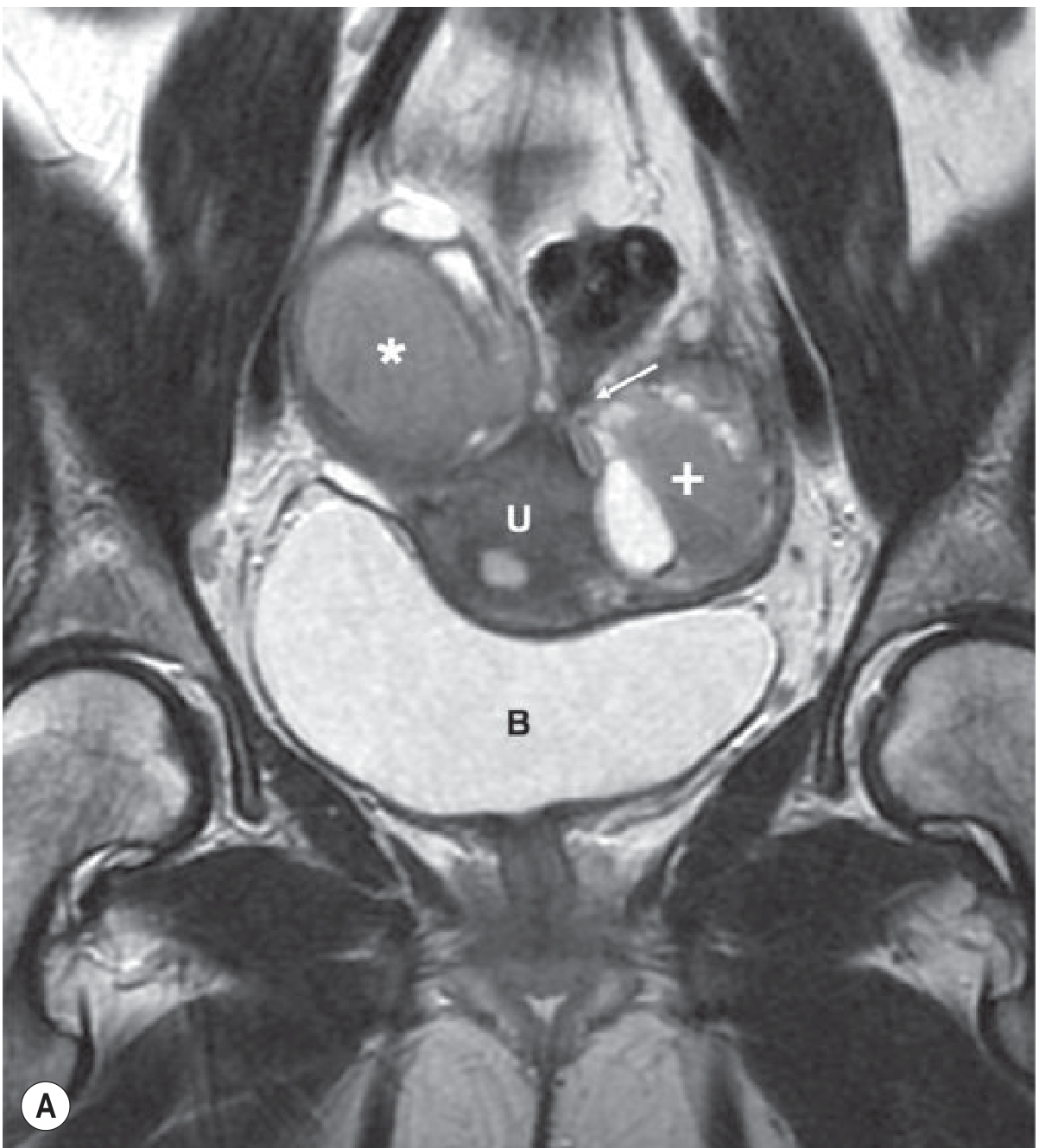
Key MRI criterion: junctional zone (JZ) thickness on T2-weighted images
| JZ Thickness | Interpretation |
|---|---|
| ≥ 12 mm | Predicts adenomyosis with high accuracy |
| 8-12 mm | Indeterminate - use ancillary criteria |
| ≤ 8 mm | Excludes adenomyosis with high accuracy |
Ancillary MRI findings:
- High T2 signal foci within low-signal myometrium (islands of ectopic endometrium / cystically dilated glands)
- High T2 linear striations extending from endometrium into myometrium (finger-like projections of invasion)
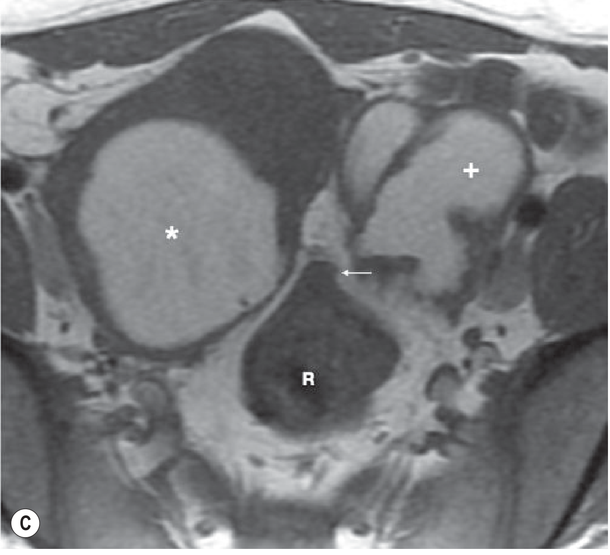
- High T1 punctate foci (small haemorrhages within ectopic tissue) - 95% positive predictive value for adenomyosis
- Cystic adenomyosis: rare; high T1 / intermediate-low T2 foci from extensive ectopic bleeding
Grainger & Allison's Diagnostic Radiology, p. 897-898
MRI of adenomyosis - T2-weighted coronal view (U = uterus, B = bladder):
MRI - axial view showing adenomyosis (R = rectum, * = leiomyoma, + = adenomyosis):
Pathology (Histology)
On microscopic examination:
- Irregular nests of endometrial stroma, with or without glands, embedded deep within myometrium between muscle bundles
- The ectopic tissue is interposed between smooth-muscle fascicles
- Secondary reactive smooth-muscle hypertrophy surrounds the deposits
- Results in an enlarged, globular uterus with a thickened uterine wall
- Focal adenomyosis can form a discrete mass = adenomyoma
Robbins & Kumar Basic Pathology, p. 689; Robbins, Cotran & Kumar, p. 931
Management
Management is guided by age, symptom severity, and desire for future fertility.
Medical (first-line, uterus-sparing)
| Agent | Mechanism / Notes |
|---|---|
| NSAIDs | Symptomatic dysmenorrhea relief |
| Combined oral contraceptives (COCs) | Menstrual suppression |
| Progestins (oral, injectable, or subdermal implant) | Suppress endometrial tissue; IUS (levonorgestrel-IUD) is highly effective |
| GnRH agonists | Induce pseudo-menopause; effective but limited to 6 months (bone loss); use add-back therapy |
A 2024 systematic review (PMID 38426337) found that progestin subdermal implants reduce dysmenorrhea and pelvic pain in adenomyosis.
Interventional / Minimally Invasive
- Uterine artery embolization (UAE) - effective, uterus-preserving option
- Uterus-preserving surgery: wedge resections, modified reductive surgery, double-flap adenomyomectomy for focal disease
Definitive
- Hysterectomy - the only cure; reserved for completed childbearing or failure of conservative management
- Also provides the definitive histological diagnosis
Berek & Novak's Gynecology, p. 581; Bailey and Love's Surgery
Key Distinctions: Adenomyosis vs. Endometriosis
| Feature | Adenomyosis | Endometriosis |
|---|---|---|
| Location of ectopic tissue | Within myometrium | Outside uterus |
| Continuity | Contiguous with endometrium | No continuity |
| Uterine size | Enlarged, globular | Usually normal size |
| Uterine mobility | Preserved | May be restricted (adhesions) |
| Adnexal involvement | Absent | Common (endometriomas) |
| Infertility | Less prominent | Common presenting complaint |
| Malignant potential | Very low | Peritoneal/ovarian forms: precursor to endometrioid/clear cell carcinoma |
Summary (Key Concepts)
- Adenomyosis = endometrial tissue within myometrium with secondary smooth-muscle hypertrophy
- Classic presentation: secondary dysmenorrhea + menorrhagia + globular tender uterus in a multiparous woman in her late reproductive years
- Diagnosis is clinical and imaging-supported; histology at hysterectomy is definitive
- MRI junctional zone ≥12 mm is the imaging hallmark
- Treatment follows the same protocol as endometriosis; hysterectomy is curative
- Classified as AUB-A in the PALM-COEIN system
Sources: Berek & Novak's Gynecology; Robbins & Kumar Basic Pathology; Robbins, Cotran & Kumar Pathologic Basis of Disease; Grainger & Allison's Diagnostic Radiology; Sabiston Textbook of Surgery
This is a shared conversation. Sign in to Orris to start your own chat.