
Stroke
Reading File
The query "Stroke" is broad. Let me clarify what you need before diving in.
Asking for Preferences
What aspect of stroke are you looking for? · What level of detail do you need?
I'll provide a comprehensive overview of stroke covering types, pathophysiology, clinical features, diagnosis, and management.
Searching sources...
Finding Sources
Searching PubMed
"stroke" AND "management"
Good - Bradley and Daroff's Neurology is the primary source. Let me pull the key stroke sections in parallel.
Finding Sources
Reading File
Let me pull the main stroke chapters from Bradley & Daroff's and Rosen's Emergency Medicine in parallel.
Reading File
Reading File
Excellent content. Let me now pull the pathophysiology and management sections in parallel.
Reading File
Reading File
Good clinical content. Let me now pull the management section including hemorrhagic stroke from Rosen's.
Reading File
Finding Sources
Reading File
Reading File
Now let me get the treatment/management section from Rosen's:
Reading File
I now have comprehensive content from multiple authoritative sources. Let me also fetch two of the key CT/imaging images to include in the answer.
Loading Image
Loading Image
I now have all the information needed for a comprehensive overview. Here is the full clinical summary:
Stroke - Comprehensive Overview
Definition
A stroke is defined as a sudden neurological deficit caused by a disruption of cerebral blood flow, either from occlusion (ischemic) or rupture (hemorrhagic) of a blood vessel. The underlying pathology determines management, so rapid classification is essential.
Classification
| Type | Proportion | Mechanism |
|---|---|---|
| Ischemic | ~87% | Thrombotic, embolic, or lacunar occlusion |
| Intracerebral hemorrhage (ICH) | ~10% | Rupture of small vessels within brain parenchyma |
| Subarachnoid hemorrhage (SAH) | ~3% | Rupture of an aneurysm or AVM into subarachnoid space |
1. Ischemic Stroke
Subtypes & Causes
- Thrombotic (~33%): Clot forming at an atherosclerotic plaque, typically at vessel bifurcations (ICA, MCA). Turbulent flow at >90% stenosis triggers platelet adhesion and occlusion.
- Lacunar (~25%): Small-vessel occlusions in deep structures (basal ganglia, thalamus, pons, internal capsule). Strongly associated with hypertension and diabetes. The underlying process is lipohyalinosis of small penetrating arteries.
- Cardioembolic (~25%): Mural thrombus embolizing from the heart. Atrial fibrillation is the most common cause - AF confers a ~5x increased stroke risk.
- Cryptogenic (>33%): No identified cause despite full workup.
- Artery-to-artery embolism: e.g., amaurosis fugax from carotid plaque embolizing to the ophthalmic artery.
Causes in Young Patients (15-45 years)
Pregnancy, oral contraceptives, antiphospholipid antibodies, protein S/C deficiency, sickle cell disease, polycythemia, fibromuscular dysplasia, cocaine/amphetamines, cervical artery dissection (leading determined cause in young adults - can follow even trivial neck movements like sneezing).
2. Vascular Territory Syndromes
Anterior Circulation
- MCA territory: Contralateral hemiplegia and hemisensory loss (face and arm > leg), aphasia (dominant hemisphere), hemineglect (non-dominant), contralateral homonymous hemianopia.
- ACA territory: Contralateral leg > arm weakness, abulia, urinary incontinence.
Posterior Circulation
- Vertebrobasilar: Ipsilateral cranial nerve deficits + contralateral hemiparesis ("crossed signs").
- PCA territory: Ipsilateral CN III palsy + contralateral homonymous hemianopia.
- Wallenberg syndrome (lateral medullary syndrome): Vertigo, Horner syndrome, ipsilateral facial numbness with loss of corneal reflex, and contralateral loss of pain and temperature - a classic crossed sensory deficit.
3. Pathophysiology
Ischemic penumbra: The zone of brain tissue surrounding the infarct core that is critically hypoperfused but still viable. It is the primary therapeutic target. Thrombolysis can salvage this tissue if given within the critical time window.
Secondary injury cascade:
- Impaired cerebral metabolic rate of O2 (CMRO2) and glucose metabolism
- Blood-brain barrier disruption
- Loss of GABA receptors
- Microglial activation and inflammatory response
- Diaschisis: remote functional impairment in connected non-infarcted areas due to disruption of neural circuits
4. Diagnosis & Investigations
Immediate Evaluation
- Non-contrast CT head (first-line): Rapidly differentiates ischemic from hemorrhagic stroke, identifies >1 cm parenchymal hemorrhages, and detects >67% of early ischemic changes within 3 hours. Limited for posterior fossa.
- ECG: Identifies AF and life-threatening arrhythmias.
- Blood glucose: Hypoglycemia is the most important stroke mimic to exclude.
Early Ischemic CT Signs
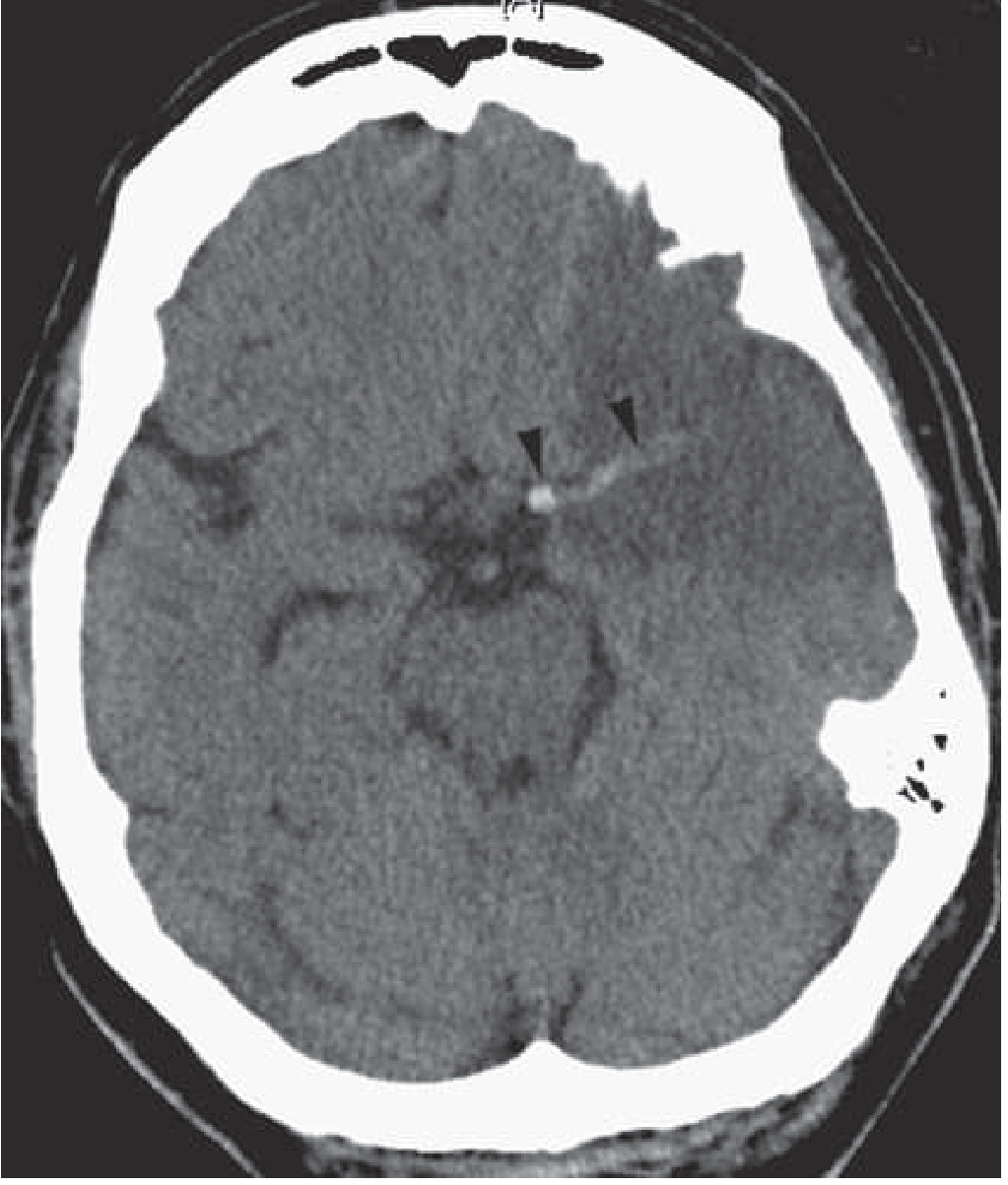
- Hyperdense MCA/artery sign (visible thrombus)
- Sulcal effacement
- Loss of gray-white interface (insular ribbon sign)
- Loss of deep gray matter contours
- Acute hypodensity (develops 6-12 hours post-onset)
CT scan showing evolving left MCA ischemic stroke with hyperdense M1 sign (arrowheads):
Advanced Imaging
- MRI DWI (diffusion-weighted imaging): Most sensitive method for acute ischemia; detects signal change within minutes of onset. ADC mapping essential.
- CT/MR angiography: Identifies large vessel occlusion (LVO), dissection, or stenosis. CTA obtained concurrently with CT head. Guides thrombectomy candidacy.
- CT perfusion (CTP): Identifies salvageable tissue in extended time windows (6-24 hours). Not typically needed in the first 6 hours.
Other Tests
- CBC, coagulation studies, renal/liver function
- Echocardiogram (if cardioembolic cause suspected)
- Carotid ultrasound / MRA neck
5. Hemorrhagic Stroke
Intracerebral Hemorrhage (ICH)
Risk factors: Hypertension (most common), older age, Black/Asian race, high alcohol consumption, low LDL and triglycerides (counterintuitively protective against ischemic but a risk for hemorrhagic stroke).
Common causes:
- Hypertensive vasculopathy (most common)
- Cerebral amyloid angiopathy (CAA) in elderly patients - lobar hemorrhages
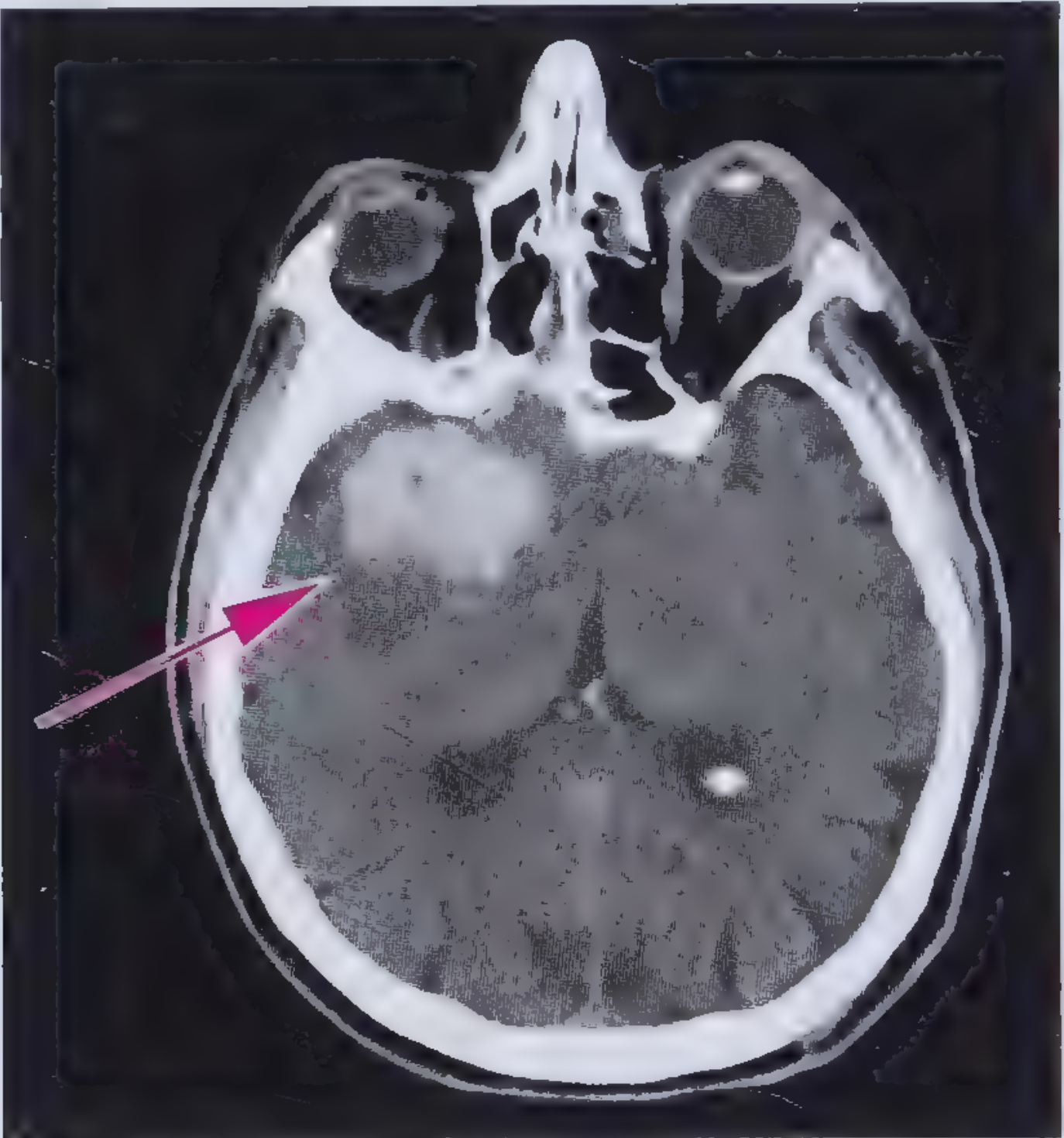
CT appearance: Round or oval hyperattenuating lesion. Initially 40-60 HU (heterogeneous), progressing to 60-80 HU within hours to days, and 80-100 HU over several days as the clot organizes.
30-day mortality: ~50%
CT showing right temporal lobe intracerebral hemorrhage (arrow pointing to hyperdensity):
6. Management
Stroke Time Targets (NINDS Protocol)
| Goal | Target |
|---|---|
| Door to doctor | 10 min |
| Door to CT completion | 25 min |
| Door to CT reading | 45 min |
| Door to treatment (t-PA) | 60 min |
| Access to neurological expertise | 15 min |
| Access to neurosurgical expertise | 2 hours |
Ischemic Stroke - Acute Treatment
A. IV Thrombolysis (t-PA / Alteplase)
- Dose: 0.9 mg/kg (max 90 mg) - 10% as IV bolus, remaining 90% over 1 hour
- Window: Within 4.5 hours of symptom onset (or last known well)
- Not recommended for mild non-disabling symptoms; recommended for mild but disabling symptoms within 4.5 hours
- Risk of symptomatic ICH post-thrombolysis: 2-7%; asymptomatic ICH (microbleeds): 30-45%
- Goal: door-to-needle time of 60 minutes or less
B. Mechanical Thrombectomy
- For large vessel occlusions (LVO)
- Window up to 24 hours from last known well (with appropriate imaging selection)
- CTA used to identify LVO and guide candidacy
- t-PA should not be delayed awaiting thrombectomy planning
C. Blood Pressure Management
- Avoid aggressive BP lowering in acute ischemic stroke (reduces perfusion pressure to penumbra)
- If thrombolytics are being given, strict BP control required to reduce hemorrhagic transformation risk
- Prognosis worsens with: fever, hypotension, hypoxia, hypo/hyperglycemia - correct these rapidly
D. Supportive Care
- Oxygen only if SpO2 < 95% (avoid in normoxic patients)
- Avoid dextrose-containing fluids (hyperglycemia worsens ischemic deficit)
- Treat fever aggressively (even mild hyperthermia worsens outcomes)
- ECG monitoring throughout
- Aspirin (antiplatelet) initiated after ruling out hemorrhage
Hemorrhagic Stroke - Acute Treatment
- Reverse coagulopathy: Vitamin K + prothrombin complex concentrate (PCC) preferred over FFP (faster INR correction, lower infection risk, smaller volume). FFP if PCC unavailable.
- Blood pressure: For SBP 150-220 mmHg, acutely lower to 140 mmHg (safe and beneficial).
- Blood glucose: Maintain normoglycemia.
- Seizure control: Antiepileptic medications if seizures occur.
- ICP management (if elevated): Intubation/sedation, head-of-bed elevation to 30°, hypertonic saline/mannitol, and hemicraniectomy if necessary.
- No thrombolytics or anticoagulants in the acute phase.
7. Stroke Mimics (Differential Diagnosis)
| Mimic | Key distinguishing feature |
|---|---|
| Hypoglycemia | Most common mimic; check BGL immediately |
| Todd's paralysis | Post-seizure, gradually resolves |
| Complex migraine | History of migraine, often with headache |
| Subdural/epidural hematoma | Trauma history, gradual progression |
| Brain tumour or abscess | Subacute onset, may have systemic signs |
| Bell's palsy | Peripheral (forehead involved), no limb weakness |
| Wernicke's encephalopathy | Ophthalmoplegia, ataxia, confusion - thiamine deficiency |
| Ménière's disease | Vertigo, hearing loss, tinnitus - no focal neurological signs |
| Air embolism | Atmospheric pressure change, IV procedures |
8. Prognosis & Rehabilitation
- Post-stroke depression and immobilization significantly worsen outcomes and are important to manage.
- Falls risk is more than doubled in stroke patients vs. age-matched controls; hip fracture risk is particularly elevated within 3 months in women.
- Motor recovery occurs via recruitment of midline premotor areas and contralateral motor cortex; recovery is associated with return to a more normal restricted activation pattern.
- Transcranial magnetic stimulation (TMS) targeting contralateral hemisphere inhibition is an emerging rehabilitation strategy.
- Early mobilization and stroke unit admission improve outcomes.
Sources: Bradley and Daroff's Neurology in Clinical Practice, ROSEN's Emergency Medicine, Frameworks for Internal Medicine
This is a shared conversation. Sign in to Orris to start your own chat.