
Near total laryngectomy whole as long question answer in detail and generate image for indications and contraindications for the procedure from standard ENT textbook
near total laryngectomy surgical technique diagram tracheopharyngeal shunt
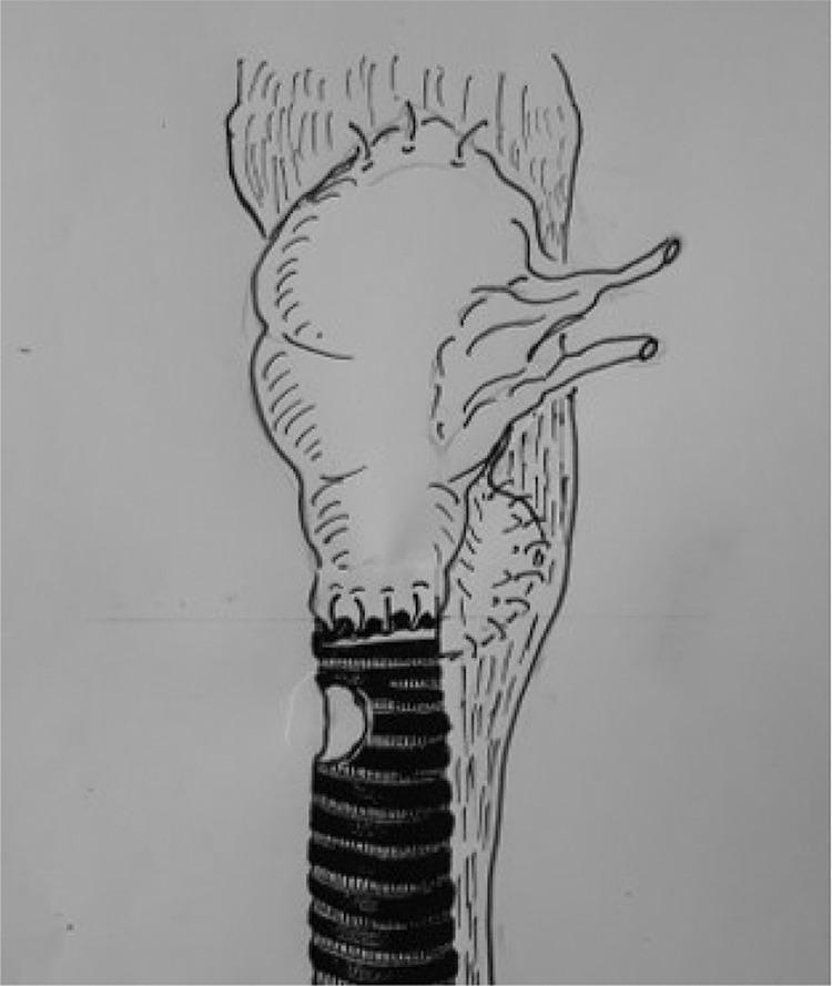
This medical illustration depicts a surgical reconstruction of the upper airway and digestive tract following a laryngectomy. The diagram shows a tracheo-pharyngeal shunt utilizing a jejunal interposition to bridge the defect between the respiratory and pharyngeal systems. Centrally, a vascularized segment of the jejunum is shown as an inflated, pouch-like structure acting as a neolarynx or air chamber. Superiorly, the jejunal segment is anastomosed to the pharynx (intestino-pharyngeal anastomosis), which serves as a neoglottis. Inferiorly, the distal end of the jejunal loop is sutured to the superior aspect of the transected trachea, identified by its characteristic horizontal cartilaginous rings. Two visible vascular pedicles extend laterally from the jejunal segment, representing the mesenteric vessels required for graft revascularization. The surrounding pharyngeal tissues are shown with vertical striations, indicating the closure of the pharyngeal defect. This technique is designed to facilitate both the closure of complex anatomical gaps and the restoration of voice function by using respiratory air redirected through the intestinal shunt.
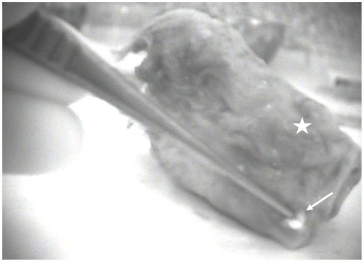
This diagnostic image demonstrates a total laryngectomy specimen (marked with a white star) captured using near-infrared (NIR) autofluorescence imaging in a lateral ex-vivo view. The specimen is an irregularly shaped, multi-contoured soft tissue mass. The focus of the clinical demonstration is a highly hyperfluorescent, bright white focal point (indicated by a white arrow) located at the periphery of the laryngeal tissue, representing an autofluorescent parathyroid gland. The NIR modality highlights the intrinsic fluorescence of the parathyroid tissue, creating a sharp contrast against the dimmer, more uniform background of the surrounding laryngeal and connective tissues. This imaging technique is utilized in surgical oncology and endocrine surgery to facilitate the identification and preservation or auto-transplantation of parathyroid glands during major neck dissections, such as total laryngectomy for laryngeal cancer, where devascularization of these glands is a common surgical risk.
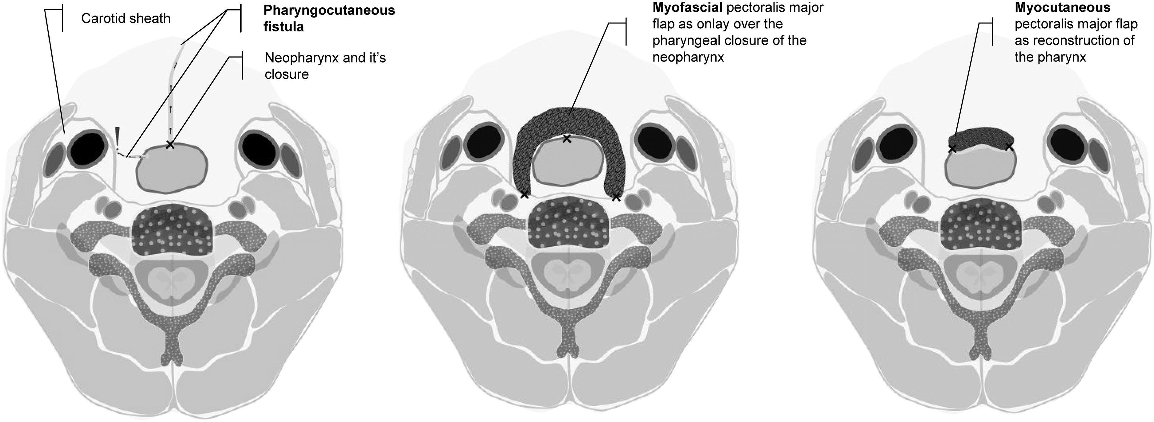
This medical illustration comprises three axial-view diagrams of the neck, demonstrating different stages and methods of surgical reconstruction following a total laryngectomy. The leftmost diagram highlights a common postoperative complication: a pharyngocutaneous fistula (PCF). It depicts the anatomical proximity of the neopharynx closure to the carotid sheath and illustrates the saliva-leaking pathway from the neopharyngeal lumen to the skin surface. The center diagram shows the prophylactic use of a myofascial pectoralis major flap (PMMF) applied as an 'onlay' reinforcement over the primary pharyngeal closure to prevent fistula formation. The rightmost diagram illustrates a more extensive reconstruction using a myocutaneous pectoralis major flap (PMMC) where the flap itself forms a significant portion of the reconstructed pharynx. Key anatomical structures visible in all views include the cervical vertebrae, spinal canal, and bilateral carotid sheaths containing the carotid artery and internal jugular vein. This comparison provides clinical insight into laryngeal cancer surgical sequelae and the use of vascularized muscle flaps in reconstructive head and neck surgery.
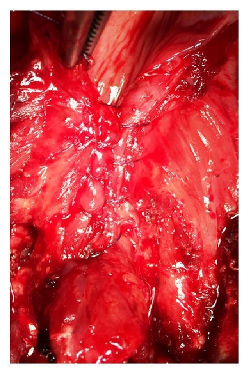
Intraoperative clinical photograph showing a close-up view of the surgical field during a total laryngectomy. The image focuses on the vertical reconstruction of the neopharynx through the closure of the pharyngoesophageal segment. The visible pathology involves the exposed muscular and mucosal layers of the neck and pharyngeal region, showing expected surgical hemorrhage and tissue manipulation. A Cushing-type continuous inverting suture technique is demonstrated using a 3/0 resorbable polyglactin 910 suture, creating a tight, linear seal. A surgical instrument, likely a hemostatic clamp or forceps, is partially visible at the superior margin of the field. This image serves as a clinical illustration of head and neck surgical techniques, specifically pharyngeal closure methods following radical laryngeal resection.
Medical educational poster titled "Near-Total Laryngectomy (NTL): Indications and Contraindications" for ENT students. Two clear columns: LEFT column titled "INDICATIONS" with green header, listing: 1. Advanced but lateralized hypopharyngeal cancer (T3/T4a), 2. Contralateral vocal cord and arytenoid spared (not involved by tumor), 3. Post-cricoid mucosa and interarytenoid region uninvolved, 4. Compromised laryngeal function or framework, 5. Salvage surgery post-radiotherapy (relative), 6. Good general performance status (PS), 7. ~20% of advanced operable pyriform sinus cancers. RIGHT column titled "CONTRAINDICATIONS" with red header, listing: 1. Interarytenoid or post-cricoid mucosal involvement, 2. Contralateral cord involvement >1/3 length, 3. Poor general condition/performance status, 4. Prevertebral involvement (no laryngeal crackle, fixed neck), 5. Superior extension into oropharynx, 6. Lateral extension to carotid artery, 7. Metastatic/distant disease, 8. Prior radiation therapy (relative contraindication - tissue edema). Clean, professional medical poster layout with anatomical larynx diagram at top center. Source: Scott-Brown's Otorhinolaryngology & Cummings Otolaryngology.
Near-Total Laryngectomy (NTL) - Comprehensive Long Answer
1. DEFINITION AND CONCEPT
2. HISTORICAL BACKGROUND
3. INDICATIONS AND CONTRAINDICATIONS
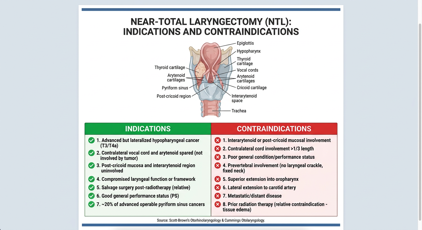
3A. Indications
- Good general condition / performance status
- Compromised laryngeal function
- Involved laryngeal framework (T4a)
- Salvage surgery following prior radiation or concurrent chemoradiation
- Advanced but lateralized hypopharyngeal cancer - typically T3/T4a pyriform sinus carcinoma where the lesion is predominantly on one side
- Contralateral vocal cord and arytenoid are free of disease - the contralateral arytenoid must be preservable and used to form the biological shunt
- Post-cricoid mucosa is uninvolved - tumor must not extend to the post-cricoid region
- Interarytenoid region is uninvolved - the interarytenoid space must be free to allow safe contralateral arytenoid preservation
- Advanced T3/T4 glottic SCC unsuitable for other conservation procedures (Pearson's series, Cummings)
- ~20% of advanced operable pyriform sinus cancers are suitable candidates
3B. Contraindications
- Interarytenoid or post-cricoid mucosal involvement - makes preservation of the contralateral arytenoid oncologically unsafe
- Contralateral cord mucosal involvement > one-third its length - does not allow preservation of sufficient laryngeal remnant for shunt formation
- Prevertebral involvement - presents with inability to extend the neck and absence of laryngeal crackle on palpation
- Superior extension into the oropharynx
- Lateral extension involving the carotid artery
- Root of neck involvement
- Metastatic (distant) disease
- Prior radiation therapy - a relative contraindication, especially if tissues are oedematous
4. SURGICAL ANATOMY AND PRINCIPLE
- The contralateral arytenoid (preserved with its innervation intact)
- A preserved strip of the contralateral vocal cord mucosa (no more than 1/3 its length involved by tumor)
- A portion of the ipsilateral cricoid cartilage
- Is too small to serve as a breathing airway (hence permanent tracheostome needed)
- Is large enough to act as a dynamic, sphincteric shunt - diverting air from the trachea to the neopharynx for speech
- Has sphincteric action due to preserved arytenoid muscle/nerve innervation, which protects the airway from aspiration
- Active sphincter closure to prevent aspiration during swallowing
- Dynamic regulation of the airway during vocalization
5. SURGICAL TECHNIQUE (Step by Step)
Pre-operative Planning
- Multidisciplinary team assessment (surgical, radiation, medical oncology + SLT)
- Nasoendoscopy and direct laryngoscopy to confirm interarytenoid and post-cricoid mucosa are free
- CT/MRI neck and chest to assess cartilage, prevertebral space, and nodal disease
- Patient counselling: permanent tracheostome, speech shunt mechanics, swallowing rehabilitation
Operative Steps
-
Patient positioning - Supine, neck extended, shoulder roll
-
Incision - Standard apron/Gluck-Sorensen incision with tracheostomy planned inferiorly
-
Neck dissection - Bilateral levels II-V (with nodal disease) or II-IV (advanced primary without nodes)
-
Laryngeal framework exposure - Strap muscles divided; thyroid lobe on the tumor side removed; external perichondrium of thyroid cartilage incised
-
Division of the trachea - At an appropriate level inferiorly, trachea divided obliquely; the tracheostome is matured to the skin
-
Pharyngotomy and tumor resection - Pharynx opened on the contralateral (healthy) side first; tumor resected with the majority of the larynx, ipsilateral pyriform sinus, and involved hypopharyngeal mucosa; margins sent for frozen section
-
Shunt construction (key step):
- The contralateral arytenoid, its mucosal cover, and a strip of contralateral true cord mucosa are preserved
- A strip of cricoid cartilage is preserved on the same side
- This mucosal/cartilaginous remnant is tubularized over a small catheter using fine absorbable sutures to create the voice shunt
- The superior end of the shunt is sutured to the neopharynx
- The inferior end is sutured to the superior aspect of the trachea
-
Pharyngeal closure - The pharyngeal defect is closed primarily if ≥3 cm unstretched pharyngeal mucosa remains; if insufficient, reconstruction is planned (see below)
-
Tracheostome maturation - Tracheostoma is matured as a permanent end tracheostomy
-
Wound closure - Suction drains placed, wound closed in layers
6. NECK MANAGEMENT
- N+ disease (manifest neck nodes): Bilateral neck dissection levels II-V
- N0 with advanced primary (T3/T4): Bilateral levels II-IV elective neck dissection
- The paratracheal nodes (level VI) are included given the subglottic/pyriform location
7. PHARYNGEAL RECONSTRUCTION
| Defect Type | Options |
|---|---|
| Partial pharyngeal defect (>3 cm mucosa preserved) | Primary closure |
| Partial defect requiring augmentation | Radial artery free flap, Anterolateral thigh (ALT) free flap, Pectoralis major myocutaneous (PMMC) pedicled flap |
| Circumferential defect | Free jejunal graft (preferred), Tubed radial forearm free flap, Tubed PMMC (poor alternative) |
| Post-TLPE defect | Gastric pull-up (GPU) - single-stage vascularized reconstruction, anastomosis in neck |
8. VOICE OUTCOMES
- The voice shunt allows intelligible, lung-powered speech in the vast majority of patients
- Published series report success rates ranging from 74% to 100%
- Almost every patient will speak unless there is shunt breakdown or stenosis
- Shunt stenosis is the commonest cause of speech failure or straining during speech - correctable by dilatation using gum elastic bougies
- In NTL specifically: "Almost every patient will speak following this procedure unless there is a shunt breakdown or stenosis, which is uncommon and is usually technique related" - Scott-Brown's, p.329
- Speech restoration with tracheo-oesophageal prosthesis (in total laryngectomy cases) is successful in >90% when pharyngeal remnant is preserved
9. SWALLOWING OUTCOMES
- Swallowing in NTLP is comparable to total laryngopharyngectomy
- Pharyngeal stenosis rate: ~10% (caused by narrow pharyngeal remnant or radiation-induced stenosis)
- Pharyngeal muscle closure technique affects swallowing quality:
- Muscle approximation → better bolus propulsion
- Mucosal-only closure → better bolus volume
- Vascularized patch reconstruction does not significantly alter swallowing function; the key variable remains the amount of functional pharyngeal remnant with preserved peristalsis
10. ONCOLOGICAL OUTCOMES
- In properly selected cases, locoregional control rates with NTL are comparable to total laryngectomy
- Scott-Brown's own series (predominantly T3/T4): local recurrence < 7%, median survival 57%
- UK national analysis of >1500 patients: 5-year survival of 57% for pharyngolaryngectomy
- Cummings cites Pearson's series showing NTL provides excellent control for T3/T4 glottic tumors unsuitable for other conservation procedures
11. MORBIDITY
| Complication | Details |
|---|---|
| Salivary/pharyngocutaneous fistula | Most common; higher in post-irradiated patients (~50%); risk reduced by 1/3 with vascularized tissue reconstruction |
| Pharyngeal stenosis | ~10% rate; treat with dilatation |
| NTL shunt stenosis | Commonest cause of speech failure; treat with gum elastic bougie dilatation |
| Aspiration | Protected against by innervated sphincteric shunt |
| Hypothyroidism | Hemithyroidectomy + RT; monitor TFTs every 1-2 months |
| Mortality | Overall hospital mortality ~6%; gastric pull-up highest (11%); reconstructive failure carries odds ratio 6:2 for in-hospital death |
12. ADJUVANT THERAPY
- Advanced primary (T3/T4)
- Close or positive cut margins
- Perineural invasion (PNI)
- Lymphovascular emboli (LVE)
- Cartilage/muscle/soft tissue invasion
- Multiple nodal involvement
- Extracapsular spread (ECS) in nodes - concurrent chemotherapy with RT is conclusively indicated for positive margins + ECS
13. COMPARISON WITH OTHER PROCEDURES
| Procedure | Voice | Nasal Breathing | Indication |
|---|---|---|---|
| Voice conservation (SCPL, VPL) | Preserved | Preserved | T1-T3 selected |
| Near-Total Laryngectomy (NTL) | Preserved (lung-powered shunt) | Lost (permanent tracheostome) | Advanced lateralized T3/T4 |
| Total laryngectomy | Lost (TEP prosthesis) | Lost | T4a, bilateral disease |
| Total laryngopharyngectomy | Lost | Lost | Post-cricoid / circumferential involvement |
Summary Points
- NTL = preservation of voice but not nasal respiration via an innervated biological shunt
- Key eligibility: lateralized tumor, contralateral arytenoid free, post-cricoid and interarytenoid mucosa uninvolved
- Absolute contraindications for NTL itself: interarytenoid/post-cricoid involvement and contralateral cord involvement > 1/3 length
- Suitable in ~20% of advanced operable pyriform sinus cancers
- Speech success rate: 74-100%; failure almost always due to shunt stenosis (treatable)
- Oncological control equivalent to total laryngectomy when properly selected
- Adjuvant chemoradiotherapy is mandatory for most stage III/IV cases
- Scott-Brown's Otorhinolaryngology Head & Neck Surgery, Chapter on Surgery for Advanced Hypopharyngeal Cancers, pp. 328-330
- Cummings Otolaryngology Head and Neck Surgery, Treatment of Advanced Glottic SCC section, p. 1994; NTL acronym and technique sections