
Causes of anemia in pregnancy 10 marks answer
Reading File
Now let me search the medical library for comprehensive content on anemia in pregnancy.
Finding Sources
Finding Sources
Reading File
Reading File
Reading File
Finding Sources
Reading File
Finding Sources
Reading File
Finding Sources
Reading File
Finding Sources
I now have comprehensive information from Creasy & Resnik's Maternal-Fetal Medicine and Harrison's. Let me fetch the key diagram and compile the full answer.
Loading Image
Reading File
I now have excellent, comprehensive content. Let me compile the full 10-mark answer.
Causes of Anemia in Pregnancy
Definition
Anemia in pregnancy is defined by the CDC as a hemoglobin (Hb) concentration <11 g/dL in the first and third trimesters or <10.5 g/dL in the second trimester, owing to the physiological hemodilution of pregnancy. Globally, approximately 38% of pregnant women are anemic, and in ~50% of cases, iron deficiency is the cause.
(Creasy & Resnik's Maternal-Fetal Medicine, p. 1329)
Classification of Causes
Anemia in pregnancy is best understood using a pathophysiologic classification:
I. PHYSIOLOGICAL (DILUTIONAL) ANEMIA
This is the most common type and is not a pathological condition.
- During pregnancy, total blood volume increases by 40-45% (maximum at 34 weeks' gestation).
- Plasma volume increases by 47%, but red blood cell (RBC) mass increases by only 17%.
- This disproportionate expansion causes hemodilution, lowering the Hb, hematocrit, and RBC count - but the MCV and MCHC remain unchanged.
- The maximum dilutional effect occurs between 28-34 weeks of gestation.
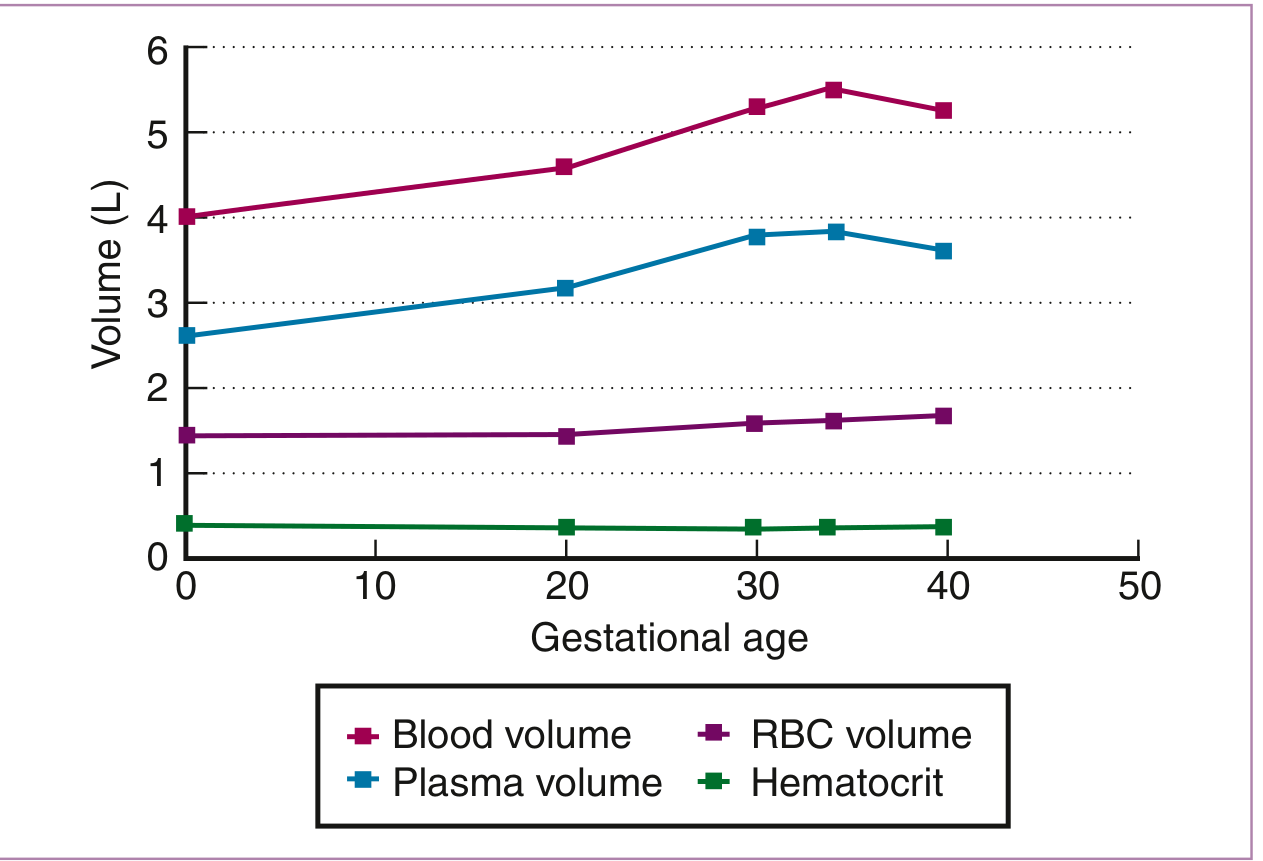
Figure: Hematologic changes during pregnancy - plasma volume expansion exceeds RBC mass rise, leading to physiologic anemia. (From Creasy & Resnik's Maternal-Fetal Medicine)
II. NUTRITIONAL DEFICIENCY ANEMIAS (Most Common Pathological Causes)
1. Iron Deficiency Anemia (IDA) - Most Common
Iron deficiency is the single most common correctable cause of anemia in pregnancy, accounting for ~50% of all cases globally.
Causes of Iron Deficiency in Pregnancy:
- Increased demand: The fetus and placenta require iron (~300 mg), expanding RBC mass requires ~500 mg, and obligatory losses account for ~200 mg - a total of ~1000 mg is needed in a typical pregnancy.
- Inadequate pre-pregnancy stores: ~50% of healthy primigravidas have minimal iron stores in the first trimester; ~two-thirds of healthy young women have minimal bone marrow iron stores.
- Poor dietary intake: Especially in developing countries; only 10% of dietary iron is absorbed.
- Increased menstrual losses before conception depleting stores.
- Multiparity: Repeated pregnancies deplete iron stores.
- Hookworm infestation (common in tropical regions): Chronic blood loss from intestinal parasites.
- Malabsorption: Celiac disease, post-gastrectomy states.
- Ferritin <20 μg/L before pregnancy: ~60% of such women become anemic by 20 weeks' gestation.
Morphology: Microcytic, hypochromic anemia; low serum iron, raised TIBC, low ferritin.
(Creasy & Resnik's Maternal-Fetal Medicine, pp. 1129-1133)
2. Folate Deficiency Anemia - Second Most Common
- Folate requirements increase 5- to 10-fold during pregnancy due to:
- Rapidly dividing fetal and placental cells
- Expanded maternal RBC mass production
- Increased renal clearance of folate
- Dietary deficiency (poor intake of leafy vegetables, legumes)
- Malabsorption (sprue, inflammatory bowel disease)
- Anti-epileptic drugs (phenytoin, phenobarbitone) - induce folate metabolism
- Hemolytic anemias that increase folate turnover
- Multiple pregnancy - doubles the folate demand
Morphology: Macrocytic (megaloblastic) anemia; hypersegmented neutrophils; low serum and RBC folate.
3. Vitamin B12 Deficiency
- Less common than folate deficiency in pregnancy; most commonly seen in:
- Strict vegetarians/vegans (no animal products)
- Pernicious anemia: Autoimmune destruction of intrinsic factor (anti-parietal cell and anti-intrinsic factor antibodies)
- Post-gastrectomy (loss of intrinsic factor-secreting parietal cells)
- Malabsorption syndromes (Crohn's disease, terminal ileal disease)
- Causes megaloblastic anemia indistinguishable from folate deficiency morphologically.
- Critical: B12 deficiency also causes subacute combined degeneration of the spinal cord - treating with folate alone can worsen neuropathy.
(Creasy & Resnik's Maternal-Fetal Medicine, p. 1251)
III. HEMOLYTIC ANEMIAS
4. Hemoglobinopathies
- Sickle Cell Disease (HbSS): Chronic hemolysis is worsened in pregnancy; risk of sickling crises increases due to relative hypoxia, acidosis, and dehydration. Particularly high-risk in pregnancy with increased maternal and fetal morbidity.
- Thalassemia (α and β): Ineffective erythropoiesis and chronic hemolysis; iron overload (from transfusions) complicates management. Pregnancy exacerbates anemia.
- HbC, HbE and other variants.
5. Hereditary Spherocytosis (HS)
- Autosomal dominant (75%); defects in spectrin/ankyrin proteins.
- Pregnancy may precipitate hemolytic crises, especially in women who have not had splenectomy.
- Increased splenic blood flow in pregnancy enhances RBC destruction.
- Requires folate supplementation and monitoring for crises.
6. Glucose-6-Phosphate Dehydrogenase (G6PD) Deficiency
- X-linked enzyme deficiency; RBCs vulnerable to oxidant stress.
- Triggered by infections, certain drugs (antimalarials, sulfonamides), or fava beans.
- Causes acute intravascular hemolysis with Heinz bodies on smear.
7. Autoimmune Hemolytic Anemia (AIHA)
- Warm-type (IgG): Associated with connective tissue disease (SLE), lymphoma.
- Direct Coombs test positive.
- Can be triggered or worsened by pregnancy.
8. Paroxysmal Nocturnal Hemoglobinuria (PNH)
- Acquired clonal defect (PIG-A gene) with complement-mediated intravascular hemolysis.
- Pregnancy increases thrombotic risk and worsens hemolysis.
- Detected by flow cytometry (GPI-anchor protein loss on RBCs).
(Creasy & Resnik's Maternal-Fetal Medicine, BOX 55.1, pp. 1009-1084)
IV. ANEMIA DUE TO DECREASED RBC PRODUCTION
9. Anemia of Chronic Disease / Inflammation
- Associated with: chronic renal disease, chronic liver disease, chronic infection (tuberculosis, HIV, malaria), chronic inflammatory conditions (rheumatoid arthritis, SLE).
- Mechanism: Hepcidin-mediated sequestration of iron in macrophages; reduced erythropoietin response.
- Normocytic or mildly microcytic anemia; serum ferritin normal or elevated; TIBC low.
10. Aplastic Anemia
- Rare but serious; bone marrow hypoplasia results in pancytopenia.
- Causes in pregnancy: Idiopathic (most), drugs, viral infections (parvovirus B19, hepatitis viruses), toxins.
- Pregnancy itself can precipitate or worsen aplastic anemia.
- High maternal and fetal mortality if untreated.
11. Anemia of Renal Disease
- Reduced erythropoietin production from damaged kidneys.
- Seen in women with pre-existing chronic kidney disease.
V. BLOOD LOSS ANEMIAS (Hemorrhagic)
12. Antepartum Hemorrhage
- Placenta previa: Painless, recurrent bleeding.
- Abruptio placentae: Painful concealed or revealed hemorrhage.
- Causes acute normocytic anemia; reticulocytosis follows after 3-5 days.
13. Ectopic Pregnancy / Abortion
- Acute hemorrhage causing rapid onset normocytic anemia.
Summary Table
| Cause | Type of Anemia | Key Feature |
|---|---|---|
| Physiological dilution | Normocytic, normochromic | MCV/MCHC unchanged |
| Iron deficiency | Microcytic, hypochromic | Low ferritin, raised TIBC |
| Folate deficiency | Macrocytic (megaloblastic) | Hypersegmented neutrophils |
| B12 deficiency | Macrocytic (megaloblastic) | Neurological features |
| Sickle cell / Thalassemia | Microcytic / hemolytic | HPLC diagnosis |
| Hereditary spherocytosis | Normocytic hemolytic | Raised MCHC, spherocytes |
| G6PD deficiency | Hemolytic (episodic) | Triggered by oxidants |
| Aplastic anemia | Normocytic, pancytopenia | Hypocellular marrow |
| Chronic disease | Normocytic (or mild microcytic) | Raised ferritin, low TIBC |
| Hemorrhage (APH) | Normocytic, acute | Reticulocytosis follows |
Clinical Note
- Screening: CBC is recommended at the first antenatal visit and at 24-28 weeks' gestation. Serum ferritin is the most accurate test for early iron deficiency, before anemia manifests.
- Most common conditions associated with anemia in pregnancy: malnutrition, multiple pregnancy, trophoblastic disease, chronic renal disease, chronic liver disease, and chronic infection.
(Harrison's Principles of Internal Medicine 22E, p. 3945; Creasy & Resnik's Maternal-Fetal Medicine, p. 1329)
Anemia fetal and maternal complications
Anemia in pregnancy causes significant maternal complications (cardiac failure, increased infection risk, poor wound healing, PPH susceptibility) and fetal complications (IUGR, preterm birth, low birth weight, stillbirth). Severe anemia (Hb <7 g/dL) carries the highest risk for both.
Searching sources...
Finding Sources
Finding Sources
Finding Sources
Finding Sources
Reading File
Finding Sources
Reading File
Finding Sources
Reading File
Finding Sources
Finding Sources
Finding Sources
Reading File
I now have rich, detailed content. Let me compile the comprehensive answer.
Anemia in Pregnancy: Fetal and Maternal Complications
MATERNAL COMPLICATIONS
1. Symptoms of Tissue Hypoxia and Cardiovascular Compensation
Even mild-to-moderate anemia produces symptoms through two main mechanisms:
- Tissue hypoxia: fatigue, lightheadedness, weakness, exertional dyspnea, headache, palpitations, tinnitus.
- Cardiovascular compensation (hyperdynamic circulation): tachycardia, systolic flow murmur, wide pulse pressure - prolonged severe anemia eventually leads to high-output cardiac failure.
2. Increased Risk of Cardiac Failure
- In severe anemia (Hb <5 g/dL), the heart compensates through increased cardiac output. Over time this causes cardiac dilatation and high-output congestive cardiac failure, which may be precipitated or worsened by the already increased cardiac demands of normal pregnancy (cardiac output rises ~40% in pregnancy).
- Women with underlying rheumatic or valvular heart disease are particularly vulnerable to decompensation.
3. Maternal Mortality
- In Africa, Asia, and Latin America, severe anemia (Hb <5 g/dL) is associated with a relative risk of maternal mortality of 3.5 (95% CI, 2.05-6.0).
- In a case series of 130 women with Hb <5 g/dL in the third trimester in India, 8 women died (approximately 6%).
- The WHO estimates 18% of maternal mortality globally is associated with anemia, the majority due to iron deficiency anemia.
- Moderate anemia (Hb <7 g/dL) was not independently associated with increased maternal mortality in this dataset, though confounders such as hemorrhage at delivery and access to care complicate interpretation.
(Creasy & Resnik's Maternal-Fetal Medicine, p. 1334)
4. Increased Susceptibility to Infection / Puerperal Sepsis
- Anemia impairs leukocyte function and reduces immune defenses.
- Anemic women are more susceptible to puerperal sepsis, urinary tract infections, and wound infections following operative delivery.
- In the case series of severe anemia in India, more than 25% had postpartum sepsis.
5. Postpartum Hemorrhage (PPH)
- Anemic women tolerate blood loss poorly. A woman with a baseline Hb of 8 g/dL who loses 500 mL of blood at delivery is in far greater danger than a woman with a Hb of 12 g/dL.
- The uterus in severe anemia may also be hypoxic and more prone to atony, increasing risk of PPH.
- Over 25% of the severe anemia case series had postpartum hemorrhage.
6. Preterm Birth
- In the same case series of Hb <5 g/dL, more than half (>50%) had preterm birth.
- Anemia-related placental hypoxia triggers prostaglandin release, which can initiate preterm labor.
7. Cardiac Surgery / Anesthetic Risk
- Severe anemia increases operative and anesthetic risk. Intraoperative hypotension is less tolerated; anemia reduces the oxygen reserve during any complication.
8. Poor Wound Healing and Reduced Lactation
- Iron deficiency and protein-energy malnutrition (often coexistent) impair collagen synthesis and wound healing post-delivery.
- Severe anemia reduces breast milk production and quality, with lower micronutrient content.
9. Pre-eclampsia Consideration
- An elevated maternal Hb early in pregnancy (>14.5 g/dL) has paradoxically been associated with poor perinatal outcomes including stillbirth and SGA neonates - thought to reflect inadequate plasma volume expansion rather than a benefit. This is not a complication of anemia per se but illustrates the importance of the physiological plasma volume increase.
10. Complications Specific to Cause
| Cause | Additional Maternal Complications |
|---|---|
| Sickle cell disease | Vaso-occlusive crises, ACS, infections, stroke |
| Folate deficiency | Neural tube defects in fetus (if periconceptional) |
| B12 deficiency | Subacute combined degeneration of the cord (neuropathy) |
| Aplastic anemia | Pancytopenia, bleeding, sepsis - high mortality |
| Hereditary spherocytosis | Hemolytic crises (precipitated by pregnancy itself) |
| PNH | Thrombosis (DVT/PE), Budd-Chiari syndrome |
FETAL AND NEONATAL COMPLICATIONS
1. Intrauterine Growth Restriction (IUGR) / Small for Gestational Age (SGA)
- Severe maternal anemia reduces oxygen delivery to the placenta and fetus, causing fetal hypoxia and IUGR.
- In a randomized trial, 17% of placebo-treated (non-supplemented) women had low-birth-weight neonates (<2500 g) vs. only 4% of iron-supplemented women (p <0.05).
- Similarly, 18% vs. 7% had SGA infants, respectively.
- The fetal compartment does preferentially obtain iron from the mother - placental transferrin receptors upregulate when maternal iron is low to maximize fetal uptake. Despite this compensatory mechanism, severe maternal depletion still affects the fetus.
(Creasy & Resnik's Maternal-Fetal Medicine, p. 1218)
2. Preterm Birth
- Severe anemia (Hb <5 g/dL) was associated with preterm birth in >50% of cases in the Indian case series.
- Anemia from malaria in endemic areas is a significant cause of preterm birth, low birth weight, and stillbirth globally - in 2018, an estimated 872,000 infants were born with low birth weight attributable to malaria-related anemia.
3. Low Birth Weight (LBW) / Prematurity
- Both preterm delivery and IUGR contribute to LBW.
- Neonates born to severely anemic mothers have lower birth weights, higher rates of respiratory distress, and higher neonatal ICU admission rates.
4. Stillbirth
- Severe anemia is associated with stillbirth due to placental hypoxia and insufficiency.
- Maternal Hb was significantly lower in mothers of stillborns in multiple studies.
- Malaria-associated anemia is a leading cause of stillbirth in endemic regions.
5. Neonatal Iron Deficiency
- Although the fetal compartment takes iron preferentially, cord blood ferritin levels are significantly reduced in infants of iron-deficient mothers, compared with controls.
- While most neonates of mildly anemic mothers are not themselves anemic at birth (ferritin not in iron-deficient range), at 1 year of age these infants had a 5.7-fold increased risk of anemia compared to infants of non-anemic mothers - even after controlling for feeding practices and socioeconomic status.
- This has long-term implications for infant neurodevelopment: iron is required for myelination, neurotransmitter synthesis, and hippocampal function. Iron deficiency in infancy is associated with impaired cognitive and motor development.
(Creasy & Resnik's Maternal-Fetal Medicine, p. 1216-1218)
6. Neonatal Folate/B12 Deficiency
- Despite the fetal compartment's priority, severe maternal deficiency can eventually affect the fetus.
- Folate deficiency in the periconceptional period causes neural tube defects (spina bifida, anencephaly) - the most important preventable fetal complication.
- Neonates of severely folate-deficient mothers may have low folate stores.
7. Fetal Hypoxia and Neurodevelopmental Sequelae
- Chronic intrauterine hypoxia from severe anemia can cause fetal distress, meconium passage, and neonatal encephalopathy.
- Long-term consequences include impaired cognitive development, poor school performance, and behavioral problems in children born to severely iron-deficient mothers.
8. Complications Specific to Cause
| Cause | Fetal/Neonatal Complications |
|---|---|
| Sickle cell disease | Preterm delivery, LBW, increased stillbirth |
| Thalassemia (β-major in fetus) | Hydrops fetalis (Hb Bart's), intrauterine death |
| Folate deficiency | Neural tube defects (periconceptional deficiency) |
| Parvovirus B19 | Fetal hydrops, aplastic crisis in fetus |
| Malaria | LBW, stillbirth, prematurity - massive contributor globally |
Summary Table
| Complication | Maternal | Fetal/Neonatal |
|---|---|---|
| Mortality | Yes (Hb <5 g/dL, RR 3.5) | Stillbirth |
| Cardiac | High-output failure | Fetal hypoxia |
| Infection | Puerperal sepsis, UTI | Neonatal sepsis |
| Hemorrhage | PPH (poorly tolerated) | Birth asphyxia |
| Growth | Poor wound healing | IUGR, SGA, LBW |
| Delivery | Preterm labor | Prematurity |
| Nutritional | Poor lactation | Neonatal iron/folate deficiency |
| Neurological | B12 neuropathy (maternal) | Impaired cognitive development |
| Specific | Hemolytic crises, thrombosis | NTDs, hydrops fetalis |
Key Threshold Concept: The margin of safety appears large with mild-to-moderate anemia in well-resourced settings. The truly dangerous threshold is Hb <7 g/dL for adverse outcomes, and Hb <5 g/dL for significantly elevated maternal mortality. In low-income countries where severe anemia is prevalent, it remains a major driver of maternal and perinatal death.
(Creasy & Resnik's Maternal-Fetal Medicine, p. 1334; Harrison's Principles of Internal Medicine 22E, p. 3945)
Management of severe anemia in labour
Severe anemia in labour (Hb <7 g/dL) is a medical emergency. Key principles include blood transfusion to raise Hb to a safe level (>8 g/dL), continuous fetal monitoring, supplemental oxygen, minimising blood loss at delivery, and treating any precipitating cause. Active management of the third stage is mandatory.
Searching sources...
Finding Sources
Finding Sources
Finding Sources
Finding Sources
Finding Sources
Finding Sources
Finding Sources
Finding Sources
Finding Sources
Searching the Web
management severe anemia in labour obstetrics guidelines WHO blood transfusion
Reading Web Page
https://pmc.ncbi.nlm.nih.gov/articles/PMC5885006
Finding Sources
Searching the Web
severe anemia in labour management congestive cardiac failure exchange transfusion diuretic obstetrics
I now have comprehensive, guideline-backed content. Here is the full structured answer:
Management of Severe Anemia in Labour
Definition
Severe anemia in pregnancy is defined as Hb <7 g/dL (WHO). When a woman with severe anemia presents in labour, it constitutes an obstetric emergency requiring immediate, structured management.
IMMEDIATE ASSESSMENT
Before specific treatment, a rapid assessment must be made:
- Vital signs: Pulse rate, BP, respiratory rate, SpO₂ - to detect cardiovascular compromise
- Clinical signs of cardiac failure: Tachycardia, raised JVP, basal crepitations, gallop rhythm, pulmonary oedema
- Haemoglobin / haematocrit: CBC urgently
- Cross-match: Group and screen / type and cross-match immediately
- Fetal wellbeing: Continuous CTG (cardiotocography) - the fetus is at high risk of hypoxia
- Assess cause: Is there active bleeding? Precipitating infection? Underlying hemoglobinopathy?
- IV access: Two large-bore IV lines; send baseline investigations (FBC, coagulation, LFTs, renal function, blood group)
A. GENERAL MEASURES
1. Hospitalization and Facility
- All women with severe anemia should be delivered in a hospital with access to blood transfusion, IV facilities, and consultant-led obstetric and anaesthetic care - not in a peripheral health facility.
- Facilities for intensive care should be available in case of decompensation.
2. Supplemental Oxygen
- Administer high-flow oxygen (10-15 L/min via face mask) continuously throughout labour.
- Improves oxygen delivery to both mother and fetus, partially compensating for reduced Hb-carrying capacity.
- Monitor with pulse oximetry - target SpO₂ ≥ 95%.
3. Posture
- Nurse in left lateral / upright position - reduces aortocaval compression, maximises venous return and cardiac output.
- Avoid supine hypotension syndrome which worsens an already compromised circulation.
4. IV Access and Fluid Management
- Two large-bore IV cannulae (14-16G).
- Avoid aggressive IV fluid loading - anemic women are already prone to pulmonary oedema and cardiac overload; fluids must be used carefully unless there is active haemorrhage.
B. BLOOD TRANSFUSION - The Cornerstone of Management
Indications for Transfusion in Labour (Guidelines)
| Situation | Transfusion Threshold |
|---|---|
| In labour (not actively bleeding) | Hb <7 g/dL - decision individualized based on symptoms and history |
| Cardiac failure / haemodynamic compromise | Transfuse regardless of exact Hb level |
| Pre-existing cardiovascular disease | Hb <8 g/dL threshold (AABB/RCOG) |
| Active haemorrhage + haemodynamic instability | Transfuse immediately without waiting for Hb |
(RCOG Green-top Guideline 47; AABB guidelines; FOGSI recommendations)
Type of Blood Product
- Packed Red Blood Cells (PRBCs) are preferred over whole blood - reduces volume overload, lowers risk of transfusion reactions.
- Single unit transfusion followed by reassessment is recommended before transfusing additional units (to avoid overload).
- Pre-warmed blood; use a filter; transfuse slowly (over 3-4 hours per unit) unless haemodynamically unstable.
Transfusion Target
- Aim to raise Hb to ≥8-9 g/dL before or during delivery.
- Each unit of PRBCs raises Hb by approximately 1 g/dL.
Transfusion with Diuretic Cover (Cardiac Failure Cases)
- If the woman presents with severe anemia + congestive cardiac failure, there is a real risk of precipitating pulmonary oedema with transfusion.
- Give IV Furosemide (frusemide) 20-40 mg IV before or during transfusion to initiate a brisk diuresis and reduce circulating volume.
- Transfuse slowly (3-4 hours per unit minimum).
- Partial exchange transfusion - bleeding off some of the patient's blood while transfusing PRBCs - is an alternative technique to improve Hb without increasing total volume, but has not been shown to be superior to simple transfusion with diuretic cover and is less commonly used now.
C. MONITORING DURING LABOUR
Maternal Monitoring
- Continuous pulse, BP, respiratory rate, SpO₂ - at least every 15-30 minutes.
- Watch for signs of fluid overload / pulmonary oedema: rising respiratory rate, worsening SpO₂, basal crepitations, frothy sputum.
- Urine output: Insert urinary catheter; target ≥30 mL/hour - oliguria indicates poor perfusion.
- Cardiac monitoring (ECG) if signs of compromise.
Fetal Monitoring
- Continuous electronic fetal monitoring (CTG) throughout labour - mandatory.
- Severe maternal anemia causes reduced uteroplacental oxygen delivery; fetus is at high risk of fetal distress, late decelerations, and hypoxia.
- Low threshold for emergency caesarean section if fetal compromise develops.
D. MANAGEMENT OF LABOUR ITSELF
Mode of Delivery
- Vaginal delivery is preferred if possible - lower blood loss than caesarean section in most circumstances.
- Assisted (instrumental) delivery - forceps or vacuum - may be considered to shorten the second stage and reduce the maternal effort/oxygen demand.
- Caesarean section may be necessary for obstetric indications but carries higher blood loss risks in an already anemic woman; an experienced surgeon and anaesthetist are essential.
Analgesia and Anaesthesia
- Epidural/regional anaesthesia is preferred over general anaesthesia where possible - avoids the risks of airway management in a compromised patient and provides good pain relief (reducing oxygen demand).
- Caution: epidural can cause hypotension, which is particularly dangerous in severe anemia; careful fluid balance and vasopressor support if needed.
- Note: increased use of regional anaesthesia, upright positioning during delivery, manual removal of placenta, and episiotomy all tend to increase blood loss and must be borne in mind.
Avoid Prolonged Labour
- Prolonged labour increases oxygen consumption and exhaustion in an anemic woman.
- Augment labour with oxytocin if progress is inadequate.
- Low threshold for operative delivery if progress stalls.
E. ACTIVE MANAGEMENT OF THE THIRD STAGE (AMTSL) - Mandatory
This is non-negotiable in any anemic woman - evidence from multiple RCTs confirms it significantly reduces postpartum blood loss.
AMTSL consists of:
- Prophylactic uterotonic - Oxytocin 10 IU IM (or 5 IU IV slowly) immediately after delivery of the baby.
- If oxytocin unavailable: Misoprostol 600 mcg sublingually is effective.
- Ergometrine is avoided if there is hypertension or cardiovascular compromise.
- Controlled cord traction (Brandt-Andrews manoeuvre) to deliver the placenta.
- Uterine massage after delivery of placenta to promote contraction.
"In all anemic patients with pregnancy, active management of third stage of labor with oxytocin or misoprostol is effective in reducing blood loss and should be practised." - (FOGSI / Indian IDA Guidelines)
Cross-matched blood should be reserved and immediately available.
F. MANAGEMENT OF CONGESTIVE CARDIAC FAILURE (CCF) with Severe Anemia
This is a specific and dangerous scenario:
- Prop the patient upright (45-60° or sitting).
- High-flow O₂ via face mask.
- IV Furosemide 40-80 mg stat - to offload volume.
- Digoxin if atrial fibrillation or severe heart failure.
- Morphine (with caution) for anxiety and preload reduction.
- Once heart failure is stabilized: slow PRBC transfusion with furosemide cover (give furosemide 20 mg IV mid-transfusion or before each unit).
- Consider intra-arterial BP monitoring and HDU/ICU care.
- Expedite delivery once stabilized.
G. MANAGEMENT OF SPECIFIC CAUSES
| Cause | Specific Management in Labour |
|---|---|
| Iron deficiency anemia | PRBC transfusion; IV iron post-delivery to replenish stores |
| Sickle cell disease | Aggressive hydration, oxygen, analgesia for crises; avoid hypothermia; exchange transfusion for ACS or severe crisis |
| Malaria-associated anemia | Antimalarials (artesunate IV for severe malaria) + transfusion |
| Aplastic anemia | Transfusion, platelets if thrombocytopenic; haematology input |
| Folate/B12 deficiency | PRBC if severe; parenteral folate/B12 |
H. POSTPARTUM MANAGEMENT
- Continue close monitoring for 24-48 hours postpartum - risk of PPH and infection highest immediately after delivery.
- Check Hb 24-48 hours post-delivery.
- If Hb <7 g/dL postpartum and no ongoing bleeding: individualized decision on further transfusion.
- Start oral iron (ferrous sulphate 200 mg TDS) or IV iron as soon as tolerated to rebuild stores.
- Prophylactic antibiotics (e.g., co-amoxiclav or cefuroxime) - anemic women are at high risk of puerperal sepsis; especially after operative delivery.
- Breastfeeding support: Correct anemia promptly as it affects milk quality.
- Delayed cord clamping (60-120 seconds) at delivery transfers additional iron to the neonate.
I. WOMEN DECLINING BLOOD PRODUCTS
- Consent/refusal should ideally be discussed and documented antenatally.
- Alternatives: Intraoperative cell salvage, recombinant erythropoietin (rHuEPO, haematologist-led), iron infusion, antifibrinolytics (tranexamic acid to reduce blood loss), meticulous surgical haemostasis.
- Hb should be optimised prior to delivery to prevent reaching a crisis point.
(RCOG Green-top Guideline 47 on Blood Transfusion in Obstetrics)
Summary Algorithm
Severe Anemia in Labour (Hb <7 g/dL)
          |
    ┌─────┴─────┐
No CCF         CCF Present
    |               |
PRBC transfusion   Sit upright + O₂ + IV Furosemide
(1 unit at a time)  → Stabilise → Slow transfusion with diuretics
    |
Continuous CTG + Maternal obs
    |
Active Mgmt 3rd Stage (Oxytocin mandatory)
    |
Watch for PPH → Cross-matched blood available
    |
Postpartum: Oral/IV iron + Antibiotics + Repeat Hb
Key Principle: The decision to transfuse in labour is individualized - Hb <7 g/dL is the threshold, but the patient's symptoms, cardiovascular status, rate of Hb fall, likely blood loss at delivery, and comorbidities must all be weighed. No anemic woman should be delivered in a facility without blood transfusion access.
(RCOG Green-top Guideline 47; FOGSI Blood Transfusion in Obstetrics 2024; PMC5885006 - Management of IDA in Pregnancy in India)
This is a shared conversation. Sign in to Orris to start your own chat.