
1. Define, classify, and discuss the pathology and pathogenesis of hypersensitivity reactions with examples and charts as necessary. 2. Discuss in detail, definition, classification, pathogenesis and properties of amyloidosis in various organs, such as in kidney, spleen, etc. 3. What is SLE. Describe etiopathogenesis and morphology of SLE. 4. Graft Vs host diseases 5. Role of MHC in disease 6. Protein misfolding diseases
hypersensitivity reactions types I II III IV mechanism diagram
This anatomical diagram and series of diagnostic-style renders illustrate the variability of sulcal and gyral patterns in the human right orbitofrontal cortex (OFC). The image classifies five distinct pattern types: TYPE I, TYPE II 'H', TYPE II 'X', TYPE III, and TYPE IV. Each panel includes a superior view highlighting major sulci: the medial orbital sulcus (magenta), lateral orbital sulcus (green), transverse orbital sulcus (cyan), and intermediate orbital sulcus (yellow). Below each sulcal pattern, corresponding cytoarchitectonic maps delineate four distinct cortical areas: Fo4 (yellow), Fo5 (orange), Fo6 (red), and Fo7 (purple). TYPE I shows a segmented medial sulcus, while TYPE II 'H' and 'X' demonstrate variations where the transverse sulcus forms a bridge or intersection. TYPE III displays a prominent transverse sulcus dividing the posterior region, and TYPE IV features a continuous medial sulcus with a fragmented lateral sulcus. This resource demonstrates inter-individual neuroanatomical variability in the lateral OFC, which is critical for understanding functional localization and surgical planning in the ventral prefrontal cortex.
A pathophysiology schematic illustrating the catalytic mechanism of a cerium-based nanocatalyst (CeAY) for degrading organic pollutants under acidic conditions, relevant to toxicology and environmental health. The diagram is divided into two phases: Section I (Ce(III) Generation) shows the reduction of CeO2 nanospheres from a Ce(IV) to a Ce(III) oxidation state using a sodium citrate reducing agent. Section II (Oxidase degradation) depicts the catalytic cycle triggered by the addition of HCl and H2SO4 at pH < 2. In this phase, an electron transfer occurs from Ce(III) back to Ce(IV), facilitating a reactive oxygen species (ROS) cascade. This biochemical pathway starts with molecular oxygen (O2) converting into superoxide radicals (•O2−), then hydrogen peroxide (H2O2), and finally hydroxyl radicals (•OH). These highly reactive species target organic pollutants, represented by a benzene-like molecular structure, resulting in their complete oxidation into non-toxic products, carbon dioxide (CO2) and water (H2O). Chemical equations at the top define the specific redox transitions and ROS generation steps.
This medical illustration depicts the Gross-LOUW classification system for intestinal atresia, a congenital malformation of the small bowel. The diagram presents six distinct morphological types: 'Stenosis' shows a narrowing of the bowel without loss of continuity. 'Type I' depicts an internal diaphragm or web with intact bowel wall and mesentery. 'Type II' illustrates two blind ends connected by a fibrous cord. 'Type III(a)' shows a complete separation of bowel ends with a V-shaped mesenteric defect. 'Type III(b)' (Apple Peel/Christmas Tree atresia) demonstrates a proximal atresia with the distal bowel spiraling around a single perfusing artery due to an extensive mesenteric vascular insult. 'Type IV' indicates multiple 'string-of-sausage' atresias. Each panel highlights the relationship between the proximal dilated segment, the distal collapsed segment, and the integrity of the associated mesenteric blood supply, serving as a critical diagnostic guide for pediatric surgical pathology.
amyloidosis kidney Congo red apple green birefringence histology
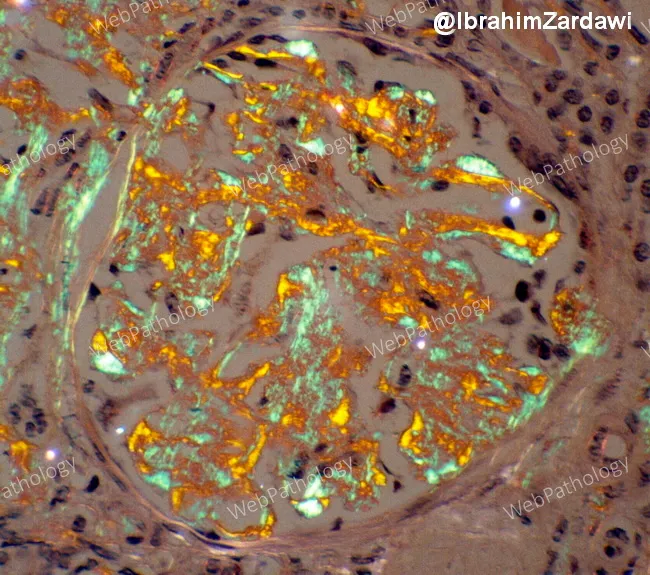
Renal biopsy histology of a patient with multiple myeloma showing AL-type amyloid deposition within the kidney. Congo red–stained sections reveal extracellular, eosinophilic, acellular deposits in the glomerular mesangium and throughout the tubulointerstitium; these deposits are salmon-pink on routine light microscopy. When viewed under polarized light, the Congo red positive material displays the diagnostic apple-green birefringence, confirming amyloid. The deposits disrupt normal glomerular architecture, with mesangial expansion, thickened capillary loops, and variable involvement of the tubules and interstitium; vascular involvement may be appreciated as deposition in arterioles. The lamina appears to be produced by a monoclonal immunoglobulin light chain (AL type) associated with underlying plasma cell dyscrasia; in the setting of myeloma, AL amyloidosis accounts for this pattern. Clinically, such renal amyloid deposition commonly correlates with nephrotic-range proteinuria, hypoalbuminemia, edema, and progressive renal insufficiency leading to chronic kidney disease if untreated. The image illustrates Congo red positivity and apple-green birefringence, the gold-standard histologic features used to diagnose systemic AL amyloidosis. Confirmatory typing by immunohistochemistry or mass spectrometry is often pursued. This histology is critical for prognosis and guides therapy targeting the plasma cell clone rather than only symptomatic management. This pattern supports systemic evaluation for underlying plasma cell dyscrasia.
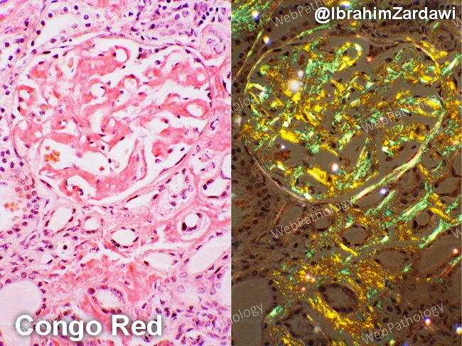
This renal cortical histology specimen demonstrates AL-type amyloidosis with kidney involvement in a patient with multiple myeloma. The left panel shows hematoxylin and eosin–stained tissue with amorphous, eosinophilic extracellular deposits within the glomerular mesangium and extending into the tubulointerstitium, causing mesangial expansion and variable basement membrane thickening. The right panel uses Congo Red staining to highlight the amyloid deposits, which appear salmon-pink on bright-field examination. Under polarized light, the same deposits display the diagnostic apple-green birefringence, a hallmark of Congo Red–positive amyloid. The deposits are diffuse, involving multiple glomeruli and extending into the tubulointerstitium, sometimes outlining capillary walls. In AL amyloidosis associated with plasma cell dyscrasia, the deposited light-chain–derived proteins disrupt normal renal architecture and contribute to proteinuria and progressive renal impairment. Differential considerations include AA (secondary) amyloidosis and other renal amyloidoses, but the combination of a known plasma cell disorder, Congo Red positivity, and apple-green birefringence strongly supports AL-type amyloidosis. This image serves as a valuable teaching reference for renal pathology, nephrology, and hematology-oncology, illustrating classic Congo Red histology, amyloid distribution patterns within glomeruli and interstitium, and the polarizing light signature. Correlates with proteinuria and renal dysfunction in many myeloma patients, guiding targeted therapy and monitoring of clonal plasma cell burden.
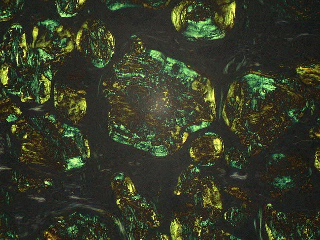
Light microscopy image of a lymph node biopsy stained with Congo Red and examined under polarized light. The tissue reveals extracellular, amorphous eosinophilic deposits distributed throughout nodal architecture consistent with amyloid. The deposits assume a dense, granular to plaque-like appearance on routine brightfield, and demonstrate distinctive apple-green birefringence when illuminated with polarized light, confirming the beta-pleated sheet rich configuration of amyloid fibrils. Congo Red positivity with birefringence distinguishes amyloid from other PAS-positive glycoprotein material and helps differentiate from non-amyloid nodal deposits. The beta-pleated sheet conformation accounts for the characteristic staining behavior and optical properties used in diagnosis. Clinically, identification of nodal amyloid supports systemic or localized amyloidosis and prompts workup for light-chain (AL) or serum amyloid A (AA) subtypes, including serum free light chains, immunofixation, and mass spectrometry-based typing. This image is relevant for educational illustrations of amyloid histology, pathophysiology, and diagnostic pathology workflows, including differential diagnosis with glycoprotein-rich PAS-positive deposits that are Congo Red negative. Potential clinical scenarios include workup of lymphadenopathy in patients with chronic inflammatory states, plasma cell dyscrasias, and lymphoproliferative disorders. High quality images like this support interdepartmental communication, teaching, and reproducible reporting of amyloid histology for junior pathologists and medical trainees in pathology education programs.
SLE systemic lupus erythematosus butterfly malar rash morphology
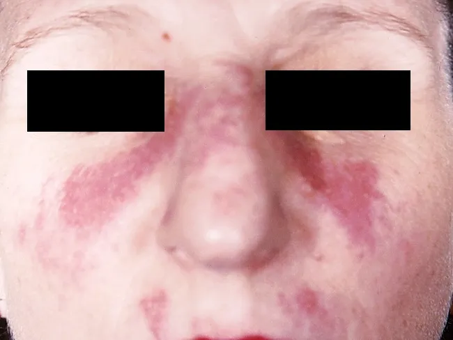
This clinical dermatology photograph displays the classic malar or butterfly rash of systemic lupus erythematosus (SLE) on the central face. Modality is clinical photography using standard white-light illumination; frontal/anterior view; color-balanced, high-resolution capture to depict superficial erythema and patchy hyperemia across the malar eminences and nasal bridge. The rash forms a bilateral, 'butterfly' distribution that typically reaches the cheeks and bridge of the nose while sparing the nasolabial folds. The observed features include confluent to patchy, erythematous macules and plaques with uniform erythema, mild perivascular edema, and subtle textural change without overt crusting or scaling in this image. The clinical morphology is characteristic for acute cutaneous lupus erythematosus; photosensitivity may exacerbate lesions. This cutaneous finding is one of the diagnostic criteria for SLE when aligned with serologic abnormalities (ANA, anti-dsDNA) and systemic features; its presence increases diagnostic probability in a compatible patient. Differential considerations include rosacea, seborrheic dermatitis, contact dermatitis, and dermatomyositis rash; however, the malar distribution and nasal bridge involvement help distinguish lupus. Clinically, this image supports SLE workup and educational reference for recognizing lupus-associated facial rash in medical students, residents, and researchers; useful for pattern-recognition training and multimodal data repository indexing. This image emphasizes clinical-context interpretation and education.
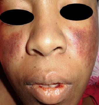
A clinical photograph of a patient's face demonstrating dermatological and mucosal manifestations of Systemic Lupus Erythematosus (SLE). A classic malar rash (butterfly rash) is present, characterized by symmetric, erythematous-to-violaceous patchy lesions over the malar eminences and the bridge of the nose, notably sparing the nasolabial folds. Additionally, the perioral region exhibits significant erythema and mucosal involvement. The lips show evidence of hemorrhagic lesions, with visible blood crusting and a small fissure on the lower lip, suggestive of vasculitis or active systemic inflammation. These visual findings are key diagnostic indicators for SLE, particularly when associated with hematological abnormalities such as thrombocytopenia. The photograph provides a clear example of cutaneous lupus manifestations for clinical diagnosis and medical education.
graft versus host disease acute skin rash histology
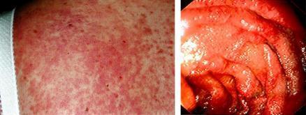
This dual-panel clinical image illustrates manifestations of acute graft-versus-host disease (GVHD) in the skin and gastrointestinal tract. The left panel shows a clinical photograph of a widespread, diffuse erythematous maculopapular rash on the skin. The lesions consist of numerous small, red macules and papules that are confluent in some areas, creating a mottled appearance. The right panel is an endoscopic image of the upper intestinal mucosa. It reveals severe mucosal inflammation characterized by intense erythema, edema (swelling), and friability. The normally distinct mucosal folds appear thickened and rounded, with surface irregularities and a glistening appearance suggestive of increased mucus production or exudate. Together, these panels demonstrate the multisystemic nature of acute GVHD following hematopoietic cell transplantation, highlighting the visual diagnostic criteria for skin and mucosal involvement.
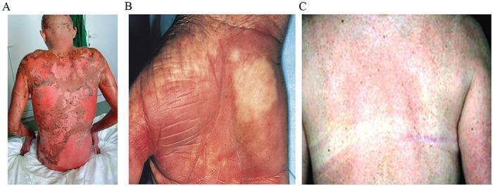
Clinical photograph set illustrating dermatological manifestations of Graft-Versus-Host Disease (GVHD). Figure A displays acute stage IV GVHD on a patient's back, characterized by diffuse, confluent erythema and extensive epidermal sloughing (desquamation) resembling toxic epidermal necrolysis, indicative of severe mucosal and skin involvement following myeloablative conditioning. Figure B shows chronic skin GVHD on a hand, featuring palmar erythema, areas of hypopigmentation, and sclerodermatous changes with accentuated skin markings and fine wrinkling, suggesting loss of elasticity and chronic dermal remodeling. Figure C demonstrates chronic GVHD on the torso with a widespread maculopapular rash, mottled pigmentation, and less severe inflammatory response compared to the acute stage. This comparison serves as an educational tool for distinguishing between the inflammatory, life-threatening presentation of acute GVHD and the fibrotic or pigmentary changes typical of chronic GVHD in hematopoietic stem cell transplant recipients.
amyloid spleen sago lardaceous gross pathology
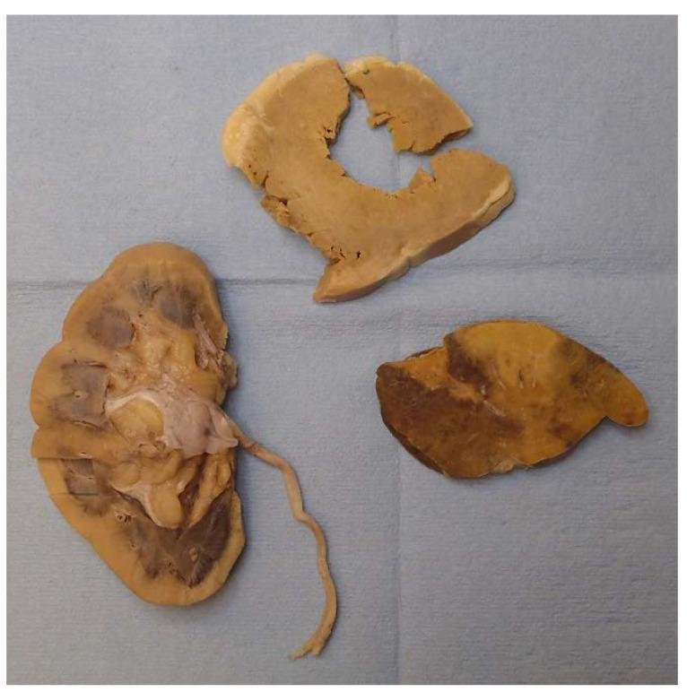
This clinical photograph displays a gross pathological examination of three formalin-fixed tissue specimens from a systemic amyloidosis case, arranged on a blue surgical drape. The specimens include the right kidney (bottom left), the left ventricle of the heart (top center), and a section of the spleen (bottom right). All tissues exhibit a characteristic firm, waxy texture and a diffuse pale-yellow discoloration indicative of extensive amyloid deposition. The kidney specimen, shown in longitudinal section, reveals pale cortical involvement and prominent renal pyramids. The heart specimen shows myocardial thickening with a uniform tan-yellow hue. The spleen specimen demonstrates a 'lardaceous' pattern, characterized by diffuse, map-like pale infiltrates throughout the parenchyma, where amyloid replaces the red pulp. This gross presentation is a hallmark of systemic amyloidosis, where extracellular protein fibrils accumulate, leading to organomegaly and eventual organ failure. This visual is intended for medical education in pathology and internal medicine to illustrate the macroscopic findings of amyloid infiltration across different organ systems.
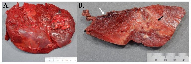
Gross anatomical specimens of a human spleen demonstrating spontaneous rupture and underlying amyloidosis. (A) External view of the splenic capsule, exhibiting a dark red, congested appearance with significant surface irregularities. A large longitudinal posterior laceration (approximately 13.5 cm) is visible, indicative of splenic rupture, along with multiple fibrous adhesions. (B) A cut section of the same spleen reveals heterogeneous parenchyma. A superior subcapsular hemorrhagic zone, measuring approximately 10.5 x 7 x 4 cm, is identified by a white arrow and covered by a thin, tan rind. Additionally, multiple ill-defined, firm, white-yellow infiltrative areas (black arrow) occupy roughly 10% of the cut surface, representing amyloid deposition. The remaining parenchyma is firm and dark red. These images illustrate the gross pathology of amyloid-related splenic friability leading to spontaneous rupture and hematoma formation. Targeted educational concepts include gross organ pathology, hematologic complications of amyloidosis, and surgical specimen evaluation in splenectomy.
Comprehensive Pathology Notes
1. Hypersensitivity Reactions
Definition
Classification (Gell and Coombs)
Summary Chart
| Feature | Type I (IgE) | Type II (Cytotoxic) | Type III (Immune Complex) | Type IV (Delayed/Cell-Mediated) |
|---|---|---|---|---|
| Mediator | IgE + mast cells | IgG/IgM antibody | IgG antibody complexes | T lymphocytes |
| Antigen | Soluble exogenous | Cell-surface/matrix | Soluble (circulating) | Cell-associated/intracellular |
| Onset | Minutes | Minutes-hours | Hours | 48-72 hours |
| Mechanism | Mast cell degranulation | Complement + opsonization | Complement + neutrophil recruitment | Cytokine release + macrophage activation |
| Examples | Asthma, anaphylaxis, urticaria | Hemolytic anemia, Goodpasture's, Rh incompatibility | Serum sickness, SLE, post-streptococcal GN | TB skin test, contact dermatitis, graft rejection |
Type I - Immediate Hypersensitivity (IgE-mediated)
Pathogenesis
- Sensitization phase: First exposure to antigen (allergen) drives Th2 differentiation, which produces IL-4 and IL-13, promoting isotype switching of B cells to produce IgE.
- IgE binding: IgE binds via its Fc region to FcεRI (high-affinity Fc receptor) on mast cells and basophils, "arming" them.
- Re-exposure and cross-linking: Second exposure to the same allergen cross-links adjacent surface IgE molecules, triggering mast cell activation.
- Two-phase response:
- Immediate phase (within minutes): Degranulation releases preformed mediators - histamine, tryptase, heparin, chymase. These cause vasodilation, increased vascular permeability, smooth muscle contraction, and mucus secretion.
- Late phase (4-12 hours): Newly synthesized mediators include leukotrienes (LTC4, LTD4), prostaglandins, PAF, and cytokines (IL-3, IL-5, GM-CSF, TNF-α). These recruit eosinophils and basophils, causing sustained inflammation and tissue damage.
Mediators and Their Effects
| Mediator | Source | Effect |
|---|---|---|
| Histamine | Preformed (granules) | Bronchoconstriction, vasodilation, pruritus |
| Leukotrienes C4, D4 | Newly synthesized | Prolonged bronchoconstriction, mucus secretion |
| Prostaglandin D2 | Newly synthesized | Bronchoconstriction, vasodilation |
| PAF | Newly synthesized | Platelet aggregation, bronchoconstriction |
| IL-5 | Cytokine | Eosinophil recruitment and activation |
| TNF-α | Cytokine | Neutrophil influx, tissue damage |
Examples
- Anaphylaxis: Systemic, life-threatening - bee sting, peanut allergy
- Asthma: Airway bronchoconstriction
- Allergic rhinitis (hay fever): Nasal congestion, sneezing
- Urticaria/angioedema: Skin wheal-and-flare
- Atopic dermatitis: Chronic skin inflammation
Type II - Cytotoxic/Antibody-Mediated Hypersensitivity
Pathogenesis
-
Complement-mediated lysis: Antibody binds to cell surface → activates classical complement pathway → formation of membrane attack complex (MAC) → cell lysis. Example: Transfusion reactions (ABO mismatch), Rh hemolytic disease of the newborn.
-
Antibody-dependent cell-mediated cytotoxicity (ADCC): IgG-coated target cells are killed by NK cells, eosinophils, or macrophages via Fc receptors.
-
Complement/Fc receptor-mediated phagocytosis (opsonization): Antibody-coated cells are recognized by macrophages in spleen/liver → phagocytosis and destruction. Example: Autoimmune hemolytic anemia, idiopathic thrombocytopenic purpura (ITP).
-
Functional alteration without cell destruction: Antibodies against receptors block or mimic receptor function.
- Blocking: Myasthenia gravis (anti-ACh receptor antibodies block neuromuscular transmission)
- Stimulating: Graves' disease (anti-TSH receptor antibodies stimulate thyroid)
Examples
| Disease | Target Antigen | Mechanism |
|---|---|---|
| Transfusion reaction | RBC blood group antigens (ABO) | Complement lysis |
| Hemolytic disease of newborn | Rh antigen | ADCC + opsonization |
| Autoimmune hemolytic anemia | RBC membrane antigens | Opsonization/lysis |
| Goodpasture's syndrome | Type IV collagen (GBM) | Complement activation |
| Pemphigus vulgaris | Desmoglein (epidermal adhesion) | Disruption of cell junctions |
| Myasthenia gravis | Acetylcholine receptor | Receptor blockade |
| Graves' disease | TSH receptor | Receptor stimulation |
Type III - Immune Complex-Mediated Hypersensitivity
Pathogenesis
-
Immune complex formation: Antibodies (IgG) bind to circulating soluble antigens, forming antigen-antibody complexes. Under normal circumstances, these are cleared by the mononuclear phagocyte system.
-
Deposition: When complexes are formed in excess antigen, they become small and soluble, evading clearance, and deposit in vessel walls, glomeruli, joint spaces, and skin.
-
Complement activation: Deposited complexes activate the complement cascade → C3a and C5a anaphylatoxins → mast cell degranulation (increases vascular permeability) → C5a also acts as a potent neutrophil chemoattractant.
-
Neutrophil-mediated injury: Neutrophils attempt to phagocytose fixed complexes but cannot; they instead release lysosomal enzymes, reactive oxygen species → fibrinoid necrosis of vessel walls (vasculitis) and tissue damage.
Histological Features
- Fibrinoid necrosis: Pale pink amorphous material replacing vessel walls
- Neutrophilic infiltrate
- Immune complex deposits visible by immunofluorescence (granular/lumpy-bumpy pattern)
Examples
| Disease | Antigen | Location of Deposition |
|---|---|---|
| Serum sickness | Foreign proteins (horse antithymocyte globulin) | Systemic vasculature |
| Post-streptococcal glomerulonephritis | Streptococcal antigens | Glomeruli |
| SLE | dsDNA, histones, nucleoproteins | Glomeruli, skin, joints |
| Polyarteritis nodosa | Hepatitis B surface antigen | Small-medium arteries |
| Arthus reaction (local) | Subcutaneous injected antigen | Local vessel walls |
Type IV - Delayed-Type/Cell-Mediated Hypersensitivity
Pathogenesis
- Antigen is presented by APC (macrophage) to CD4+ Th1 cells via MHC II.
- Sensitized Th1 cells, on re-exposure, secrete cytokines: IFN-γ (activates macrophages), TNF (promotes inflammation), IL-2 (T cell proliferation).
- Activated macrophages become "angry macrophages" - release more TNF, IL-12, reactive oxygen species.
- Result: granuloma formation and tissue destruction.
- CD8+ T cells recognize antigen presented by MHC class I on target cells.
- Direct killing via perforin/granzyme pathway or Fas-FasL interaction.
- Example: viral infections, transplant rejection, killing of tumor cells.
Examples
| Disease/Condition | Antigen | Effector |
|---|---|---|
| Tuberculin skin test (Mantoux) | PPD (mycobacterial proteins) | CD4+ Th1 |
| Contact dermatitis | Poison ivy (urushiol), nickel | CD4+ + CD8+ |
| Transplant rejection (acute) | Alloantigens | CD8+ CTL |
| Type 1 diabetes mellitus | Pancreatic beta-cell antigens | CD8+ CTL |
| Multiple sclerosis | Myelin antigens | Th1/Th17 |
| Granulomatous diseases | Persistent antigens (TB, fungi) | Th1 + macrophages |
2. Amyloidosis
Definition
Chemical Composition of All Amyloids
- Protein-derived amyloid fibers - the component that differs among subtypes
- Amyloid P component (SAP) - a 25-kDa glycoprotein, a member of the pentraxin family, present in ALL forms
- Ground substance - sulfated glycosaminoglycans (heparan sulfate) and proteoglycans
Staining Properties
- Congo red: Salmon-pink on light microscopy; apple-green birefringence under polarized light - the diagnostic gold standard
- Thioflavin T: Fluorescent staining
- PAS: Weakly positive, diastase resistant
- Crystal violet: Purple (metachromatic)
- Electron microscopy: Straight, non-branching, non-anastomosing fibrils 8-12 nm in diameter
Classification
Chart 1: Biochemical Classification of Major Amyloids
| Type | Precursor Protein | Clinical Setting |
|---|---|---|
| AL (primary) | Immunoglobulin light chains (variable region) | Multiple myeloma, plasma cell dyscrasias |
| AA (secondary) | Serum amyloid A (SAA) protein | Chronic inflammatory diseases (RA, TB, IBD, FMF) |
| ATTR | Transthyretin (TTR) | Familial (mutant TTR) or senile cardiac (wild-type TTR) |
| Aβ2M | Beta-2 microglobulin | Long-term dialysis patients (carpal tunnel) |
| Aβ | Amyloid precursor protein (APP) | Alzheimer's disease (cerebral plaques) |
| AIAPP | Islet amyloid polypeptide | Type 2 diabetes (pancreatic islets) |
| ApoA-I, ApoA-II | Apolipoproteins | Hereditary forms |
| Afib | Fibrinogen alpha chain | Hereditary renal amyloidosis |
| ALECT2 | Leukocyte chemotactic factor 2 | Common in Latinos, Native Americans (renal) |
Chart 2: Clinical Classification
AMYLOIDOSIS
├── SYSTEMIC
│   ├── Primary (AL) - plasma cell disorder
│   ├── Secondary (AA) - chronic inflammation
│   ├── Dialysis-related (Aβ2M)
│   ├── Heredofamilial (ATTR, Afib, ApoA-I...)
│   └── Senile systemic (wild-type ATTR - cardiac)
│
└── LOCALIZED
    ├── Cutaneous (macular, lichen, nodular)
    ├── Cerebral (Alzheimer's - Aβ plaques)
    ├── Pancreatic (type 2 DM - AIAPP)
    └── Medullary thyroid carcinoma (calcitonin)
Pathogenesis
- A predisposing condition leads to overproduction of an amyloidogenic precursor protein (e.g., plasma cell clone producing light chains in myeloma; elevated SAA in chronic inflammation).
- The protein adopts an abnormal beta-pleated sheet conformation - cofactors such as SAP, glycosaminoglycans, and seeding by existing fibrils promote this misfolding.
- Oligomeric intermediates (soluble, cytotoxic) form first, then polymerize into insoluble fibrils.
- SAP binds to all amyloid fibrils - stabilizes deposits and protects them from proteolysis.
- Fibrils accumulate extracellularly → compress and replace normal parenchyma → organ dysfunction.
- Deposits exist in a dynamic state and can regress if the source is eliminated (demonstrated by SAP scintigraphy). - Brenner and Rector's The Kidney
Pathology in Specific Organs
Kidney (Most Common Clinically Significant Site)
- Gross: Enlarged, pale, waxy, firm kidneys
- Light microscopy (H&E): Amorphous eosinophilic material expanding the mesangium in glomeruli; may involve capillary walls, interstitium, and blood vessels
- Congo red: Apple-green birefringence under polarized light
- Immunofluorescence: Deposits labeled with anti-lambda or anti-kappa (AL); anti-SAA (AA)
- Electron microscopy: 8-12 nm non-branching fibrils in mesangium, GBM (subepithelial, intramembranous, subendothelial)
- Clinical: Nephrotic syndrome (massive proteinuria, hypoalbuminemia, edema) → progressive renal failure
- Predominantly AL (86%) and AA (7%) in renal biopsy series - Brenner and Rector's The Kidney
Spleen
- Sago spleen: Deposits limited to follicles/white pulp → translucent sago grain-like nodules on cut section. Most common initially.
- Lardaceous (diffuse) spleen: Diffuse involvement of red pulp → large, firm, pale-yellow, waxy appearance ("lard-like"). Seen in advanced disease.
Liver
- Gross: Hepatomegaly; pale, waxy cut surface
- Micro: Deposits in space of Disse (between sinusoidal lining cells and hepatocytes) → progressive hepatocyte atrophy
- May cause cholestasis, mildly elevated liver enzymes
- Rarely causes liver failure
Heart
- Gross: Firm, rubbery, enlarged heart
- Micro: Deposits between myocardial fibers
- Types: ATTR (senile/wild-type - most common cause of cardiac amyloid in elderly), AL
- Echocardiogram: "Sparkling" or "granular sparkling" appearance of myocardium; concentric hypertrophy
- Clinical: Restrictive cardiomyopathy, heart failure, arrhythmias, conduction defects. Cardiac involvement is the primary cause of death in amyloidosis.
Other Organs
| Organ | Findings | Clinical Manifestation |
|---|---|---|
| Tongue | Macroglossia | Difficulty swallowing/speaking (AL) |
| Peripheral nerves | Endoneurial deposits | Polyneuropathy (ATTR, AL) |
| Joints/Carpal tunnel | Tendon sheath deposits | Carpal tunnel syndrome (Aβ2M in dialysis) |
| Adrenal glands | Cortical deposits | Adrenal insufficiency |
| GI tract | Submucosal deposits | Constipation, malabsorption, bleeding |
| Skin | Periorbital purpura (pinch purpura), waxy papules | Classic in AL amyloidosis |
3. Systemic Lupus Erythematosus (SLE)
Definition
Autoantibodies in SLE
| Antibody | Significance |
|---|---|
| Anti-dsDNA | Most specific for SLE; correlates with disease activity (especially nephritis); score = 6 points in classification |
| Anti-Smith (anti-Sm) | Highly specific for SLE |
| ANA (antinuclear) | Most sensitive (~99%); required as entry criterion |
| Anti-histone | Drug-induced lupus |
| Anti-Ro/SSA, Anti-La/SSB | Neonatal lupus, subacute cutaneous lupus, Sjögren's overlap |
| Antiphospholipid | Antiphospholipid syndrome - thrombosis, recurrent pregnancy loss |
| Anti-RBC | Hemolytic anemia |
| Anti-platelet | Thrombocytopenia |
Etiopathogenesis
Genetic Factors
- HLA associations: HLA-DR2 or HLA-DR3 each confer odds ratio of 2-3; if both present, odds ratio ~5
- Complement deficiencies: C1q, C2, C4 deficiencies in ~5% of SLE patients → impaired clearance of immune complexes and apoptotic cells → failure of B-cell tolerance
- FcγRIIB polymorphism: Defective inhibitory Fc receptor → inadequate control of B-cell activation
- PTPN22 polymorphism: Phosphatase involved in lymphocyte signaling
- TREX1 mutations: DNase that degrades intracellular DNA; mutations lead to accumulation of nuclear material
Environmental Factors
- Ultraviolet (UV) light: Induces apoptosis of skin cells → release of nuclear antigens (nucleosomes, dsDNA, ribonucleoproteins) that normally remain intracellular
- Drugs: Procainamide, hydralazine, isoniazid → drug-induced lupus (anti-histone antibodies; usually ANA+ but anti-dsDNA-)
- Infections: Molecular mimicry, TLR activation by microbial nucleic acids
- Estrogen: Explains female predominance; estrogen enhances autoimmune responses
Pathogenesis Model (IFN-α Loop)
UV light / Apoptosis
        ↓
Release of nuclear antigens (dsDNA, nucleosomes)
        ↓
Inadequate clearance (complement deficiency, TREX1 mutation)
        ↓
Activation of plasmacytoid dendritic cells (pDCs) via TLR7/TLR9
        ↓
Massive IFN-α production ("interferon signature")
        ↓
Activation of autoreactive B cells and T cells
        ↓
Production of anti-dsDNA, anti-Smith autoantibodies
        ↓
Immune complex formation and deposition
        ↓
Complement activation → Inflammation → Tissue damage
Why Autoantibodies Cause Disease
- Immune complexes: Anti-dsDNA + dsDNA complexes deposit in glomeruli, skin, joints, choroid plexus → complement activation → neutrophil recruitment → tissue damage (Type III hypersensitivity)
- Direct cell targeting: Anti-RBC → hemolytic anemia; anti-platelet → thrombocytopenia (Type II hypersensitivity)
Classification Criteria (EULAR/ACR 2019)
| Domain | Criteria | Points |
|---|---|---|
| Constitutional | Fever | 2 |
| Neuropsychiatric | Seizure | 5; Psychosis 3; Delirium 2 |
| Mucocutaneous | Acute cutaneous lupus | 6; Discoid lupus 4; Oral ulcers 2; Alopecia 2 |
| Musculoskeletal | Joint involvement | 6 |
| Serosal | Acute pericarditis | 6; Effusion 5 |
| Hematologic | Autoimmune hemolysis 4; Thrombocytopenia 4; Leukopenia 3 | |
| Renal | Class III/IV LN = 10; Class II/V LN = 8; Proteinuria >0.5g/24h = 4 | |
| Antiphospholipid Ab | Anticardiolipin / anti-β2GPI | 2 |
| Complement | Low C3 AND C4 | 4; Either alone 3 |
| SLE-specific Ab | Anti-dsDNA | 6 |
Morphology (Pathological Findings)
Skin (Malar/Butterfly Rash)
- Acute cutaneous lupus: Butterfly (malar) rash - erythematous macular-papular eruption over cheeks and nasal bridge, sparing nasolabial folds; photosensitive
- Discoid lupus: Erythematous plaques with central scarring and follicular plugging; heals with scarring alopecia
- Histology: Vacuolar degeneration of basal layer, thickening of basement membrane, perivascular lymphocytic infiltrate
- Immunofluorescence: "Lupus band test" - granular deposits of IgG, IgM, C3 at DEJ (positive in both lesional and non-lesional skin in SLE, unlike in discoid lupus)
Kidney (Lupus Nephritis)
- Class I: Minimal mesangial (normal LM, deposits by IF/EM)
- Class II: Mesangial proliferative (mesangial deposits + hypercellularity)
- Class III: Focal proliferative (<50% glomeruli affected) - active lesions
- Class IV: Diffuse proliferative (≥50% glomeruli) - most common and most severe; "wire-loop" lesions (massive subendothelial deposits), hyaline thrombi
- Class V: Membranous (diffuse subepithelial deposits, resembles idiopathic MN)
- Class VI: Advanced sclerosing (≥90% global sclerosis)
Heart
- Libman-Sacks endocarditis: Non-bacterial verrucous endocarditis - small, irregular, sterile vegetations on both surfaces of the mitral valve (most commonly) and tricuspid valve. Distinctive because they occur on both sides of valve leaflets (unlike rheumatic fever, which is only on atrial surface of mitral valve).
- Pericarditis (fibrinous)
- Myocarditis (less common)
Blood Vessels
- Vasculitis: Immune-complex deposition in small vessels → leukocytoclastic vasculitis
- Libman-Sacks endocarditis (as above)
- Antiphospholipid syndrome in some: thrombosis (paradoxically) → increased risk of MI, stroke
Joints
- Non-erosive synovitis (in contrast to rheumatoid arthritis, which is erosive)
- Arthralgia and arthritis without joint deformity
Spleen
- Onion-skin lesion (periarteriolar fibrosis): Concentric fibrosis around splenic arterioles - characteristic of SLE on histology
CNS/Neuropsychiatric
- Small vessel vasculopathy (non-inflammatory), microthrombi (from antiphospholipid antibodies)
- Cognitive dysfunction, psychosis, seizures
4. Graft Versus Host Disease (GVHD)
Definition
Prerequisites (Three Requirements)
- The transplanted cells must be immunologically competent (contain mature T lymphocytes)
- The recipient must express tissue antigens not present in the donor (HLA mismatch - either major or minor histocompatibility differences)
- The recipient must be unable to reject the transplanted cells (immunosuppressed, as in HSCT recipients)
Pathogenesis
- The preconditioning regimen (chemotherapy + total body irradiation) damages rapidly dividing tissues (gut, skin, liver) → activates host dendritic cells (APCs) → they upregulate HLA molecules and other minor histocompatibility antigens.
- Host APCs present recipient alloantigens to donor T cells (both CD4+ and CD8+).
- Donor T cells become activated and expand → produce cytokines, especially IL-2, TNF-α, and IFN-γ, which amplify the host-donor immune interaction.
- Cytokine storm leads to inflammation and destruction of target organs: skin, liver, gastrointestinal tract (the classic triad).
- Some degree of immunologic competence of transplanted cells is actually desired for the graft-versus-tumor (GVT) effect - donor cells attack residual malignancy.
Classification
Acute GVHD
- Classic: Within 100 days of transplant; can occur up to 1 year with tapering of immunosuppression
- Onset: Typically days 14-42 after HSCT, peak at day 30
- Skin: Erythematous morbiliform eruption of face and trunk → may become confluent → exfoliative erythroderma; bullae in severe cases. Starts with follicular/eccrine duct involvement (looks like keratosis pilaris). More monomorphous than drug eruption; involves upper back.
- Liver: Cholestatic jaundice (elevated bilirubin, alkaline phosphatase)
- GI tract: Profuse watery or bloody diarrhea, abdominal pain, nausea/vomiting
| Stage | Skin | Liver (Bilirubin) | GI (Diarrhea/day) |
|---|---|---|---|
| 1 | Rash <25% BSA | 2-3 mg/dL | 500-1000 mL |
| 2 | Rash 25-50% BSA | 3-6 mg/dL | 1000-1500 mL |
| 3 | Rash >50% BSA | 6-15 mg/dL | >1500 mL |
| 4 | Erythroderma + bullae | >15 mg/dL | Severe pain/ileus |
Chronic GVHD
- Occurs later (>100 days or with features distinct from acute GVHD)
- Resembles autoimmune/connective tissue diseases (Sjögren's, scleroderma, lichen planus, primary biliary cirrhosis)
- Skin: Lichenoid or sclerodermoid changes, dyspigmentation, scleroderma-like fibrosis
- Eyes: Sicca syndrome (dry eyes)
- Oral cavity: Lichenoid changes, xerostomia
- Lung: Bronchiolitis obliterans (obstructive lung disease)
- Liver: Chronic cholestatic disease
Target Organs
| Organ | Acute GVHD | Chronic GVHD |
|---|---|---|
| Skin | Maculopapular rash → erythroderma | Lichenoid/sclerodermoid fibrosis |
| Liver | Cholestatic jaundice | Cirrhosis-like changes |
| GI tract | Diarrhea, enteritis | Malabsorption, dysmotility |
| Lung | - | Bronchiolitis obliterans |
| Eyes | - | Sicca syndrome |
Treatment
- Prevention: HLA-matching, T-cell depletion of graft, calcineurin inhibitors (cyclosporine, tacrolimus) + methotrexate
- Acute GVHD: Systemic corticosteroids (first-line); refractory: ruxolitinib (JAK1/2 inhibitor), ibrutinib
- Chronic GVHD: Steroids + cyclosporine; photopheresis; rituximab
5. Role of MHC in Disease
Background
MHC Classes
| Feature | MHC Class I | MHC Class II |
|---|---|---|
| Genes | HLA-A, -B, -C | HLA-DR, -DQ, -DP |
| Structure | α chain + β2-microglobulin | α + β heterodimer |
| Expressed on | All nucleated cells | Professional APCs (DC, macrophage, B cell) |
| Peptide source | Intracellular (endogenous) | Extracellular (exogenous) |
| Presents to | CD8+ cytotoxic T cells | CD4+ helper T cells |
Mechanisms by Which MHC Alleles Confer Disease Susceptibility
1. Differential Peptide Binding (Molecular Mimicry)
- Different MHC alleles have different peptide-binding clefts with unique amino acid compositions.
- A particular allele may bind and present self-peptides that would not otherwise be presented → activates autoreactive T cells.
- Example: The DQβ chain polymorphism at a critical position in the peptide-binding groove correlates most strongly with susceptibility to type 1 diabetes.
2. Failure of Negative Selection
- If a self-peptide/MHC complex is not present in the thymus, self-reactive T cells specific for that combination are not deleted → escape into periphery as autoreactive cells.
- Some alleles may fail to present critical self-antigens in the thymus, allowing escape of potentially autoreactive clones.
3. Defective T-Regulatory Cell Function
- Certain HLA alleles may affect the efficiency of Treg generation or function, impairing peripheral tolerance.
4. Protective Alleles
- Some alleles are protective because they bind self-peptides so strongly that autoreactive T cells are deleted in thymus.
- Example: HLA-DQ6 (DQB1*0602) is strongly protective against type 1 diabetes (relative risk = 0.02).
HLA Disease Associations (Key Table)
| Disease | HLA Allele | Relative Risk | Notes |
|---|---|---|---|
| Ankylosing spondylitis | B27 | 87.4 | Strongest known HLA-disease association |
| Type 1 diabetes | DQ2 and DQ8 | ~25 | DQ6 is protective (RR 0.02) |
| Goodpasture's syndrome | DR2 | 15.9 | Anti-GBM disease |
| Pemphigus vulgaris | DR4 | 14.4 | Blistering skin disease |
| Autoimmune uveitis | B27 | 10 | |
| Psoriasis vulgaris | CW6 | 7 | |
| SLE | DR3 | 5.8 | Female predominance 10-20:1 |
| Addison's disease | DR3 | 5 | |
| Multiple sclerosis | DR2 | 4.8 | |
| Rheumatoid arthritis | DR4 | 4.2 | "Shared epitope" on DRβ1 |
| Graves' disease | DR3 | 3.7 | |
| Hashimoto's thyroiditis | DR5 | 3.2 | |
| Myasthenia gravis | DR3 | 2.5 |
How MHC Contributes: The Arthritis Example (HLA-B27)
- Ankylosing spondylitis is 87x more common in HLA-B27+ individuals.
- Proposed mechanisms include:
- Arthritogenic peptide hypothesis: HLA-B27 presents specific peptides (possibly from gut bacteria) that activate cytotoxic T cells attacking the spine/sacroiliac joints.
- Molecular mimicry: HLA-B27 resembles antigens of Klebsiella pneumoniae; anti-Klebsiella antibodies cross-react with HLA-B27.
- Misfolding: HLA-B27 tends to misfold in the ER → triggers ER stress and unfolded protein response → inflammatory cytokine production.
MHC Beyond Autoimmune Disease
- Transplantation: Degree of MHC mismatch determines the intensity of allograft rejection
- Susceptibility to infectious disease: Some HLA alleles affect ability to mount immune responses against pathogens (e.g., HLA-B57 is protective against HIV progression)
- Drug hypersensitivity: HLA-B5701 - abacavir hypersensitivity; HLA-B1502 - carbamazepine Stevens-Johnson syndrome in Han Chinese
6. Protein Misfolding Diseases
Definition and Concept
The Prion Principle
- Rich in beta-sheet structure (vs. alpha-helix of PrP^C)
- Resistant to protease digestion (and harsh physical/chemical denaturing)
- Self-propagating: It acts as a template to convert newly synthesized PrP^C → PrP^Sc
- Transmissible (at least in classic prion diseases)
Classification of Protein Misfolding Diseases
Chart: Major Categories
| Category | Disease | Misfolded Protein | Site of Deposition |
|---|---|---|---|
| Classic Prion diseases | CJD, vCJD, Fatal Familial Insomnia, Kuru, GSS | PrP^Sc | Brain (spongiform encephalopathy) |
| Neurodegenerative | Alzheimer's disease | Aβ + tau | Amyloid plaques + neurofibrillary tangles |
| Neurodegenerative | Parkinson's disease | α-Synuclein | Lewy bodies (substantia nigra) |
| Neurodegenerative | ALS / FTLD | TDP-43, FUS, SOD1 | Motor neurons / frontal cortex |
| Neurodegenerative | Huntington's disease | Huntingtin (polyglutamine) | Striatum, cortex |
| Neurodegenerative | Multiple system atrophy | α-Synuclein | Glial cytoplasmic inclusions |
| Systemic amyloidosis | AL amyloidosis | Ig light chains | Kidney, heart, liver |
| Systemic amyloidosis | AA amyloidosis | Serum amyloid A | Kidney, spleen, liver |
| Systemic amyloidosis | ATTR (transthyretin) | Transthyretin | Heart, peripheral nerves |
| Endocrine | Type 2 diabetes | IAPP (amylin) | Pancreatic islets |
| Lung | Alpha-1 antitrypsin deficiency | AAT polymers | Hepatocyte ER (liver) + lung injury |
| Hereditary | Serpinopathies | Serpins | Various |
Shared Molecular Mechanisms
1. Normal Protein Folding vs. Misfolding
- Proteins fold into their native 3D conformation aided by molecular chaperones (HSP70, HSP90, GRP78/BiP).
- Mutations, oxidative stress, aging, or overwhelming chaperone capacity can cause misfolding.
- Misfolded proteins expose hydrophobic regions normally buried → prone to aggregation.
2. Beta-Sheet Conversion and Fibrillization
- Many misfolded proteins convert from alpha-helical or random-coil to beta-sheet rich structures.
- These oligomerize → protofibrils → mature insoluble fibrils.
- Toxic species: Soluble oligomeric intermediates (NOT the end-stage fibrils) are generally more toxic to cells.
3. Seeding and Templated Misfolding (Prion-Like Spreading)
- Pre-existing misfolded aggregates act as seeds that catalyze the misfolding of normal protein.
- Evidence for cell-to-cell spreading in Alzheimer's (tau/Aβ), Parkinson's (α-synuclein transplantation studies), ALS (TDP-43).
- In PD: After dopaminergic transplantation (1990s), healthy fetal grafts showed Lewy bodies 11-16 years later, indicating transmission of α-synuclein pathology from host to graft. - Bradley and Daroff's Neurology in Clinical Practice
4. Cellular Toxicity Mechanisms
- Proteasome dysfunction: Misfolded proteins overwhelm the ubiquitin-proteasome system
- Mitochondrial dysfunction: Aggregates damage mitochondria → oxidative stress
- ER stress / Unfolded Protein Response (UPR): Accumulation of misfolded proteins in ER activates UPR; if prolonged → apoptosis
- Membrane disruption: Oligomeric intermediates can perforate membranes
- Synaptic dysfunction: In neurons, aggregates disrupt synaptic vesicle trafficking
5. Degradation Pathways
- Ubiquitin-proteasome system (UPS): Primary route for misfolded cytoplasmic proteins
- Autophagy-lysosomal pathway: Handles aggregates too large for proteasome
- Failure of either system leads to aggregate accumulation
Key Examples in Detail
Alzheimer's Disease
- Aβ amyloid: From amyloid precursor protein (APP) cleaved by β and γ-secretase → Aβ40/Aβ42 peptides → form senile plaques (extracellular)
- Tau: Hyperphosphorylated tau dissociates from microtubules → forms neurofibrillary tangles (intracellular)
- Both pathologies spread through the brain in a prion-like manner
Parkinson's Disease
- α-Synuclein misfolds → forms Lewy bodies (intracytoplasmic eosinophilic inclusions, halo appearance)
- Braak staging: pathology starts in olfactory bulb/gut enteric nervous system → ascends to substantia nigra → cortex
- Substantia nigra dopaminergic neuron loss → dopamine deficiency → motor symptoms
Prion Diseases
- CJD: Sporadic (most common, ~85%), Familial (PRNP mutations, ~15%), Acquired (iatrogenic via contaminated tissue/instruments, vCJD via BSE)
- Pathology: Spongiform encephalopathy - vacuolation of neuropil, neuronal loss, astrogliosis, NO inflammatory infiltrate
- vCJD: Florid (daisy-like) PrP plaques surrounded by vacuoles; tonsil biopsy positive for PrP^Sc
Therapeutic Implications
| Strategy | Target | Example |
|---|---|---|
| Reduce production | APP, tau expression | BACE1 inhibitors (Alzheimer's) |
| Prevent aggregation | Oligomer formation | Anti-Aβ antibodies (lecanemab, donanemab) |
| Clear aggregates | Phagocytosis, autophagy | Immunotherapy |
| Stabilize native fold | Prevent misfolding | Tafamidis (stabilizes TTR tetramer in ATTR amyloidosis) |
| Enhance degradation | Proteasome, autophagy | mTOR inhibitors (increase autophagy) |
| Topic | Core Concept | Key Mechanism | Clinical Importance |
|---|---|---|---|
| Hypersensitivity | Exaggerated immune response | IgE/IgG/T-cell mediated | Allergy, autoimmunity, transplant rejection |
| Amyloidosis | β-pleated sheet protein deposits | Misfolding + fibril formation | Nephrotic syndrome, restrictive cardiomyopathy |
| SLE | Anti-dsDNA autoimmunity | IFN-α loop + immune complex disease | Nephritis, butterfly rash, Libman-Sacks endocarditis |
| GVHD | Donor T cells attack host | Alloantigen recognition + cytokine storm | Post-HSCT complication; skin/liver/GI triad |
| MHC in disease | Allele-specific peptide binding | Escape of autoreactive T cells | Strongest: HLA-B27 (AS), DQ2/8 (T1DM) |
| Protein misfolding | Templated conformational change | β-sheet aggregation; prion-like spreading | Alzheimer's, Parkinson's, prion diseases, amyloidosis |