
Simple microscopy of 1)microcytic hypochromic anemia 2) macrocytic hyperchromic anemia 3)eosinophilia 4)neutrophilic leucocytosis 5)sickle cell anemia
Reading File
Finding Sources
Finding Sources
Finding Sources
Searching PubMed
anemia blood smear
Searching the Web
Merck Manual peripheral blood smear microcytic macrocytic anemia eosinophilia neutrophilia sickle cells
Searching Images
peripheral blood smear microcytic hypochromic anemia iron deficiency
Searching Images
peripheral blood smear megaloblastic macrocytic anemia macro ovalocytes hypersegmented neutrophils
Searching Images
peripheral blood smear eosinophilia eosinophils
Searching Images
peripheral blood smear neutrophilic leukocytosis neutrophils left shift
Searching Images
Wright stained peripheral blood smear sickle cell anemia sickled erythrocytes target cells
Peripheral blood smear findings
| Condition | Simple microscopy findings |
|---|---|
| 1. Microcytic hypochromic anemia | RBCs are small and pale, with an increased central pallor occupying more than one-third of the cell diameter. There may be anisocytosis and poikilocytosis. In iron-deficiency anemia, pencil cells or elliptocytes may be seen. |
| 2. Macrocytic hyperchromic anemia | RBCs are large, oval-shaped macro-ovalocytes, often with anisopoikilocytosis. The central pallor is reduced or absent, giving an apparent hyperchromic look. Hypersegmented neutrophils are characteristic in megaloblastic anemia due to vitamin B12 or folate deficiency. Technically, MCHC is usually not truly increased. Merck's smear description |
| 3. Eosinophilia | Increased number of eosinophils. Eosinophils have a bilobed nucleus and cytoplasm packed with large, coarse orange-red granules. |
| 4. Neutrophilic leucocytosis | Increased number of neutrophils. Mature neutrophils show a multilobed nucleus with fine cytoplasmic granules. In acute bacterial infection, a left shift may be present, with increased band forms and occasionally metamyelocytes. Toxic granulation, Döhle bodies, and cytoplasmic vacuoles may occur in severe infection. |
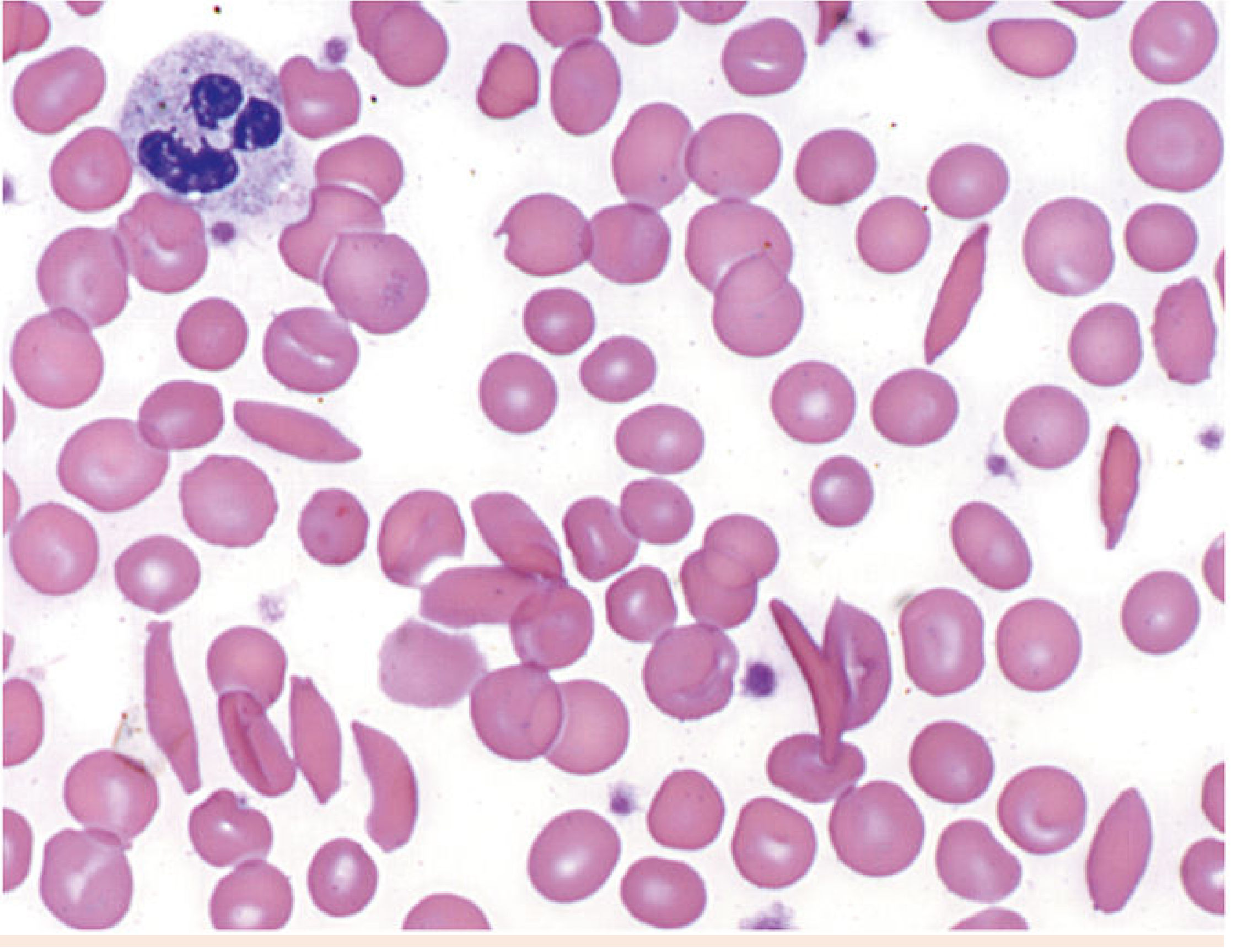
| 5. Sickle-cell anemia | Numerous sickle-shaped or crescent-shaped RBCs with pointed ends are seen. Other possible findings include target cells, polychromasia due to reticulocytosis, anisopoikilocytosis, and Howell-Jolly bodies, especially with functional asplenia. The Merck anemia review also notes sickled cells and reticulocytosis on peripheral smear. |
One-line practical identification
- Microcytic hypochromic: small pale RBCs
- Macrocytic hyperchromic: large oval RBCs with hypersegmented neutrophils
- Eosinophilia: excess orange-red granular cells
- Neutrophilic leucocytosis: excess multilobed neutrophils, possibly band forms
- Sickle-cell anemia: crescent-shaped RBCs
This is a shared conversation. Sign in to Orris to start your own chat.