
Preterm pediatrics
Reading File
The query "Preterm pediatrics" is broad. Let me clarify what you need before diving in.
Asking for Preferences
What aspect of preterm infants are you looking for? · Who is this for?
Searching sources...
Finding Sources
Finding Sources
Searching PubMed
"preterm infant" AND management
Reading File
Finding Sources
Finding Sources
Reading File
Finding Sources
Finding Sources
Finding Sources
Finding Sources
Reading File
Reading File
Reading File
Loading Image
Reading File
I now have excellent content from the textbooks. Let me compile the comprehensive exam-prep guide.Here is a comprehensive, exam-focused guide on preterm infants.
Preterm Infants - Exam-Prep Guide
1. Definitions and Classification
Preterm birth = delivery before 37 completed weeks' gestational age (GA).
| Category | GA |
|---|---|
| Late preterm | 34-36+6 weeks |
| Moderately preterm | 32-33+6 weeks |
| Very preterm | 28-31+6 weeks |
| Extremely preterm | < 28 weeks |
| Periviable | ~22-25 weeks |
Birth weight categories:
- Low birth weight (LBW): < 2500 g
- Very low birth weight (VLBW): < 1500 g
- Extremely low birth weight (ELBW): < 1000 g
Epidemiology: Extremely preterm infants (<32 weeks or <1500 g) comprise 1-2% of all deliveries; late preterm (34-36 weeks) account for 8-9% of all births and represent the fastest-growing segment.
2. Mortality Risk
The chart below (from NICHD Neonatal Research Network data) shows how mortality risk is determined jointly by gestational age AND birth weight. Notably, females consistently have lower mortality than males at the same GA/weight:
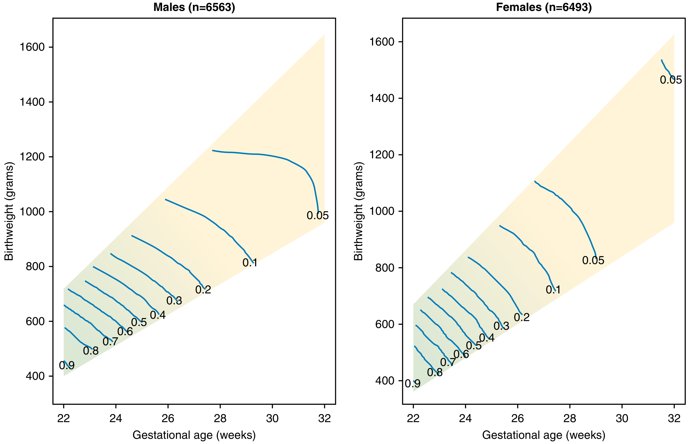
Mortality risk contours (0.05 = 5% mortality) for NICHD NRN singleton infants 22-32 weeks. - Creasy & Resnik's Maternal-Fetal Medicine, Fig. 73.1
Key point: mortality risk roughly doubles with each gestational week lost before 25 weeks.
3. Complications by Organ System
| System | Complication |
|---|---|
| Pulmonary | RDS, BPD, pulmonary hypoplasia, apnea of prematurity |
| Cardiovascular | Patent ductus arteriosus (PDA), apnea + bradycardia, hypotension |
| GI/Liver | NEC, dysmotility/reflux, feeding difficulties, hypoglycemia |
| CNS | IVH, periventricular leukomalacia (PVL), cerebral palsy, attention deficit disorders |
| Eyes | Retinopathy of prematurity (ROP) |
| Skin | Excess insensible water loss, hypothermia |
| Immune/Hematologic | Sepsis/meningitis, anemia of prematurity |
Incidence by prematurity group (exam favorite):
| Complication | Early preterm (<32 wks) | Late preterm (34-36 wks) |
|---|---|---|
| RDS | 10-80% (varies with GA and steroid use) | <5% |
| BPD | ~22% of <1500 g infants | Uncommon |
| ROP | ~20% of <1500 g infants | Rare |
| IVH (with dilation/parenchymal involvement) | ~12% of <1500 g infants | Rare |
Source: Creasy & Resnik's Maternal-Fetal Medicine, Table 73.4
4. Key Complications - Detailed Review
A. Respiratory Distress Syndrome (RDS) / Hyaline Membrane Disease
Pathophysiology: Surfactant deficiency → alveolar collapse → decreased compliance → V/Q mismatch → hypoxemia.
Risk factors for RDS:
- Prematurity (incidence: ~80% at 28 weeks, ~90% at 24 weeks)
- Cesarean delivery without labor
- Maternal diabetes
- Perinatal asphyxia
- Male sex, Caucasian race, surfactant protein B deficiency
Protective factors: Female sex, chronic intrauterine stress (IUGR), prolonged ROM, antenatal steroids
Clinical signs: Tachypnea, grunting, nasal flaring, subcostal/intercostal retractions - typically evident at delivery or shortly after
CXR: Diminished lung volumes, diffuse "ground glass" infiltrates, air bronchograms, indistinct cardiothymic silhouette - due to atelectasis from surfactant deficiency (contrasted with TTN which shows perihilar densities with hyperinflation)
Treatment:
- Antenatal betamethasone/dexamethasone (24-34 weeks; also now 34-37 weeks for late preterm)
- Exogenous surfactant replacement therapy (postnatal)
- CPAP or mechanical ventilation
- Supplemental O2 to target SpO2 (in <32 weeks, titrate with O2 blender)
B. Bronchopulmonary Dysplasia (BPD)
Definition: Need for supplemental oxygen in a premature (<32 weeks) infant for >28 days. Also called chronic lung disease of prematurity.
Pathophysiology: Results from mechanical ventilation + O2 therapy for RDS. Barotrauma, volutrauma, and oxygen toxicity disrupt alveolarization.
Outcome: Some infants gradually wean off respiratory support; others progress to severe respiratory failure and pulmonary hypertension (high mortality risk).
Risk factors: Chorioamnionitis increases BPD risk.
C. Intraventricular Hemorrhage (IVH)
Pathophysiology: Bleeding from the fragile germinal matrix (subependymal) into the ventricular system. The germinal matrix involutes by ~32-34 weeks, so IVH is almost exclusively a complication of very/extremely preterm infants.
Papile Grading System (exam high-yield):
| Grade | Description |
|---|---|
| I | Subependymal/germinal matrix hemorrhage only |
| II | IVH without ventricular dilation |
| III | IVH with ventricular dilation |
| IV | IVH with parenchymal involvement (periventricular hemorrhagic infarction) |
Key fact: Grades III and IV carry serious neurodevelopmental prognosis (cerebral palsy, cognitive impairment). Grade IV is sometimes termed "periventricular hemorrhagic infarction" and is not simply "Grade 4 IVH" by newer nomenclature.
Screening: Cranial ultrasound is the standard imaging modality in the NICU.
Prevention: Antenatal corticosteroids and magnesium sulfate (neuroprotection).
D. Necrotizing Enterocolitis (NEC)
Definition: Intestinal necrosis of uncertain etiology, predominantly in preterm infants.
Pathophysiology: Combination of intestinal immaturity, altered microbiome, formula feeding (vs. breast milk), and ischemia/infection triggers inflammatory cascade → bowel wall necrosis → perforation.
Clinical features: Feeding intolerance, abdominal distension, bloody stools, temperature instability.
Imaging: Pneumatosis intestinalis (gas in bowel wall) is pathognomonic; free air indicates perforation.
Bell Staging:
- Stage I: Suspected NEC
- Stage II: Definite NEC (pneumatosis on imaging)
- Stage III: Advanced NEC (perforation, shock)
Treatment: NPO, bowel decompression, IV antibiotics; surgery (peritoneal drain or laparotomy) for perforation.
Prevention: Breast milk (reduces NEC risk significantly), judicious use of antibiotics, possibly probiotics (evidence evolving - a 2025 Cochrane review continues to examine this).
E. Retinopathy of Prematurity (ROP)
Pathophysiology: Immature retinal vasculature responds abnormally to extrauterine oxygen exposure → neovascularization → possible retinal detachment.
Screening criteria (Harriet Lane Handbook):
- Born ≤27 weeks: first exam at 31 weeks postmenstrual age
- Born ≥28 weeks: first exam at 4 weeks chronologic age
- Born <25 weeks: consider earlier exam at 6 weeks chronologic age (to detect aggressive posterior ROP)
ROP Staging:
| Stage | Finding |
|---|---|
| 1 | Demarcation line separates avascular from vascularized retina |
| 2 | Ridge forms along demarcation line |
| 3 | Extraretinal fibrovascular proliferation on ridge |
| 4 | Partial retinal detachment |
| 5 | Total retinal detachment |
Plus disease = vascular dilation and tortuosity; indicates active, aggressive disease requiring urgent treatment.
Treatment: Laser photocoagulation or anti-VEGF injection (bevacizumab/ranibizumab) for threshold/type 1 ROP.
F. Apnea of Prematurity
Definition: Cessation of breathing for ≥20 seconds, or shorter if accompanied by bradycardia or desaturation.
Types: Central (no respiratory effort), obstructive (effort without airflow), mixed (most common).
Treatment: Caffeine citrate (methylxanthine) - reduces apnea episodes and also reduces BPD risk; CPAP; gentle tactile stimulation.
G. Patent Ductus Arteriosus (PDA)
Physiology: Ductus normally closes in response to increased PaO2 and decreased prostaglandins after birth. In preterm infants, the ductus remains patent more often and longer.
Hemodynamic consequence: Left-to-right shunt → pulmonary overcirculation → increased work of breathing, worsening RDS, NEC risk.
Treatment options:
- Pharmacologic closure: Indomethacin (COX inhibitor, reduces PGE2) or ibuprofen; acetaminophen also used
- Surgical ligation if pharmacologic treatment fails
- Conservative/expectant management (many PDAs close spontaneously - evidence has shifted toward less aggressive treatment)
5. Key Prenatal Interventions
| Intervention | Purpose |
|---|---|
| Antenatal corticosteroids (betamethasone/dexamethasone) | Accelerate lung maturity, reduce RDS, IVH, NEC; given at 24-34 wks (single rescue course if >7 days since previous course) |
| Magnesium sulfate | Neuroprotection - reduces IVH and cerebral palsy in <32 weeks |
| Antibiotics in PPROM | Prolong latency; note amoxicillin-clavulanate associated with increased NEC risk |
| Tocolytics | Delay delivery to allow steroid administration |
| Group B Strep prophylaxis | Reduce early-onset neonatal sepsis |
6. Postnatal General Management Principles
- Thermoregulation: Plastic wrap/polyethylene bag immediately at delivery; servo-controlled incubator; preterm infants have large body surface area/weight ratio with minimal brown fat.
- Fluid management: Limited IV fluids initially; preterm infants have excess insensible water losses (thin skin, no vernix).
- Nutrition: Early parenteral nutrition (PN) followed by enteral feeds; breast milk preferred (reduces NEC, improves neurodevelopment); human milk fortification improves protein/caloric density for VLBW infants.
- Infection control: VLBW infants are profoundly immunocompromised; strict hand hygiene, aseptic line care.
- Neurodevelopmental follow-up: Standard for all infants <32 weeks or <1500 g.
7. Late Preterm Infants (34-36+6 weeks) - High-Yield Points
Often underestimated clinically but carry significantly higher morbidity than term infants:
- 3x higher infant mortality rate than those born at 40 weeks
- At 35 weeks, brain volume is approximately half that of a term infant
- Increased risk of: hypoglycemia, hypothermia, jaundice (hyperbilirubinemia), poor feeding, sepsis, readmission
- Long-term: learning disabilities, attention deficit disorders
- IVH rare after 32 weeks, but white matter myelination is still rapidly occurring through the 3rd trimester
8. Viability and Ethics
- Current threshold of viability: approximately 22-23 weeks GA
- At 22-23 weeks: outcomes are uncertain; shared decision-making with parents is standard
- NRP protocols apply but experienced neonatologist should assess gestational age, weight, and response to resuscitation
- Parents are primary surrogate decision-makers
- Moral distress is common among NICU staff; ethics consultation available when providers and parents conflict
Key sources: Creasy & Resnik's Maternal-Fetal Medicine (9th ed.) Ch. 73; The Harriet Lane Handbook (23rd ed.) Ch. 18; Bradley and Daroff's Neurology in Clinical Practice; Barash Clinical Anesthesia (9th ed.)
Recent evidence note: A 2025 Cochrane review (PMID: 40292760) is examining lutein/zeaxanthin for reducing preterm morbidity/mortality - currently insufficient evidence for routine use. A 2026 systematic review (PMID: 40534564) confirms human milk fortifier is superior to preterm formula for fortifying breast milk in VLBW infants.This is your complete exam-prep guide on preterm pediatrics. Here is a quick orientation to what is covered:
Definitions & Classification - GA-based categories (late/very/extremely preterm, periviable) and birth weight categories (LBW, VLBW, ELBW), with epidemiologic context.
The "Big 5" Complications (almost exclusively preterm):
- RDS - surfactant deficiency, CXR ground glass pattern, treated with antenatal steroids + surfactant replacement
- BPD - chronic lung disease from ventilation/O2 injury, defined as O2 need >28 days in <32-week infant
- IVH - germinal matrix bleeding, Papile grades I-IV, screening by cranial U/S, prevented by antenatal steroids + MgSO4
- NEC - intestinal necrosis, pneumatosis intestinalis on imaging, Bell staging, breast milk is protective
- ROP - retinal neovascularization from O2 exposure, 5 stages, screened by ophthalmology per strict GA-based protocol, treated with laser or anti-VEGF
Other key topics: PDA management, apnea of prematurity (caffeine), thermoregulation, nutrition (breast milk + human milk fortifier), antenatal corticosteroids, and late preterm infant risks.
Ask if you want any section expanded further - for example, IVH grading images, surfactant types, or NICU ventilation strategies.
Acute diarrhea disease Management algorithm
Reading File
Searching sources...
Finding Sources
Finding Sources
Searching PubMed
"acute diarrhea" AND management AND guidelines
Finding Sources
Finding Sources
Reading File
Reading File
Reading File
Reading File
Reading File
Loading Image
Finding Sources
Finding Sources
Reading File
Loading Image
Reading File
Excellent - I now have the full Harrison's algorithm, textbook treatment protocols, and pathogen-specific data. Here is the comprehensive management guide:
Acute Diarrhea Disease - Management Algorithm
Pathophysiology Classification
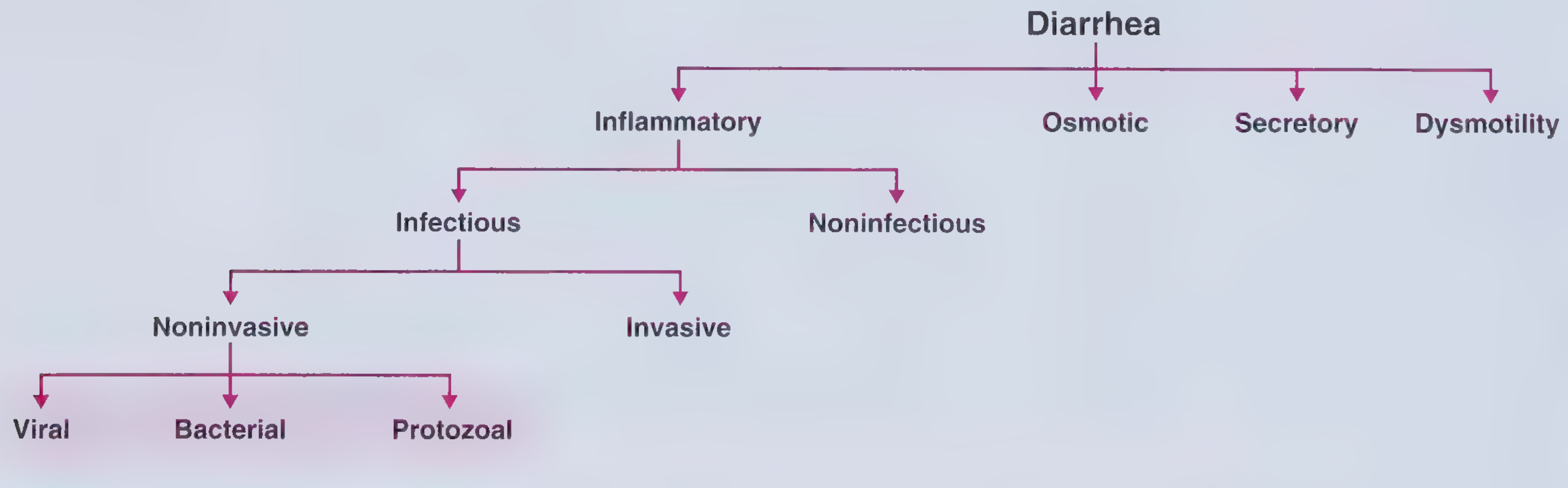
Classification of diarrhea - Frameworks for Internal Medicine
Step 1 - Initial Assessment: History and Physical
The first branch point is infectious vs. noninfectious.
Features suggesting infectious etiology:
- Acute onset (<14 days)
- Nausea, vomiting, fever
- Epidemiologic clues: recent travel, contaminated food/water, sick contacts, outbreaks (cruise ships, daycare, nursing homes)
- Recent antibiotic use (think C. difficile)
Features suggesting noninfectious etiology:
- New medication (hundreds of drugs cause diarrhea)
- Post-surgical bowel changes
- IBD flare, ischemic colitis, partial obstruction
- Radiation exposure → If likely noninfectious: evaluate and treat the underlying cause separately.
Step 2 - Severity Stratification (determines urgency)
| Severity | Description | Action |
|---|---|---|
| Mild | Unrestricted daily activity; few loose stools | Observe + oral fluids |
| Moderate | Activities altered; multiple stools; some cramping | ORS + symptomatic treatment |
| Severe | Incapacitated; profuse stool output; dehydration signs | IV rehydration + urgent workup |
Fluid and electrolyte replacement is the cornerstone at ALL severity levels.
Step 3 - Rehydration (Always First)
Oral Rehydration Therapy (ORT)
- Indicated for mild to moderate diarrhea with any dehydration
- Works by Na⁺-glucose co-transport in small bowel (intact even during acute diarrhea)
- WHO ORS formula: ½ tsp salt + 6 tsp sugar + 1 L clean water
- Commercial premixed solutions (e.g., Pedialyte, Ceralyte) are equivalent
- Zinc supplementation (10 mg/day <6 months; 20 mg/day ≥6 months) for children in resource-limited settings - reduces severity, duration, and recurrence
IV Rehydration
- Indicated for severe diarrhea with significant volume depletion, unable to tolerate oral intake, or profoundly dehydrated patients (especially infants and elderly)
- Fluids: Ringer's lactate (preferred) or normal saline
- Transition to ORS as tolerated
Step 4 - Decision: Evaluate or Observe?
Indications for stool microbiological studies (Harrison's Principles of Internal Medicine, 22e):
| Indication | Rationale |
|---|---|
| Profuse diarrhea with dehydration | Severity demands identification |
| Overtly bloody stools | Invasive pathogen; antibiotic decision needed |
| Fever ≥ 38.5°C | Systemic infection likely |
| Duration >48 h without improvement | Not self-limited |
| Recent antibiotic use | C. difficile possible |
| New community outbreak | Public health implications |
| Severe abdominal pain, age >50 | Ischemia/other pathology |
| Elderly (≥70 yr) or immunocompromised | High-risk host |
If none of the above: observe + symptomatic treatment - most acute diarrhea is self-limited viral gastroenteritis.
Diagnostic Workup (when indicated)
- First-line: Multiplex PCR stool panel - rapid, sensitive, cost-effective; identifies common bacteria, viruses, parasites simultaneously
- Stool culture - used when multiplexed tests unavailable, for outbreak investigation, or antibiotic sensitivity needed
- Fecal leukocytes / calprotectin - elevated = invasive/inflammatory etiology
- C. difficile toxin EIA or PCR - if recent antibiotics, hospitalization, or prolonged course
- Ova and parasites (O&P) - if travel history, immunocompromised, or diarrhea lasting >7-10 days
- Specific cultures - EHEC (O157:H7), Vibrio, Yersinia if clinical clues present
Step 5 - Management Algorithm (Harrison's, Fig. 49-3)
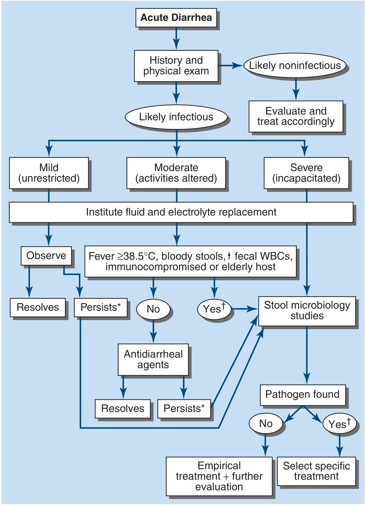
FIGURE 49-3 Algorithm for the management of acute diarrhea. [] = consider empirical metronidazole; [†] = consider empirical quinolone. - Harrison's Principles of Internal Medicine 22e*
Reading the Algorithm:
Acute Diarrhea
     ↓
History & Physical
     ↙           ↘
Likely Infectious    Likely Noninfectious → Evaluate & Treat
     ↓
Severity: Mild / Moderate / Severe
     ↓
FLUID & ELECTROLYTE REPLACEMENT (all patients)
     ↙                    ↘
Mild: Observe        Fever ≥38.5°C, bloody stools,
     ↓               ↑ fecal WBCs, immunocompromised, elderly
  Resolves?               ↙           ↘
    Yes → done         No            Yes
    No →          Antidiarrheal    Stool Microbiology Studies
             agents                      ↙             ↘
              ↓                   Pathogen         Pathogen
           Resolves?               NOT found        Found
           Yes → done               ↓                 ↓
           No → ↓          Empirical Tx +       Specific Tx
                    Stool microbiology   Further evaluation
Step 6 - Symptomatic (Non-Antibiotic) Treatment
Antimotility Agents
- Loperamide 4 mg initial dose, then 2 mg after each unformed stool (max 16 mg/day)
- Use only in nonfebrile, non-bloody diarrhea (moderate, watery)
- AVOID with:
- Febrile dysentery (prolongs illness)
- Bloody/mucoid stools
- Suspected EHEC O157:H7 or Shigella (may worsen HUS risk)
- C. difficile
Bismuth Subsalicylate
- 524 mg every 30-60 min as needed, up to 8 doses/day
- Useful for mild diarrhea and viral gastroenteritis; reduces nausea and stool frequency
- Avoid in: immunocompromised, renal impairment (bismuth encephalopathy risk)
- Contains salicylate - avoid in children (Reye syndrome risk)
Probiotics
- Not recommended as treatment for acute diarrhea (Goldman-Cecil, 2024)
Step 7 - Antibiotic Therapy
When to use antibiotics:
- Moderate-severe febrile diarrhea
- Bloody diarrhea (invasive pathogen confirmed or suspected - except EHEC)
- Traveler's diarrhea
- Immunocompromised patients (treat regardless of identified organism)
- Mechanical heart valves, recent vascular grafts, elderly
Empiric Regimens
| Indication | First-line | Alternative |
|---|---|---|
| Adults - general | Azithromycin 500 mg single dose or 500 mg/day x 3 days | Ciprofloxacin 500 mg q12h x 3-5 days |
| Traveler's diarrhea (noninvasive) | Rifaximin 200 mg TID x 3 days | Azithromycin 1000 mg single dose |
| Traveler's diarrhea (invasive/febrile) | Azithromycin (fluoroquinolone resistance is common in SE Asia) | Ciprofloxacin 500 mg q12h x 3 days (where resistance is low) |
| Suspected Giardia | Metronidazole 250 mg QID x 7 days | Nitazoxanide 500 mg bid x 3 days |
| Children - traveler's | Azithromycin 10 mg/kg (max 500 mg) single dose | - |
Pathogen-Specific Antibiotic Choices
| Pathogen | Treatment | Key Notes |
|---|---|---|
| Salmonella (non-typhoidal) | Usually self-limited; no routine antibiotics - prolongs excretion | Treat high-risk: infants <3 mo, immunocompromised, hemoglobinopathy, invasive disease → Ceftriaxone or Ciprofloxacin |
| Typhoid fever (S. Typhi) | Ceftriaxone 50-75 mg/kg/day OR Ciprofloxacin x 10-14 days | Multidrug resistance is common; check susceptibilities; Dexamethasone for delirium/shock |
| Shigella | Azithromycin 20 mg/kg/day (max 500 mg) | Ceftriaxone if unable to take oral; TMP-SMX/ampicillin often resistant |
| Campylobacter | Azithromycin 500 mg/day x 3 days | Can cause Guillain-Barré syndrome (ascending paralysis post-gastroenteritis) |
| EHEC (E. coli O157:H7) | NO ANTIBIOTICS - increases HUS risk | NO loperamide either; supportive care only |
| C. difficile | Stop offending antibiotic; Vancomycin PO or Fidaxomicin | Metronidazole for mild disease |
| Vibrio cholerae | ORS (massive losses); Doxycycline or Azithromycin single dose | IV rehydration often needed given massive secretory losses |
| Giardia | Metronidazole 250 mg QID x 7 days | Nitazoxanide as alternative |
| Cryptosporidium | Nitazoxanide 500 mg bid x 3 days | Immunocompromised: difficult to treat |
| E. histolytica (amebic dysentery) | Metronidazole + Paromomycin (to clear cysts) | Associated with liver abscesses |
Step 8 - When Diarrhea Persists (>7-14 days)
If diarrhea persists after initial evaluation/treatment:
- Reconsider stool studies: Giardia is the most common cause of persistent diarrhea
- Rule out C. difficile (especially if antibiotics recently given)
- Consider Cryptosporidium, Campylobacter, E. histolytica
- If >30 days (chronic): sigmoidoscopy/colonoscopy with biopsies and upper endoscopy with duodenal aspirates may be needed to exclude celiac disease, Crohn's disease, or microscopic colitis (do NOT scope during or immediately after acute enteritis - false positives)
Special Populations
| Population | Key Modifications |
|---|---|
| Infants/children | ORS is critical; zinc supplementation (resource-limited settings); Azithromycin preferred over fluoroquinolones; AVOID loperamide in young children |
| Elderly ≥70 years | High risk for dehydration and systemic spread; lower threshold for antibiotics and IV fluids |
| Immunocompromised | Always treat with antibiotics even without identified pathogen; consider broader workup including CMV |
| Pregnant | Azithromycin preferred; avoid fluoroquinolones; avoid bismuth subsalicylate |
| Traveler's diarrhea | Rifaximin first-line if noninvasive (not absorbed); Azithromycin if febrile/invasive; avoid TMP-SMX (widespread resistance) |
High-Yield "Avoid" Summary (Exam Favorite)
| Drug | Avoid When | Reason |
|---|---|---|
| Loperamide | EHEC, Shigella, febrile dysentery, bloody stools | Prolongs organism clearance; increases HUS risk with EHEC |
| Antibiotics | EHEC O157:H7 | Triggers Shiga toxin release → HUS |
| Bismuth subsalicylate | Immunocompromised, renal impairment, children | Encephalopathy risk; Reye syndrome risk |
| TMP-SMX | Traveler's diarrhea (empiric) | Widespread global resistance |
| Rifaximin | Invasive/febrile traveler's diarrhea | Not absorbed; no systemic activity |
Sources: Harrison's Principles of Internal Medicine 22e, Ch. 49; Goldman-Cecil Medicine (International Ed.); Goodman & Gilman's Pharmacological Basis of Therapeutics; Rosen's Emergency Medicine (Treatment of infectious diarrhea in children); Frameworks for Internal Medicine
Recent guideline note: A 2025 Chinese Pediatric Practice Guideline (PMID 40437180) for acute infectious diarrhea in children reinforces ORS as first-line with selective antibiotic use - consistent with current international standards.
This is a shared conversation. Sign in to Orris to start your own chat.