
I am suffering from hair thinning and hair fall what shall I do
hair loss alopecia treatment
androgenetic alopecia hair loss pattern
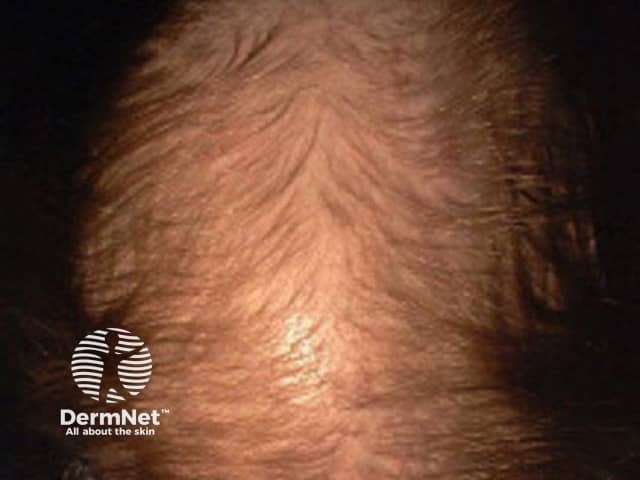
Clinical photography of the scalp in color (frontal-vertex view) illustrating female pattern alopecia. Primary subject: female pattern hair loss (androgenetic alopecia) in an adult patient. The image shows vertex-predominant thinning with the frontal hairline largely preserved. Hair density is reduced across the central scalp, with perifollicular thinning and apparent miniaturization resulting in finer, shorter hairs in affected regions. The occipital and temporal areas appear comparatively less involved. Overall pattern is typical for female pattern hair loss: diffuse, bilateral thinning with a central partition and crown involvement, often progressing gradually. The scalp skin demonstrates no scarring, inflammation, or pigmentary changes; there is no visible scarring alopecia. No magnification or dermoscopic technique is depicted. This photograph is valuable for education on androgenetic alopecia, assisting differentiation from telogen effluvium, alopecia areata, and other non-scarring alopecias when combined with history. It supports clinical diagnosis, informs prognosis, and aids in patient counseling about therapeutic options and expected outcomes. For research and teaching, the image serves as a representative example of typical FPHL pattern, enabling pattern recognition, grading of density changes, and longitudinal assessment after treatment or hormonal interventions. Any accompanying image metadata should include patient age range, photo date, and equipment used for reproducibility standards.
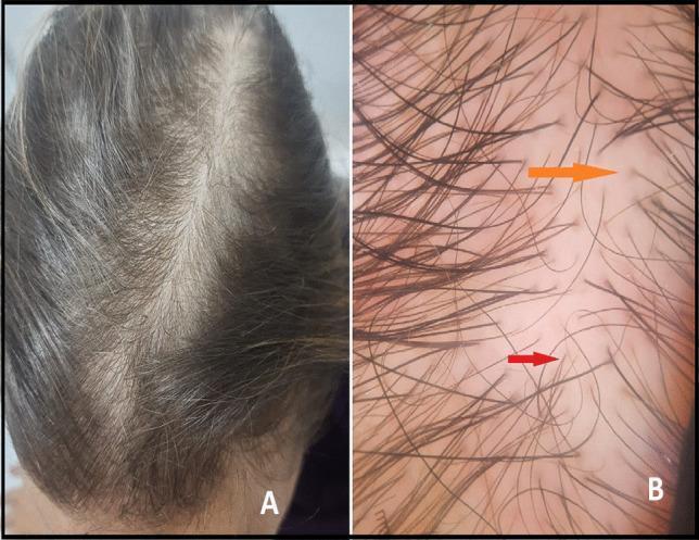
This composite educational figure illustrates the clinical and dermoscopic features of female pattern hair loss, specifically androgenetic alopecia. Image A is a clinical photograph of a 32-year-old female scalp showing a diffuse reduction in hair density. The thinning is most prominent across the vertex and mid-frontal regions, resulting in a widened central part and increased visibility of the underlying scalp. Image B is a high-magnification dermoscopy image of the same patient, highlighting diagnostic follicular signs. It demonstrates significant hair shaft diameter diversity (anisotrichosis), characterized by the coexistence of thick terminal hairs and thin vellus-like hairs. A red arrow points to a fine, non-pigmented vellus hair. An orange arrow indicates the 'peripilar sign,' which manifests as brown-gray perifollicular hyperpigmentation surrounding the hair shaft exit point. These visual markers are essential for distinguishing pattern alopecia from other forms of hair loss, such as telogen effluvium, in a clinical dermatology setting.
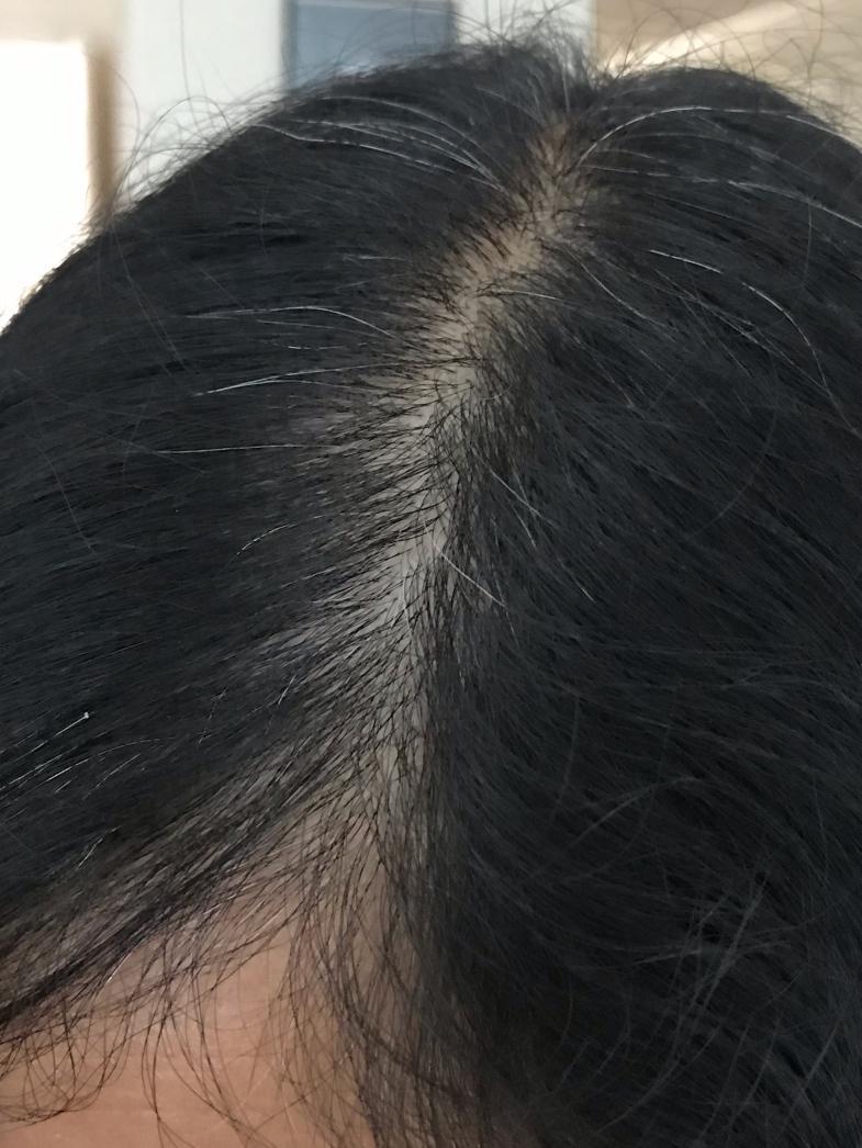
This clinical photograph shows a superior view of a human scalp and hair, demonstrating characteristic signs of female pattern hair loss (androgenetic alopecia). The primary clinical feature is a visible widening of the central part line, where there is a noticeable reduction in follicular density and thinning of hair shafts, leading to increased scalp exposure. The hair is predominantly dark with interspersed gray/white (canities) strands. The exposed scalp along the part line appears healthy, with a light skin tone and no immediate evidence of inflammation, scarring, or scaling. This visual presentation is typically used in dermatology to illustrate the progression of non-scarring alopecia and serves as a clinical reference for discussing treatment options, such as topical therapies or dietary supplements.
Hair Thinning and Hair Fall: Causes and What to Do
Understanding What's Happening
| Type | Who it affects | Pattern | Key feature |
|---|---|---|---|
| Androgenetic Alopecia (AGA) | Men & women | Patterned (temples/crown in men; central parting in women) | Genetic + hormonal |
| Telogen Effluvium (TE) | Anyone | Diffuse shedding all over | Triggered by stress, illness, diet |
| Alopecia Areata | Anyone | Patchy coin-shaped bald spots | Autoimmune |
| Traction Alopecia | More common in women | Hairline recedes where tension is applied | Tight hairstyles |
Step 1 - Find the Root Cause
- Low iron / ferritin (check iron saturation and serum ferritin)
- Hypothyroidism - one of the most overlooked causes; get TSH tested
- Protein deficiency (crash dieting is a very well-known trigger)
- Deficiencies of zinc, biotin, vitamin D
- Androgens (DHT) shrink hair follicles over time - the hallmark of AGA. The enzyme 5-alpha reductase converts testosterone to DHT, which progressively miniaturizes hair follicles (Dermatology 2-Volume Set 5e, p. 3159)
- PCOS in women
- Major surgery, high fever, childbirth, emotional shock - these cause a mass shift of hairs into "resting phase," which then shed 3-5 months later. In most cases this self-resolves within a few months once the trigger is removed.
- Blood thinners, beta-blockers, antidepressants, retinoids, some hormonal contraceptives
Step 2 - Get Tested
- CBC (complete blood count) - check for anaemia
- Serum ferritin (target >70 ng/mL for hair health)
- TSH (thyroid stimulating hormone)
- Fasting glucose / insulin (PCOS screen in women)
- Vitamin D, zinc, B12 levels
Step 3 - Evidence-Based Treatments
A. Medical / Topical Treatments
- The 5% topical solution/foam is FDA-approved for men; the 2% and 5% foam are approved for women too
- Works via vasodilatory and potassium-channel effects on hair follicles
- Also available as low-dose oral minoxidil (1-5 mg/day) - increasingly used
- Effects are modest but consistent; must be continued long-term (Goodman & Gilman's Pharmacology)
- 1 mg/day oral tablet - inhibits 5-alpha reductase, blocking DHT production
- Shown to slow hair loss and promote some regrowth
- Not for use in women of childbearing age
- Men with fewer CAG repeats in the androgen receptor gene respond better (Dermatology 5e, p. 3159)
- Spironolactone (anti-androgen) or low-dose oral minoxidil are common options
- Hormonal evaluation is important before starting
B. Nutritional Supplementation
- Correct iron deficiency with supplements (and find the source of blood loss if present)
- Ensure adequate protein intake (hair is ~95% keratin)
- Consider biotin (2500-5000 mcg/day), zinc, vitamin D if deficient
- Eat a varied, balanced diet - avoid crash dieting
C. Scalp and Hair Care Habits
- Use a mild, sulfate-free shampoo
- Avoid tight hairstyles (ponytails, braids) - these cause traction alopecia
- Minimize heat styling and chemical treatments
- Don't roughly towel-dry wet hair
- Gently massage the scalp - improves blood flow to follicles
D. Advanced / Clinic-Based Options
- Platelet-Rich Plasma (PRP): Injections of concentrated growth factors from your own blood. Evidence is mixed but some benefit shown; typically 3 monthly sessions are done
- Low-Level Laser / Photobiomodulation: A 2025 consensus review (Maghfour J et al., PMID 40253006) supports its use for hair loss
- Hair Transplantation: For men with severe AGA or women with localized loss; uses follicular unit grafts for a natural appearance. Long-term results are better when combined with minoxidil/finasteride post-op (Goldman-Cecil Medicine)
What Typical Female Pattern Hair Loss Looks Like
Step 4 - When to See a Doctor
- Hair loss is patchy (could be alopecia areata)
- The scalp is itchy, scaly, or inflamed
- Loss is rapid or accompanied by other symptoms (fatigue, weight change, menstrual irregularity)
- Hair is not recovering after 6 months of self-treatment