
Breast carcinoma
breast carcinoma histology pathology
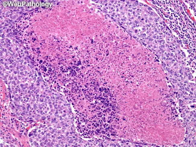
Bright-field light microscopy of an H&E-stained breast tissue section. The image captures parenchymal breast tissue with a high-grade, basal-like carcinoma component displaying geographic necrosis within tumor nests. Tumor cells are pleomorphic with hyperchromatic nuclei, conspicuous nucleoli, and scant cytoplasm arranged in solid sheets and cords; mitotic activity is brisk, contributing to the aggressive histology. The surrounding stroma shows infiltrating tumor cells at the margins, with limited luminal differentiation and a basal-type immunophenotype suggested by cytokeratin-positive basaloid cells (not shown here). This basal-like phenotype is characteristic of triple-negative breast cancers, which often lack estrogen receptor, progesterone receptor, and HER2 overexpression, and tend to behave aggressively with higher grade and poorer prognosis. The image exemplifies features used to distinguish basal-like breast carcinoma from luminal subtypes, such as geography-like necrosis and high mitotic index (>40 mitoses per 10 high-power fields) as described in published pathology resources. Knowledge of this histology supports diagnostic categorization, prognostic assessment, and therapeutic planning, including eligibility for neoadjuvant chemotherapy and enrollment in subtype-specific studies. In educational contexts, this image serves as a reference for recognizing basal-like morphology, comparing with serous ovarian tumor mutational patterns, and reinforcing the link between histology and molecular subtype stratification in breast cancer clinical practice.
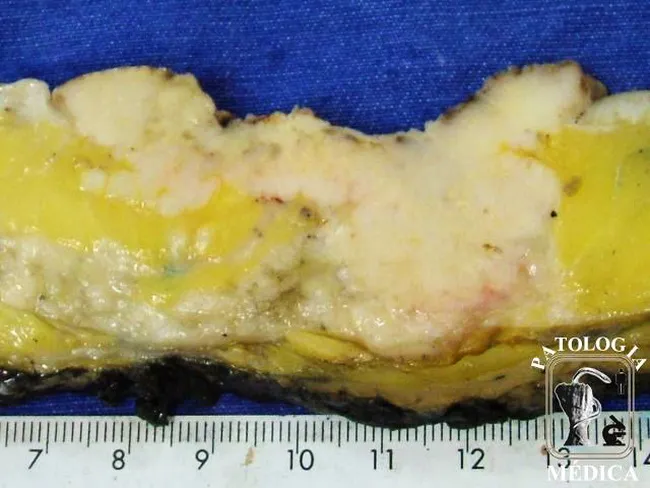
Gross pathology photograph of a human breast tissue specimen displaying a large, multifocal, high-grade invasive ductal carcinoma with extensive involvement of the overlying skin. Modality: gross pathology imaging; technique: en-face macroscopic view with scale ruler included, color in natural balance. The breast parenchyma shows multiple whitish, firm nodules interspersed within adipose tissue, with irregular, infiltrative margins. The skin over the lesion is visibly involved, exhibiting erythema and induration consistent with dermal invasion by tumor. No formal staining or contrast is applied in this gross photograph; histology would be required to confirm lymphovascular invasion. The lesion spans a substantial portion of the specimen and demonstrates heterogeneity in consistency and color, ranging from pale yellow to whitish fibrous areas. Clinically, this pattern corresponds to locally advanced breast cancer with cutaneous involvement, which has implications for staging (T4-like features), prognosis, and surgical planning (mastectomy versus breast-conserving approaches). This image serves educational purposes for trainees in pathology, oncology, breast surgery, and radiology, illustrating how invasive ductal carcinoma can present with extensive skin invasion on gross examination. Potential differential considerations include inflammatory breast carcinoma and other high-grade ductal neoplasms; correlate with imaging and biopsy for definitive diagnosis.
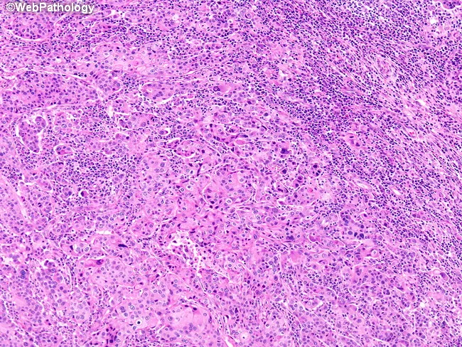
Imaging modality: Light microscopy using bright-field, Hematoxylin and Eosin (H&E) staining of a breast tissue section. Precise anatomical location: Mammary gland parenchyma, intratumoral mass within breast tissue; subtype: medullary carcinoma of the breast. Visual features: diffuse, sheet-like growth with broad cellular nests and anastomosing trabeculae; tumor cells with high-grade, pleomorphic nuclei and abundant eosinophilic cytoplasm; indistinct cytoplasmic borders producing a syncytial cytomorphology; lack of glandular differentiation and absence of an intraductal component. Microenvironment: prominent lymphoid and plasmacytic infiltrate both within and surrounding the tumor, creating a rich inflammatory milieu. Pathological interpretation: histology consistent with medullary carcinoma; margins and invasion assessed by surrounding tissue; phenotype often triple-negative with basal-like features; high cellularity contrasted by dense lymphoid infiltration. Diagnostic significance: recognition of characteristic syncytial architecture and inflammatory infiltrate supports diagnosis; prognosis is relatively favorable for medullary-type breast carcinomas compared with other triple-negative basaloid cancers; important for differential diagnosis with inflammatory/ductal carcinomas. Clinical relevance: guides treatment planning including surgery and systemic therapy; immunohistochemistry (ER/PR/HER2) and molecular profiling may clarify receptor status; imaging-pathology correlation essential for accurate classification and prognosis. In educational contexts, this image demonstrates classic medullary histology, aiding differential diagnosis awareness, grading schemes, and teaching concepts of tumor-infiltrating lymphocytes and syncytial morphology in breast pathology for students and clinicians.
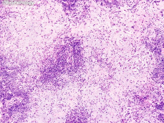
Imaging modality and technique: Light microscopy of a hematoxylin-eosin stained breast tissue section. Anatomical location: breast parenchyma; histology reveals a metaplastic carcinoma dominated by undifferentiated spindle cells arranged in fascicles with marked pleomorphism, high mitotic activity, and areas of chondrosarcomatous differentiation forming dense cartilaginous matrix. Rare foci show adenosquamous differentiation with epithelial glandular elements and squamous features embedded in the spindle stroma. The lesion demonstrates biphasic morphology comprising epithelial and mesenchymal components, characteristic of metaplastic breast carcinoma. Notable features include heterogeneous cellularity, loss of cohesion in spindle cells, abundant eosinophilic cartilaginous matrix, and invasion into surrounding breast stroma. Differential diagnosis includes primary breast sarcoma and ossifying/chondroid metaplasia vs metastatic disease. When such tumors metastasize, epithelial elements may predominate or mesenchymal elements may prevail; metastases to bone can resemble primary bone tumors, complicating diagnosis. Clinically, this entity is associated with aggressive behavior, triple-negative phenotype, and poor prognosis, often requiring comprehensive immunohistochemical profiling to identify epithelial markers (cytokeratins) within the sarcomatous areas. This image is relevant for educational purposes in surgical pathology, breast oncology, and histopathology training, illustrating the spectrum of heterologous differentiation in metaplastic breast carcinoma and its diagnostic challenges in distinguishing from primary bone tumors for pathology teaching purposes.
"breast cancer"[MeSH Terms] AND "treatment"[MeSH Terms]
Breast Carcinoma
Epidemiology
- Rare under age 25; incidence rises rapidly after age 30
- ~75% of cases are diagnosed after age 50
- Triple-negative and HER2-positive subtypes account for nearly half of breast cancers in young females, but <20% in older females
Risk Factors
| Category | Factor | Relative Risk |
|---|---|---|
| Modifiable | Obesity (BMI >30), postmenopausal | RR 1.29 |
| Nulliparity / first pregnancy >35 yr | Increased | |
| HRT use >10 years | RR 1.2 | |
| Alcohol (>4 drinks/day) | RR 1.46 | |
| Radiation exposure | RR 6 | |
| Tobacco (≥25 cig/day) | RR 1.14 | |
| Non-modifiable | Increasing age | Strong |
| Female sex | 100:1 (F:M) | |
| BRCA1/BRCA2 germline mutations | ~12% of all breast cancers | |
| Prior DCIS or LCIS | High | |
| Family history (1st degree) | RR 2-4 | |
| Early menarche / late menopause | Increased estrogenic exposure | |
| Dense breast tissue | Increased |
Classification
A. Histological Classification
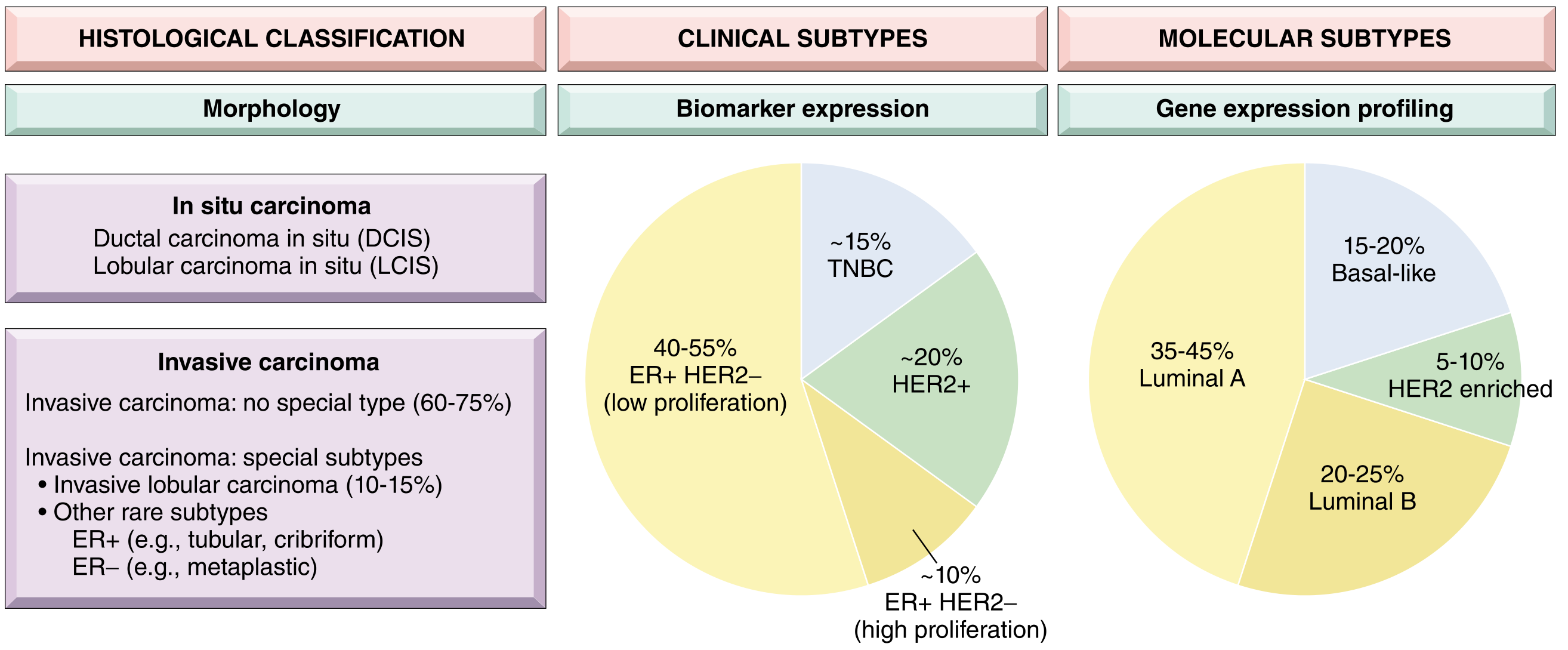
In Situ Carcinomas
- Proliferation of malignant epithelial cells confined within basement membrane
- Most often found on mammographic screening as clustered microcalcifications (seen in ~50% of non-palpable cancers)
- If untreated, ~30-50% progress to invasive ductal carcinoma in the same breast
- Subtypes: comedo, cribriform, micropapillary, papillary, solid
- A risk marker and precursor lesion; usually an incidental finding
- If carcinoma develops: 2/3 in the same breast, 1/3 in the contralateral breast
- Associated with E-cadherin loss
Invasive Carcinomas (Foote & Stewart Classification)
| Type | Frequency |
|---|---|
| Invasive ductal carcinoma - No Special Type (NST) | ~75-80% |
| Invasive lobular carcinoma | 10-15% |
| Medullary carcinoma (now reclassified as NST with medullary pattern) | ~4% |
| Mucinous (colloid) carcinoma | ~2% |
| Papillary carcinoma | ~2% |
| Tubular carcinoma | ~2% |
| Paget's disease of the nipple | Rare |
| Rare types (adenoid cystic, squamous, metaplastic, apocrine) | <1% |
- Invasive ductal carcinoma (NST): Most common; hard, irregular, radiodense mass; desmoplastic stromal reaction; characteristic grating sound on cut surface. Generally worse prognosis than special types.
- Invasive lobular carcinoma: Single-file ("Indian-file") infiltration pattern; loss of E-cadherin; bilateral risk; may be mammographically occult.
- Mucinous (colloid) carcinoma: Well-circumscribed; good prognosis; gelatinous gross appearance; pools of mucin with floating tumor cells.
- Tubular carcinoma: Well-differentiated; excellent prognosis; haphazard tubules with open lumina.
- Paget's disease of the nipple: Chronic eczematous nipple eruption; pathognomonic large pale vacuolated cells (Paget cells) in the rete pegs; associated with underlying DCIS or invasive cancer; differentiated from melanoma by CEA staining (vs S-100).
- Inflammatory breast carcinoma: Skin erythema, peau d'orange, warmth, tenderness; tumor emboli in dermal lymphatics; T4d in staging; aggressive, poor prognosis.
B. Clinical Subtypes (Biomarker-Based)
| Subtype | ER/PR | HER2 | Frequency | Characteristics |
|---|---|---|---|---|
| Luminal (ER+/HER2-) - low proliferation | + | - | 40-55% | Best prognosis; hormone-sensitive |
| Luminal (ER+/HER2-) - high proliferation | + | - | ~10% | Intermediate; Ki-67 high |
| HER2-positive | +/- | + | ~20% | Aggressive; responds to anti-HER2 therapy |
| Triple-Negative (TNBC) | - | - | ~15% | Aggressive; no targeted therapy; basal-like overlap |
C. Molecular (Intrinsic) Subtypes (Gene Expression Profiling)
| Subtype | Frequency | Key Features |
|---|---|---|
| Luminal A | 35-45% | Low grade, ER+, low Ki-67, best prognosis |
| Luminal B | 20-25% | Higher grade, ER+, higher Ki-67, worse prognosis than Luminal A |
| HER2-enriched | 5-10% | HER2 amplified, ER-, aggressive |
| Basal-like | 15-20% | ER-/PR-/HER2-, BRCA1-associated, high grade, TNBC overlap |
| Normal-like | Rare | Good prognosis |
| Claudin-low | Rare | Stem cell-like, poor prognosis |
Histologic Grading (Nottingham Score)
- Tubule formation (glandular differentiation)
- Nuclear pleomorphism
- Mitotic rate
- Grade 1 (3-5): Well differentiated - small uniform nuclei, tubular/cribriform growth
- Grade 2 (6-7): Moderately differentiated - solid clusters, greater pleomorphism
- Grade 3 (8-9): Poorly differentiated - ragged nests, enlarged irregular nuclei, necrosis
Pathology / Morphology
- Invasive ductal NST: irregular nests, cords, single cells in fibrous stroma
- Invasive lobular: single-file pattern, target-like arrangement around ducts
- Special types: defined by their characteristic architecture (see above)
Staging (AJCC 8th Edition - Anatomic)
| Stage | Tumor (T) | Nodes (N) | Metastasis (M) | 10-yr Survival |
|---|---|---|---|---|
| 0 | DCIS | No metastases | Absent | 97% |
| I | Invasive ≤2 cm | None or micrometastases | Absent | 87% |
| II | >2 cm or >2-5 cm | 1-3 positive LNs or 0-3 | Absent | 65% |
| III | >5 cm / any / skin/chest wall / inflammatory | Variable | Absent | 40% |
| IV | Any | Any | Present | ~5% |
- Supraclavicular LN metastases = Stage III (not IV), per current system
- Axillary LN involvement remains the single most important prognostic factor
Prognostic & Predictive Biomarkers
| Biomarker | Role |
|---|---|
| ER/PR | Predicts response to hormonal therapy (tamoxifen, aromatase inhibitors) |
| HER2 | Predicts response to trastuzumab, pertuzumab, T-DM1, lapatinib |
| Ki-67 | Proliferation index; distinguishes Luminal A from B |
| BRCA1/2 | Predicts PARP inhibitor response (olaparib, talazoparib) |
| PIK3CA mutation | Predicts response to PI3K inhibitors (alpelisib) in ER+/HER2- |
| PD-L1 | Predicts benefit from immune checkpoint inhibitors in TNBC |
| Multigene assays (Oncotype DX, MammaPrint) | Guide chemotherapy decisions in early-stage ER+/HER2- |
| Circulating tumor DNA (ctDNA) | Emerging: detects emerging actionable mutations |
Diagnosis & Screening
- Reduces breast cancer mortality by 20-25% in women ≥50 years
- Only 20% of non-palpable cancers have axillary LN metastases (vs 50% of palpable tumors)
- NCNN guidelines: breast exam every 3 years for women ≥20; annual mammogram from age 40
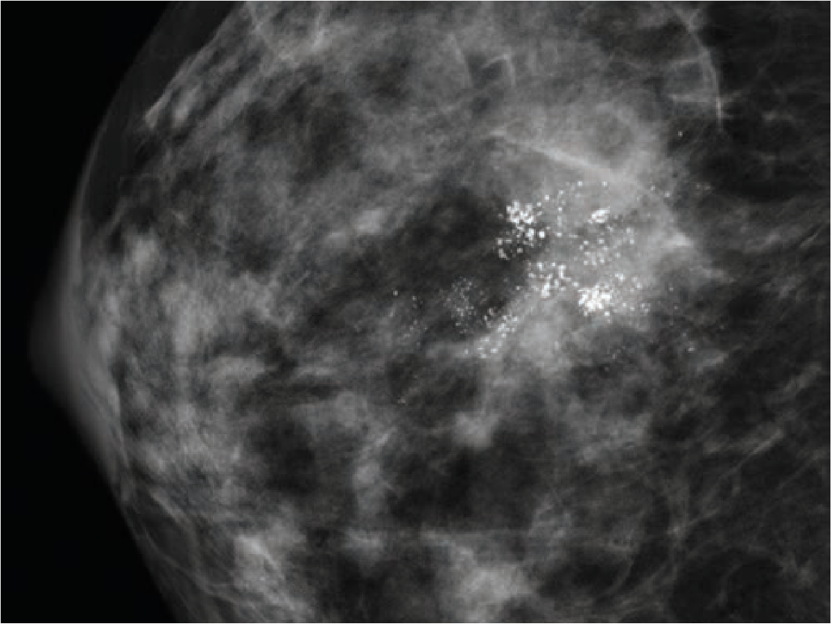
- Solid spiculated mass with or without stellate margins
- Asymmetric thickening
- Clustered microcalcifications (fine, stippled)
- Skin tethering
Management
Surgery
- Locally advanced disease
- Multiple tumors (multifocal/multicentric)
- Patient preference
- BRCA mutations (prophylactic contralateral mastectomy discussion)
- Inflammatory breast carcinoma (usually after neoadjuvant chemotherapy)
Systemic Therapy
| Subtype | Treatment |
|---|---|
| ER+/HER2- | Endocrine therapy (tamoxifen premenopausal; aromatase inhibitors postmenopausal) ± CDK4/6 inhibitors; chemotherapy if high-risk |
| HER2+ | Anti-HER2 therapy (trastuzumab, pertuzumab, T-DM1, trastuzumab deruxtecan) + chemotherapy |
| Triple-Negative | Chemotherapy (anthracyclines + taxanes); immunotherapy (pembrolizumab for PD-L1+); PARP inhibitors if BRCA mutated |
| Metastatic (all types) | Palliative systemic therapy; bisphosphonates/denosumab for bone metastases |
Radiation Therapy
- After BCS: whole breast irradiation ± regional nodal irradiation
- After mastectomy: post-mastectomy RT for high-risk features (≥4 positive LNs, T3/T4 tumors)
Neoadjuvant Therapy
- Used to downstage locally advanced or large tumors before surgery
- Pathologic complete response (pCR) after neoadjuvant therapy is a strong prognostic marker
- Inflammatory breast carcinoma is treated with neoadjuvant chemotherapy followed by mastectomy + RT
Metastatic Pattern
Special Subtypes
Male Breast Carcinoma
- <1% of all breast cancers
- Usually presents as a painless subareolar mass
- Risk factors: Klinefelter syndrome, BRCA2 mutation, exogenous estrogens, gynecomastia
- Usually ER/PR positive; treated similarly to female breast cancer with mastectomy + systemic therapy
Inflammatory Breast Carcinoma
- Rapid onset skin erythema, peau d'orange, warmth - resembles mastitis
- Dermal lymphatic tumor emboli are the pathological hallmark
- Classified as T4d regardless of tumor size; Stage III minimum
- Treated with neoadjuvant chemotherapy → modified radical mastectomy → radiation
Key Teaching Points (Summary)
- Most common malignancy in females globally; 1 in 8 lifetime risk
- BRCA1/2 mutations account for ~50% of hereditary cases; BRCA1 → basal-like/TNBC; BRCA2 → luminal
- Ductal carcinoma NST = 75-80% of invasive cancers; carries worst prognosis among common subtypes
- Three-tier clinical classification (ER+/HER2-, HER2+, TNBC) drives treatment selection
- Six molecular intrinsic subtypes by gene expression; Luminal A = best prognosis; basal-like = worst
- Nottingham grade (tubules + nuclei + mitoses) is a strong independent prognostic factor
- Axillary LN status remains the single most important prognostic factor
- AJCC 8th edition integrates anatomic + biologic staging for prognostic stage
- BCS + radiation = equivalent to mastectomy in survival; SLNB is standard for node-negative staging
- DCIS → microcalcifications on mammography; LCIS → bilateral risk marker
- Robbins, Cotran & Kumar - Pathologic Basis of Disease (Figs. 23.15, 23.20, Table 23.6)
- Robbins & Kumar Basic Pathology (p. 719)
- Schwartz's Principles of Surgery, 11th Edition (Ch. 17)
- Bailey and Love's Short Practice of Surgery, 28th Edition (Ch. 58, Table 58.3)