Here is a complete, structured overview of the Anterior Compartment of the Thigh.
Anterior Compartment of Thigh
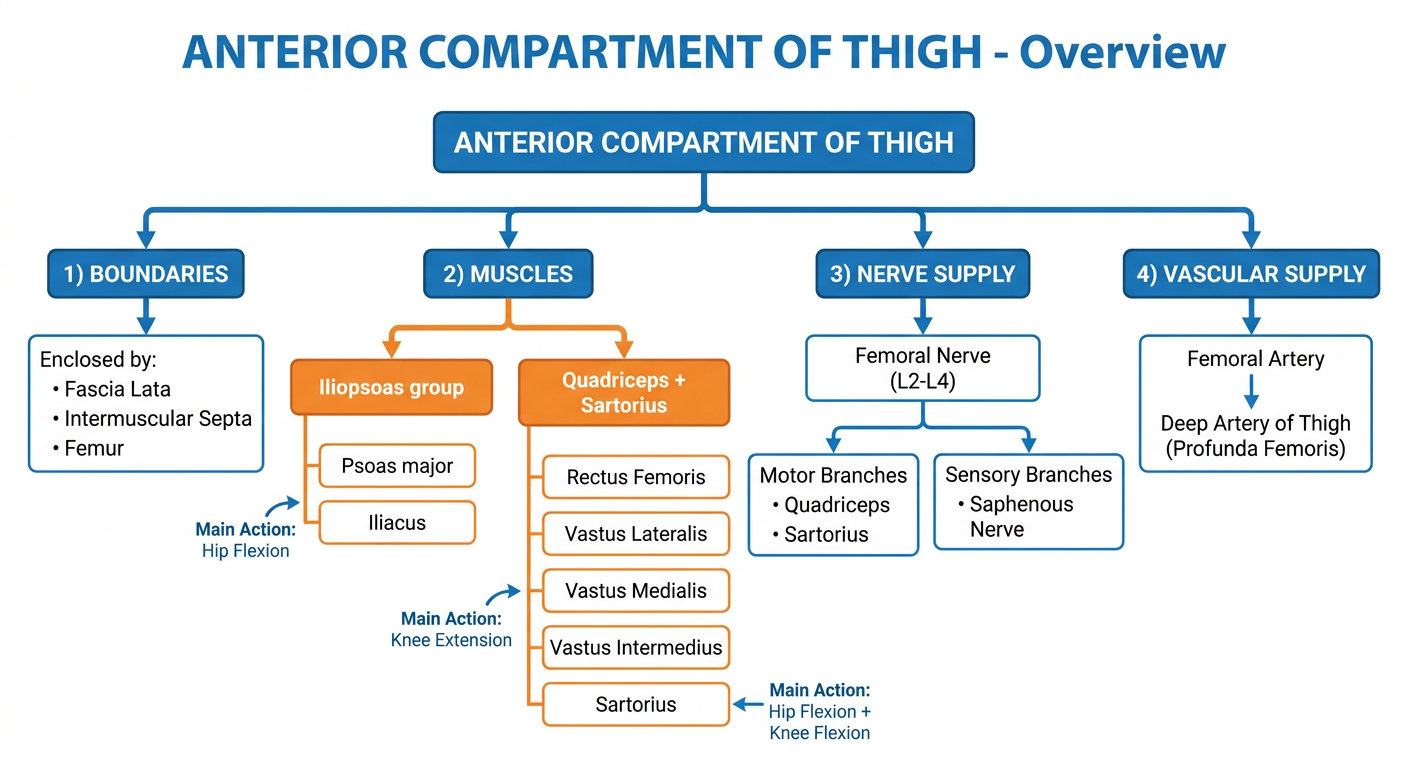
Flowchart Overview
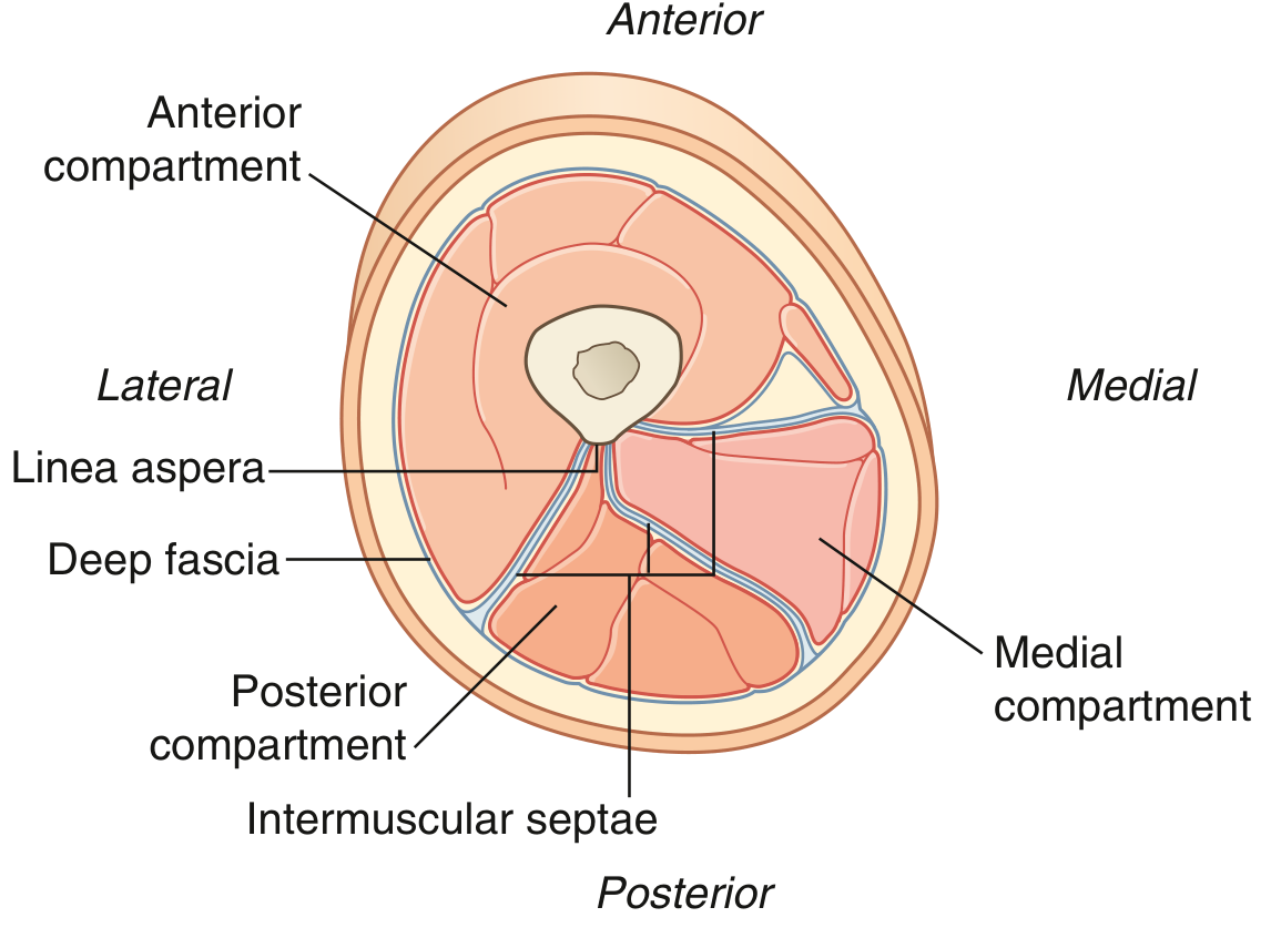
Cross-sectional Anatomy
The thigh is divided into three compartments by intermuscular septa running between the femur and the investing fascia lata:
Fig. 6.57 - Transverse Section Through the Midthigh (Gray's Anatomy for Students)
Muscles
The anterior compartment contains 6 muscles (7 if you count psoas and iliacus separately):
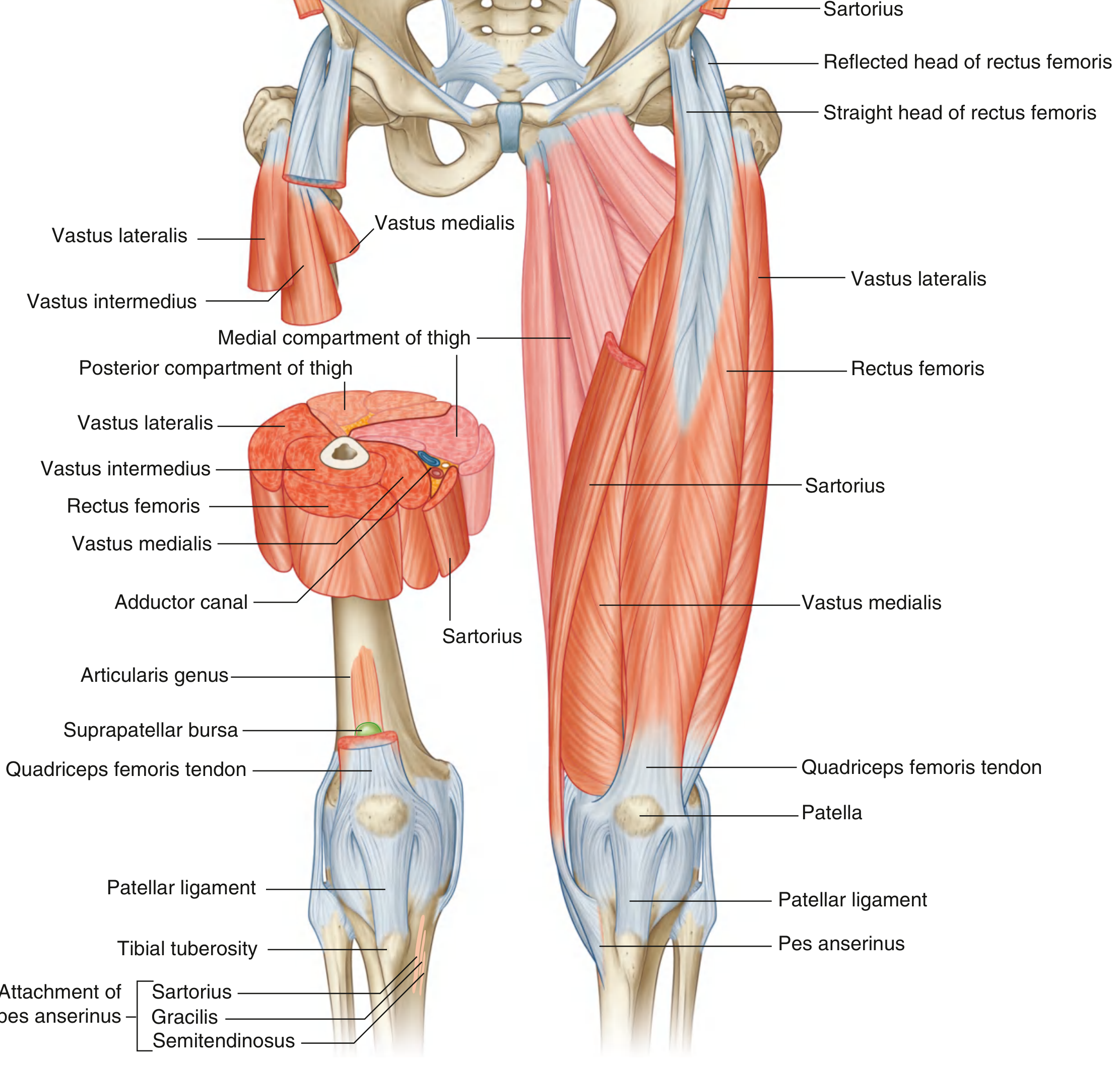
Fig. 6.59 - Muscles of the Anterior Compartment of the Thigh (Gray's Anatomy for Students)
Group 1: Iliopsoas (hip flexors entering from abdomen)
| Muscle | Origin | Insertion | Innervation | Action |
|---|
| Psoas major | Lumbar vertebrae (T12-L5), intervertebral discs, transverse processes | Lesser trochanter of femur | Anterior rami L1, L2, L3 | Flexes thigh at hip |
| Iliacus | Iliac fossa (posterior abdominal wall) | Lesser trochanter of femur | Femoral nerve (L2, L3) | Flexes thigh at hip |
These two muscles travel together under the inguinal ligament and share a common tendon - together called the iliopsoas, the primary hip flexor.
Group 2: Quadriceps Femoris (the "four-headed" extensor)
| Muscle | Origin | Insertion | Innervation | Action |
|---|
| Rectus femoris | Straight head: anterior inferior iliac spine; Reflected head: ilium above acetabulum | Quadriceps femoris tendon → patella → patellar ligament → tibial tuberosity | Femoral nerve (L2, L3, L4) | Flexes hip + extends knee |
| Vastus lateralis | Lateral intertrochanteric line, greater trochanter, lateral linea aspera | Lateral margin of patella, quadriceps tendon | Femoral nerve (L2, L3, L4) | Extends knee |
| Vastus medialis | Medial intertrochanteric line, pectineal line, medial linea aspera | Medial border of patella, quadriceps tendon | Femoral nerve (L2, L3, L4) | Extends knee; stabilizes patella |
| Vastus intermedius | Upper 2/3 of anterior and lateral femur surfaces | Deep aspect of quadriceps tendon, lateral condyle of tibia | Femoral nerve (L2, L3, L4) | Extends knee |
All four quadriceps converge on the quadriceps femoris tendon, which wraps around the patella and continues as the patellar ligament to the tibial tuberosity. Testing the patellar reflex (knee jerk) primarily tests L3-L4.
Group 3: Sartorius (the "tailor's muscle")
| Muscle | Origin | Insertion | Innervation | Action |
|---|
| Sartorius | Anterior superior iliac spine (ASIS) | Medial surface of tibia just below tibial tuberosity (pes anserinus) | Femoral nerve (L2, L3) | Flexes thigh + flexes leg at knee; assists lateral rotation of hip |
Sartorius is the longest muscle in the body. It forms the lateral border of the femoral triangle.
Nerve Supply
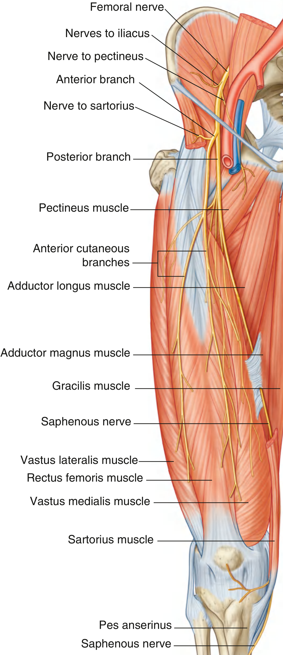
Fig. 6.69 - Femoral Nerve (Gray's Anatomy for Students)
The femoral nerve (L2-L4) is the sole nerve of the anterior compartment:
- Arises from the lumbar plexus, enters the thigh by passing under the inguinal ligament, lateral to the femoral artery (outside the femoral sheath)
- Before entering the thigh: supplies iliacus and pectineus
- Divides immediately into anterior and posterior branches:
- Anterior branch: motor to sartorius; sensory (anterior cutaneous branches) to front of thigh
- Posterior branch: motor to all four quadriceps; gives off the saphenous nerve (L3-L4) - the longest branch, supplying skin on the medial side of the leg and foot as far as the great toe
Vascular Supply
The primary arterial supply is via the femoral artery (continuation of external iliac artery below the inguinal ligament):
Femoral Artery
├── Direct muscular branches → anterior compartment muscles
└── Deep Artery of Thigh (Profunda Femoris)
├── Lateral circumflex femoral artery → vastus lateralis, rectus femoris
├── Medial circumflex femoral artery → femoral neck, hip joint
└── Perforating arteries → communicate with posterior compartment
The femoral artery passes through the femoral triangle, then travels in the adductor canal (subsartorial canal) before exiting posteriorly through the adductor hiatus as the popliteal artery.
Quick Summary Table
| Feature | Details |
|---|
| Boundary | Fascia lata + intermuscular septa + femur |
| Primary function | Extend knee; flex hip |
| Muscles | Iliopsoas (2) + Quadriceps (4) + Sartorius (1) = 7 |
| Nerve | Femoral nerve (L2-L4) |
| Artery | Femoral + Profunda femoris |
| Key reflex | Knee jerk (L3-L4) - tests quadriceps |
| Clinical note | Compartment syndrome of anterior thigh occurs with trauma/hemorrhage; requires fasciotomy via lateral incision |
Source: Gray's Anatomy for Students, Chapter 6