
Elisa
Teach me ELISA
ELISA enzyme-linked immunosorbent assay diagram types
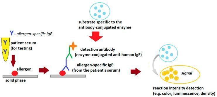
This medical diagram illustrates the general scheme of an indirect Enzyme-Linked Immunosorbent Assay (ELISA) used to detect allergen-specific IgE antibodies in human serum. The process is depicted in four sequential stages: (1) Sample Introduction: Patient serum containing allergen-specific IgE (yellow Y-shaped molecules) is added to a solid phase pre-coated with allergens (red circles). (2) Binding: The specific IgE antibodies bind to the immobilized allergens. (3) Detection: An enzyme-conjugated anti-human IgE detection antibody is introduced, forming a complex with the patient's IgE. This detection antibody is represented by a multi-colored structure topped with an orange star signifying the enzyme. (4) Signal Generation: A specific substrate is added, which the enzyme catalyzes into a detectable signal, visually represented by a transition from light blue circles to a yellow oval. The diagram highlights the clinical utility of ELISA in immunology and allergy diagnostics, specifically for quantifying reaction intensity via colorimetry, luminescence, or density changes.
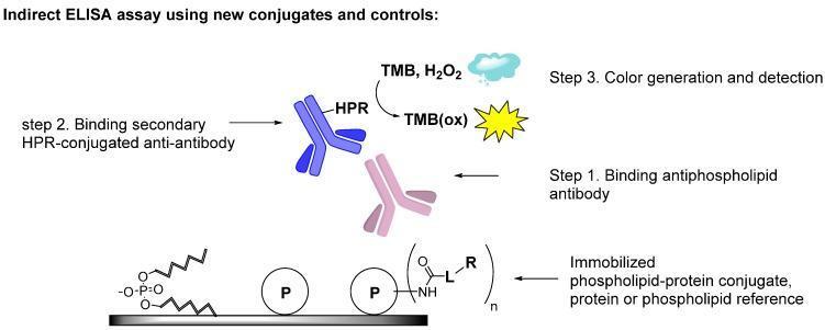
This diagram illustrates the methodology of an indirect Enzyme-Linked Immunosorbent Assay (ELISA) designed for the detection of antiphospholipid antibodies, relevant to the diagnosis of Antiphospholipid Syndrome (APS). The workflow is divided into three sequential steps. Step 1 demonstrates the immobilization of antigens on a solid surface, including phospholipids, proteins (labeled 'P'), and phospholipid-protein conjugates formed via a chemical linker ('L'). A primary antiphospholipid antibody (pink Y-shape) binds to these immobilized antigens. Step 2 shows the introduction of a secondary anti-antibody (blue Y-shape) conjugated with Horseradish Peroxidase (HPR). Step 3 depicts the signal detection phase where HPR catalyzes the reaction between TMB and H2O2 to form oxidized TMB (TMB(ox)), producing a measurable color change. This educational schematic emphasizes the use of stable covalent conjugates to improve the diagnostic sensitivity and reproducibility of assays detecting autoantibodies against complex phospholipid-protein epitopes.
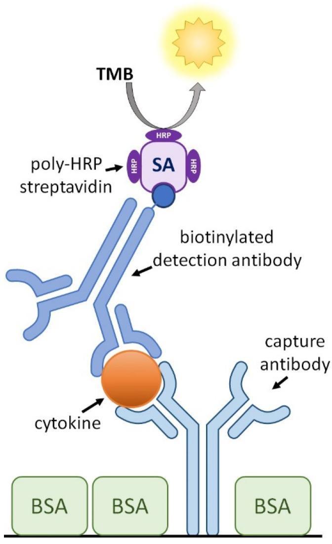
This medical diagram illustrates the schematic configuration of a cytokine capture Enzyme-Linked Immunosorbent Assay (ELISA), a diagnostic tool used in immunology. The process is depicted through a vertical assembly of molecular components. At the foundation, a capture antibody is anchored to a surface blocked by bovine serum albumin (BSA), shown as green rectangular blocks. This capture antibody is bound to a specific target molecule, a cytokine, represented as an orange sphere. Above the cytokine, a biotinylated detection antibody is attached, creating a sandwich complex. The biotin tag (dark blue circle) on the detection antibody is bound to a streptavidin (SA) molecule, which is conjugated with multiple horse radish peroxidase (HRP) enzymes (poly-HRP). At the apex, the HRP catalyzes a reaction with the substrate 3,3’,5,5’-Tetramethylbenzidine (TMB), resulting in a visual or chemical signal, depicted by a yellow light icon. This infographic effectively demonstrates the sequential binding and signal amplification logic used in laboratory medicine to quantify proteins such as IL-2, IL-6, or IFN-γ.
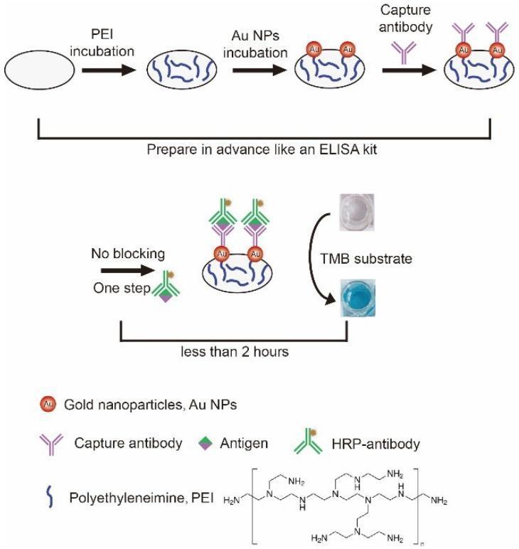
This schematic diagram illustrates a specialized, blocking-free 'sandwich' Enzyme-Linked Immunosorbent Assay (ELISA) utilizing a polyethyleneimine (PEI) and gold nanoparticle (Au NP) coated microwell plate. The process is divided into two phases: preparation and detection. In the preparation phase, a microwell undergoes PEI incubation followed by Au NP incubation to form a functionalized base layer; capture antibodies are then immobilized onto the Au NPs. In the detection phase, labeled as a 'No blocking - One step' procedure, the antigen and horseradish peroxidase (HRP)-conjugated antibody are introduced. The final step shows the addition of TMB substrate, which undergoes a visual transition from a colorless liquid to a blue-colored solution, indicating successful antigen detection. The diagram highlights that this modified protocol simplifies traditional ELISA by omitting the 2-hour blocking step, reducing total assay time to less than 2 hours. Chemical structures for PEI and icons for antigens, Au NPs, and various antibodies are included to clarify the molecular binding sequence and surface modifications.
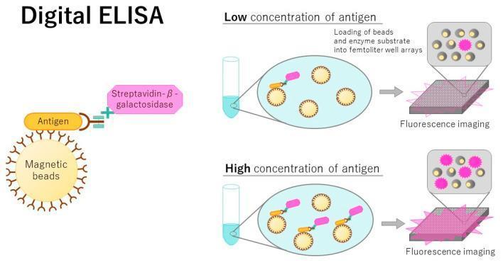
This pathophysiology diagram illustrates the principles of a Digital ELISA (Enzyme-Linked Immunosorbent Assay), an ultrasensitive diagnostic technique for single-molecule detection. The left panel details the molecular complex: an antibody-conjugated magnetic bead captures a target antigen, which is then bound by a secondary antibody labeled with Streptavidin-β-galactosidase. The right panel demonstrates the workflow and comparative analysis for low versus high antigen concentrations. Initially, mixed complexes are loaded into a femtoliter-volume microwell array designed to isolate single beads. Following the addition of a fluorogenic substrate, the array is analyzed via fluorescence imaging. In 'Low concentration' scenarios, most wells remain dark as beads lack the target antigen; in 'High concentration' scenarios, a larger percentage of wells exhibit fluorescent signals (pink bursts). The concentration of the target protein in the clinical sample is quantitatively determined by the ratio of 'on' (fluorescent) to 'off' (non-fluorescent) wells, enabling much higher sensitivity than conventional bulk ELISAs for detecting trace biomarkers.
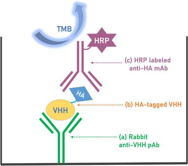
This medical laboratory diagram illustrates a sandwich Enzyme-Linked Immunosorbent Assay (ELISA) technique used for the detection of VHH (variable heavy-chain antibodies). The schematic depicts the spatial arrangement of the assay components within a microtiter plate well from bottom to top: (a) A capture layer consisting of Rabbit anti-VHH polyclonal antibodies (pAb), which are passively adsorbed to the bottom of the well. (b) The analyte, represented as an HA-tagged VHH molecule, where the VHH core binds to the capture antibody and an HA (hemagglutinin) tag is available for detection. (c) The detection layer featuring an HRP-labeled anti-HA monoclonal antibody (mAb). This antibody targets the HA tag of the analyte and is conjugated to Horseradish Peroxidase (HRP), shown as a purple star-shaped molecule. At the top, a blue curved arrow labeled TMB (3,3′,5,5′-Tetramethylbenzidine) represents the chromogenic substrate added to the well. The diagram demonstrates the biochemical principle of signal transduction where HRP catalyzes the oxidation of TMB to produce a measurable colorimetric signal, indicating the presence and concentration of the VHH analyte.
ELISA - Enzyme-Linked Immunosorbent Assay
What is ELISA?
Core Principle
- At least one reagent must be available in a pure, detectable form.
- There must be a way to separate bound from unbound labeled reagent - achieved by trapping one partner on a solid surface and washing away the rest.
Basic ELISA Steps (Direct ELISA)
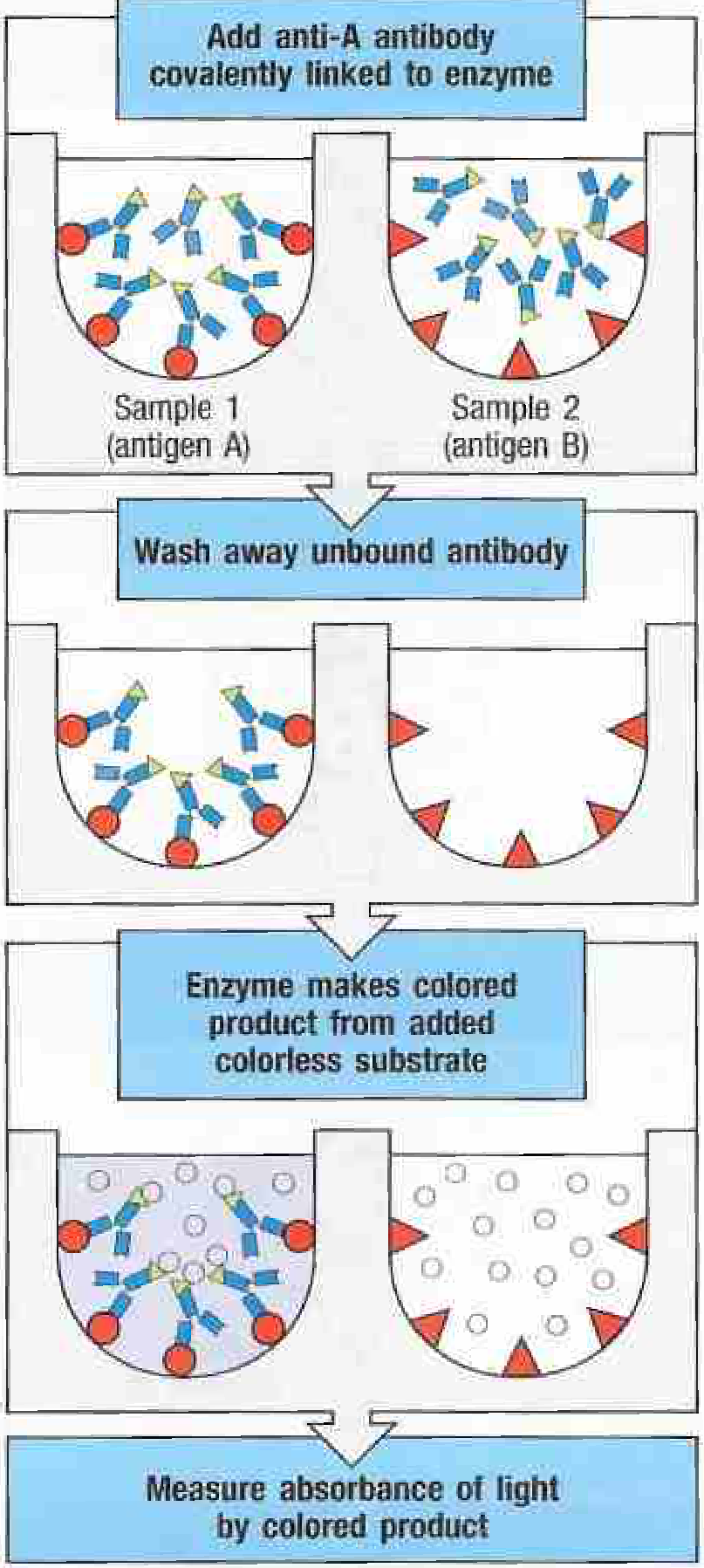
| Step | What Happens |
|---|---|
| 1. Coat the well | The test sample (antigen) is coated onto plastic microtiter well surfaces - proteins bind nonspecifically to plastic. |
| 2. Block | Irrelevant proteins are added to block remaining sticky sites on the plastic (prevents false positives). |
| 3. Add labeled antibody | A purified antibody specific for the antigen is added. This antibody is covalently linked to an enzyme (e.g., horseradish peroxidase, HRP). |
| 4. Wash | Unbound antibody is washed away. Only antibody bound to the target antigen remains. |
| 5. Add substrate | A colorless enzyme substrate is added. The enzyme converts it into a colored product. |
| 6. Read absorbance | The intensity of color is measured by a spectrophotometer - proportional to the amount of antigen present. |
Types of ELISA
1. Direct ELISA
- Antigen coated to the plate
- Enzyme-labeled antibody added directly
- Simple but less sensitive
2. Indirect ELISA
- Antigen coated to plate
- Primary (unlabeled) antibody added first, binds antigen
- Enzyme-labeled secondary anti-immunoglobulin antibody added
- Signal is amplified because multiple secondary antibodies bind each primary antibody
- Used for HIV diagnosis (detect patient's antibodies against HIV antigens)
3. Sandwich (Capture) ELISA
- Antibody (not antigen) is coated to the plate first
- Antigen from the sample binds to the captured antibody
- A second labeled antibody (recognizing a different epitope) detects bound antigen
- Excellent for detecting secreted proteins like cytokines even at very low concentrations
- Higher sensitivity than direct ELISA
4. Competitive Inhibition ELISA
- Used when you want to measure an antigen in a sample of unknown composition
- Known labeled antigen and unknown test antigen compete for the same antibody
- More test antigen present = less labeled antigen binds = less color signal
- Result is inversely proportional to test antigen concentration
Common Enzymes & Substrates
| Enzyme | Substrate | Color Produced |
|---|---|---|
| Horseradish Peroxidase (HRP) | TMB (tetramethylbenzidine) + H₂O₂ | Blue → Yellow (after stop solution) |
| Alkaline Phosphatase (AP) | p-Nitrophenyl phosphate (pNPP) | Yellow |
Key Advantages Over RIA
| Feature | ELISA | RIA |
|---|---|---|
| Label | Enzyme | Radioactive isotope (¹²⁵I) |
| Safety | No radiation hazard | Radiation hazard |
| Equipment | Simple plate reader | Gamma counter |
| Sensitivity | Very high (comparable to RIA) | Highest |
| Applications | Diagnostics, research | Hormone levels in blood |
Clinical Applications
- HIV diagnosis - detect anti-HIV antibodies in patient serum (indirect ELISA)
- Viral diagnostics - COVID-19 serology, Hepatitis B/C
- Cytokine quantification - measure IL-6, TNF-alpha (sandwich ELISA)
- Allergy testing - detect allergen-specific IgE
- Pregnancy tests - detect hCG
- Drug level monitoring - measure drug concentrations in serum
Summary Mnemonic: "CWABS"
- Coat the well
- Wash after blocking
- Add labeled antibody
- Bind (let it happen)
- Signal = color readout