
Lab Report Summary - Al-Shifa Clinical Laboratory (08 Aug 2026)
Test 1: Serum Uric Acid
| Result | 7.8 mg/dL (FLAGGED HIGH ⚠️) |
| Reference Range | 2.5 - 7.0 mg/dL |
| Status | Elevated (Hyperuricemia) |
- Uric acid crystallizes as monosodium urate and deposits in joints, potentially causing gout (acute painful arthritis, classically in the big toe)
- At levels above 7 mg/dL, the risk of gout increases, though not everyone with hyperuricemia develops gout
- Persistent elevation can lead to uric acid kidney stones or, over the long term, kidney damage
- High-purine diet (red meat, organ meats, shellfish, beer)
- High intake of sugar-sweetened beverages / fructose
- Obesity
- Dehydration
- Medications (thiazide diuretics, low-dose aspirin)
- Reduced kidney excretion
| Action | Details |
|---|---|
| Avoid | Organ meats (liver, kidney), high-fructose drinks, alcohol overuse |
| Limit | Red meat (beef, pork, lamb), shellfish, beer |
| Encourage | Low-fat dairy products, plenty of water (stay well-hydrated) |
| General | Weight loss if overweight, regular exercise, smoking cessation |
- Repeat uric acid test in 4-6 weeks after dietary changes
- The doctor may check kidney function (creatinine, eGFR) and urine uric acid
- At 7.8 mg/dL without symptoms, medication (like allopurinol) is generally not recommended yet - lifestyle changes are the first step
- If the patient develops joint pain/swelling, immediate medical evaluation is needed
Test 2: Total Calcium
| Result | 8.7 mg/dL |
| Reference Range | 8.5 - 10.5 mg/dL |
| Status | Normal ✅ |
Summary for the Patient
| Test | Result | Normal? |
|---|---|---|
| Serum Uric Acid | 7.8 mg/dL | ❌ Slightly High |
| Total Calcium | 8.7 mg/dL | ✅ Normal |
- See a doctor - share these results, especially if you have any joint pain or back/flank pain (possible kidney stones)
- Drink more water - aim for 2-3 liters daily
- Reduce purine-rich foods - cut back on red meat, organ meats, shellfish, and sugary sodas
- Avoid alcohol - especially beer, which strongly raises uric acid
- Recheck uric acid in 4-6 weeks after dietary changes
how to check kidney stones
kidney stone CT scan ureteral calculus
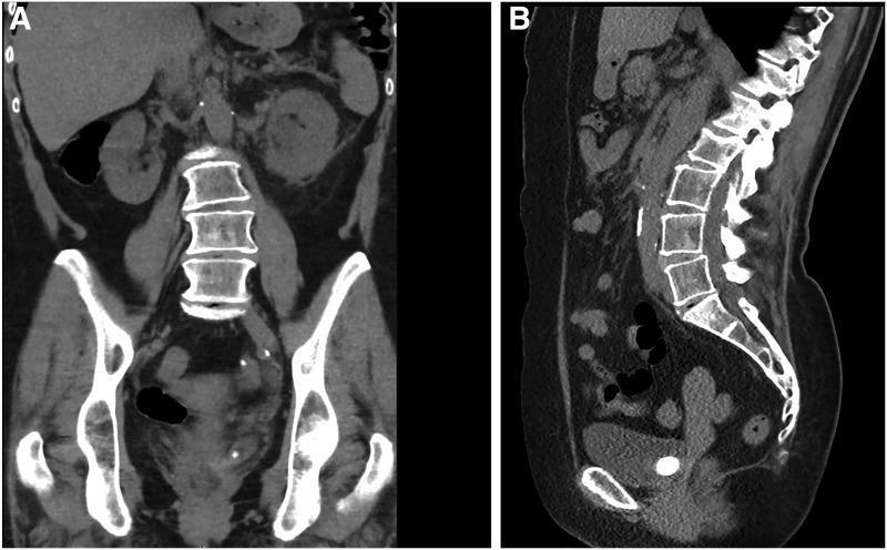
This diagnostic image consists of two panels showing a noncontrast CT scan of the abdomen and pelvis. Panel A (coronal section) displays the lumbar vertebrae, pelvic girdle, and kidneys. A distinct, hyperdense (calcified) structure is visible in the lower pelvic region near the left vesicoureteral junction. Panel B (sagittal section) provides a lateral view of the lumbar and sacral spine, illustrating normal vertebral alignment and disc spacing. A large, well-defined, radiopaque calculus is clearly demonstrated within the bladder area, specifically localized within an associated ureterocele at the vesicoureteral junction. The clinical significance of these findings is diagnostic for a large obstructing ureteral stone, which has caused marked hydronephrosis in the corresponding kidney. This image is a primary educational resource for urology and radiology, demonstrating the classic CT appearance of a distal ureteral calculus and its anatomical relationship to the bladder and ureterocele.
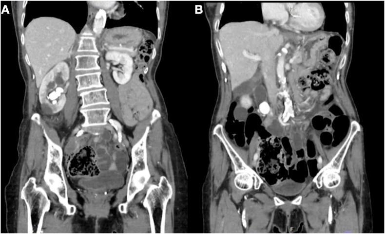
Diagnostic imaging: Two coronal sections of a non-contrast computerized tomography (CT) scan of the abdomen and pelvis. Panel A displays a large, hyperdense staghorn calculus within the right kidney. The stone occupies a significant portion of the renal collecting system, extending through the calyces and into the renal pelvis. Panel B, a different coronal plane, reveals a distinct, well-defined hyperdense opacity in the proximal right ureter, consistent with a secondary ureteric stone. There is evidence of mild hydroureter and pelvicalyceal dilatation proximal to the ureteral obstruction. The left kidney and ureter appear unremarkable in both views. This imaging demonstrates synchronous nephrolithiasis and ureterolithiasis, which is clinically significant for evaluating obstructive uropathy and planning surgical interventions such as percutaneous nephrolithotomy (PCNL).
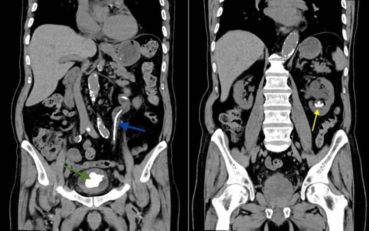
Two coronal sections of a non-contrast Computed Tomography (CT) of the Kidneys, Ureters, and Bladder (CT-KUB) demonstrate nephrolithiasis and a long-term retained urinary stent. In the left panel, a large, irregularly shaped, hyperdense bladder stone (green arrow) is visible within the urinary bladder lumen. A radiopaque double-J (DJ) stent (blue arrow) is seen following the course of the left ureter, extending from the renal pelvis down to the bladder; the distal coil appears encrusted and associated with the bladder stone. In the right panel, a different coronal slice reveals a hyperdense renal calculus (yellow arrow) located in the lower pole of the left kidney. These images illustrate the complications of a 'forgotten' or retained ureteral stent, including severe encrustation and the formation of secondary calculi within both the kidney and the bladder. The scan provides anatomical localization of the urinary tract, highlighting the kidneys, psoas muscles, vertebral column, and pelvic girdle alongside the urological pathology.
How to Check for Kidney Stones
Step 1 - Recognize the Symptoms First
| Symptom | Description |
|---|---|
| Renal colic | Sudden, severe, cramping pain in the flank (side/back), below the ribs |
| Radiating pain | Pain may travel to the lower abdomen, groin, or testicle (in males) |
| Blood in urine | Pink, red, or brown urine (hematuria) - very common |
| Nausea/vomiting | Accompanying the severe pain |
| Frequent urination | Especially when stone is near the bladder |
| Burning urination | Can mimic a urinary tract infection |
In your case (elevated uric acid at 7.8), you may have no symptoms at all - the stone risk is elevated but a stone may not have formed yet.
Step 2 - Blood & Urine Tests
| Test | What it checks |
|---|---|
| Urinalysis | Blood (RBCs), white cells, crystals in urine - most important first test |
| Urine culture | Rules out infection alongside a stone |
| Serum uric acid | Already done - yours is 7.8 (elevated, risk factor for uric acid stones) |
| Serum calcium | Already done - yours is normal (8.7) |
| Serum creatinine + BUN | Checks how well kidneys are functioning |
| Serum phosphorus + electrolytes | Full metabolic workup |
| 24-hour urine collection | The most sensitive metabolic test - measures calcium, uric acid, oxalate, citrate, sodium, pH over a full day |
Step 3 - Imaging (Most Important)
Option 1: Non-Contrast CT Scan (Gold Standard) ⭐
- Sensitivity: 94-97%, Specificity: 96-99% (highest accuracy)
- Called CT-KUB (CT of Kidneys, Ureters, Bladder) or NCCT abdomen/pelvis
- Detects stones as small as 1 mm - including uric acid stones that don't show on X-ray
- Shows exact location, size, and whether the stone is blocking urine flow
- No dye/contrast injection needed
- Slight radiation exposure
Option 2: Ultrasound (Good for Screening)
- No radiation - safe, widely available, cheaper
- Sensitivity 63-85% (misses small stones and ureteral stones)
- First choice in pregnancy, children, and for repeated follow-up checks
- Good for detecting blockage (hydronephrosis - swollen kidney)
- Recommended as the first imaging test, with CT reserved if ultrasound is inconclusive
Option 3: KUB X-Ray (Plain Abdominal X-Ray)
- Quick and cheap, but limited
- Shows calcium-containing stones (60-70% of all stones)
- Cannot detect uric acid stones (which is your risk, given high uric acid)
- Used together with ultrasound
Step 4 - What the Doctor Decides After Tests
| Finding | Meaning |
|---|---|
| Stone < 5 mm | Usually passes on its own with fluids and pain control |
| Stone 5-10 mm | 50% chance of passing; may need medication (tamsulosin) |
| Stone > 10 mm | Usually requires procedure (lithotripsy or surgery) |
| Infection + obstruction | Medical emergency - needs immediate drainage |
Your Specific Situation (Uric Acid Risk)
- Flank/back pain
- Blood in urine
- Pain while urinating
- Urinalysis
- CT-KUB (non-contrast CT scan)
actually i am a diabetic
Diabetes + Elevated Uric Acid - What You Need to Know
Why Diabetes Makes This More Serious
"Hyperuricemia and gout are associated with multiple cardiometabolic-kidney comorbidities, including obesity, hypertension, type 2 diabetes, myocardial infarction, stroke, chronic kidney disease (CKD), and urate nephrolithiasis."
- Harrison's Principles of Internal Medicine 22E
The Diabetes-Uric Acid-Kidney Stone Connection
Diabetes / Insulin Resistance
        ↓
Impaired ammonium excretion by kidneys
        ↓
Urine becomes more acidic (low pH)
        ↓
Uric acid becomes insoluble in acidic urine
        ↓
Uric acid crystals form → KIDNEY STONES
"Patients with type 2 diabetes have lower urinary pH than non-diabetics... Because individuals with diabetes have impaired ammonium excretion, they have a higher incidence of uric acid stone formation." "Uric acid stone formers have greater body weight and a higher incidence of insulin resistance and type 2 diabetes."
Your Combined Risk Picture
| Factor | Your Status | Risk |
|---|---|---|
| Age (21 Male) | Young | Moderate |
| Serum Uric Acid | 7.8 mg/dL (HIGH) | High |
| Total Calcium | 8.7 (Normal) | Low |
| Diabetes | Yes | Significantly increases uric acid stone risk |
| Urine pH (likely acidic due to diabetes) | Untested yet | Key missing piece |
What Tests You Now Specifically Need
| Test | Why it matters for you |
|---|---|
| HbA1c | Check diabetes control - poor control worsens uric acid risk |
| Urinalysis with urine pH | Diabetics typically have acidic urine (pH <5.5), the main driver of uric acid stones |
| 24-hour urine collection | Gold standard - measures uric acid, oxalate, citrate, calcium, and pH over a full day |
| Serum creatinine + eGFR | Kidney function - diabetes damages kidneys over time (diabetic nephropathy) |
| Fasting blood glucose | Assess current sugar control |
| Ultrasound KUB | Baseline check for any silent stones already in kidneys |
Medications to be Aware Of
| Drug | Effect on Uric Acid |
|---|---|
| SGLT2 inhibitors (e.g., empagliflozin, dapagliflozin) | Lower uric acid - actually beneficial for you |
| Thiazide diuretics (sometimes used for BP in diabetics) | Raise uric acid - avoid if possible |
| Low-dose aspirin | Can raise uric acid - discuss with your doctor |
Diet Advice - Specific for a Diabetic with High Uric Acid
| Avoid | Reason |
|---|---|
| Sugary drinks, fruit juices, fructose | Raises both blood sugar AND uric acid |
| Red meat, organ meats, shellfish | High purine - raises uric acid |
| Alcohol (especially beer) | Raises uric acid, worsens blood sugar |
| White rice, white bread in excess | Spikes blood sugar |
| Do This | Reason |
|---|---|
| Drink 2.5-3 liters of water daily | Dilutes uric acid, keeps urine less acidic |
| Eat low-fat dairy | Lowers uric acid, good protein source |
| Vegetables, whole grains, fibre | Controls both sugar and uric acid |
| Citrus fruits (lemon water) | Makes urine more alkaline - dissolves uric acid crystals |
Bottom Line - Priority Action for You
- Tell your diabetes doctor about the uric acid result (7.8) - these need to be managed together
- Get a urine pH test - simple dipstick test, if pH < 5.5, you are actively forming uric acid stone conditions
- Get HbA1c checked if not done recently - better diabetes control directly reduces stone risk
- Get kidney function tests (creatinine, eGFR) - diabetes + high uric acid is a double threat to kidneys
- Drink more water - the single cheapest and most effective intervention