
Is extensor retinaculum of foot and ankle are same!!?
The extensor retinaculum of the foot and ankle are not the same - they are two distinct structures. The extensor retinaculum is divided into a superior and inferior part, located at different levels.
Searching sources...
Reading File
Finding Sources
Searching the Web
extensor retinaculum foot ankle superior inferior difference anatomy
Reading File
Reading File
Let me fetch the key anatomical dissection image showing both retinacula:
Loading Image
Loading Image
No, they are NOT the same. The "extensor retinaculum" of the ankle/foot region refers to two completely separate structures - the Superior Extensor Retinaculum (SER) and the Inferior Extensor Retinaculum (IER). Here is a full breakdown:
Extensor Retinaculum of the Ankle & Foot - Two Distinct Structures
Here is a cadaveric dissection clearly showing both structures:
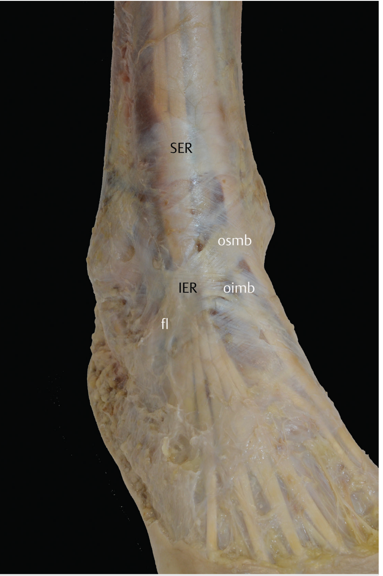
SER = Superior Extensor Retinaculum (ankle level); IER = Inferior Extensor Retinaculum (foot level); osmb = oblique superomedial band; oimb = oblique inferomedial band; fl = frondiform ligament
1. Superior Extensor Retinaculum (SER) - "Ankle" retinaculum
| Feature | Detail |
|---|---|
| Location | Just above the ankle (tibiotalar joint), lower leg |
| Shape | Rectangular / transverse band |
| Origin | Formed by thickening of the superficial crural fascia |
| Attachments | Laterally: distal fibula and lateral malleolus; Medially: distal tibia and medial malleolus |
| Function | Holds extensor tendons, prevents bowstringing across the ankle |
| Tunnels | ~25% of cases: tibialis anterior has its own tunnel formed by a split in the retinaculum |
2. Inferior Extensor Retinaculum (IER) - "Foot" retinaculum
| Feature | Detail |
|---|---|
| Location | Below the SER, anterior to the ankle joint and over the tarsus (dorsum of foot) |
| Shape | Y-shaped (classic) - the "cruciate crural ligament" |
| Three components | 1. Oblique superomedial band (osmb) 2. Oblique inferomedial band (oimb) 3. Frondiform ligament (fl) |
| Variant | In ~25% of cases a superolateral band exists, making it X-shaped instead |
| Thickest part | At the intersection of the bands, located anterior to the subtalar joint |
| Function | Stabilizes extensor tendons over the tarsus; stabilizes the subtalar joint; prevents deep fibular nerve and tendons from bowstringing |
| Clinical significance | Used in the Brostrom-Gould procedure for chronic lateral ankle instability |
Quick Memory Summary
LEG (just above ankle) → Superior Extensor Retinaculum → RECTANGULAR
↓
FOOT (over tarsus) → Inferior Extensor Retinaculum → Y-SHAPED
So when someone says "extensor retinaculum of the ankle," they typically mean the SER, and "extensor retinaculum of the foot" refers to the IER. They are at different levels, have different shapes, different attachments, and different clinical implications - but both belong to the same continuous fascial system and both restrain the same group of extensor tendons.
- Imaging Anatomy Text and Atlas, Vol. 3 - Bones, Joints, Vessels and Nerves, Superior & Inferior Extensor Retinaculum sections
This is a shared conversation. Sign in to Orris to start your own chat.