
Give a 10mark / 5 page answer on arches of foot from standard orthopaedic textbooks for orthopaedic rguhs pg exams
Reading File
Now let me search the medical library for arches of the foot content from orthopaedic textbooks.
Finding Sources
Finding Sources
Finding Sources
Reading File
Reading File
Reading File
Reading File
Reading File
Reading File
Finding Sources
Reading File
Finding Sources
Reading File
Reading File
Now let me fetch the key diagrams and get the windlass mechanism details:
Loading Image
Loading Image
Reading File
I now have comprehensive content from Gray's Anatomy for Students, the THIEME Atlas, and Imaging Anatomy (Rockwood equivalent) to write a thorough 10-mark answer. Let me also fetch the support of arch diagram:
Loading Image
Loading Image
I now have everything needed to write a comprehensive, exam-standard 10-mark answer. Let me compose it:
ARCHES OF THE FOOT
Sources: Gray's Anatomy for Students | THIEME Atlas of General Anatomy and Musculoskeletal System | Imaging Anatomy Vol. 3 (Bones, Joints, Vessels and Nerves)
INTRODUCTION
The bones of the foot do not lie in a horizontal plane. Instead, they are arranged into a series of arches that enable the foot to bear and transmit the weight of the body, absorb shock during locomotion, and adapt to uneven terrain. There are three arches: the medial longitudinal arch, the lateral longitudinal arch, and the transverse arch. From a structural engineering standpoint, the foot can be thought of as a tripod - the first ray, the fifth ray, and the calcaneus forming the three contact points - with the arches distributing compressive forces under optimal mechanical conditions.
- Gray's Anatomy for Students, p. 754; Imaging Anatomy Vol. 3, p. 929
1. MEDIAL LONGITUDINAL ARCH
Bones (9 bones)
The medial longitudinal arch is composed of:
- Calcaneus (posterior pillar)
- Talus (keystone/apex - the head rests on the plantar calcaneonavicular ligament)
- Navicular
- 3 Cuneiform bones (medial, intermediate, lateral)
- Medial 3 metatarsals (heads form the anterior pillar)
The weight-bearing elements are the calcaneal tuberosity posteriorly and the medial three metatarsal heads anteriorly.
Characteristics
- It is the higher, more pronounced arch
- The talus sits at the apex and receives the body weight transmitted from the tibia through the talocrural joint
- During stance, this weight is distributed anteriorly to the forefoot and posteriorly to the heel
- It is more elastic than the lateral arch, acting as a spring during walking
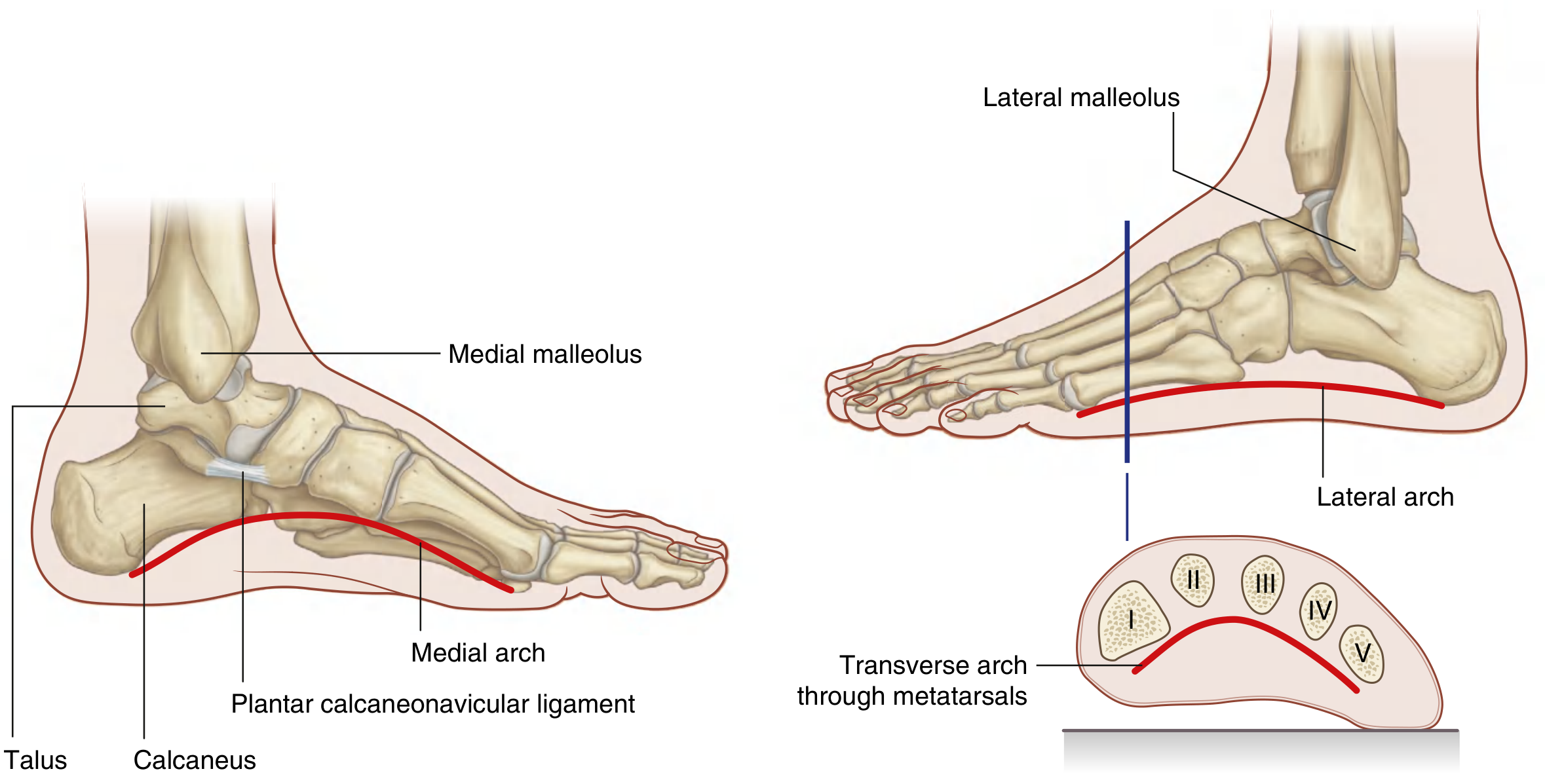
Fig: Longitudinal and transverse arches of the foot. The medial arch is taller and more curved; the lateral arch sits close to the ground. - Gray's Anatomy for Students
Ligamentous Support (Passive)
| Ligament | Role |
|---|---|
| Plantar calcaneonavicular (Spring) ligament | Supports the head of the talus - the keystone of the arch; allows some elastic motion |
| Plantar aponeurosis | Most important passive stabilizer - longest lever arm from calcaneal tuberosity to metatarsal heads |
| Long plantar ligament | Secondary support |
| Short plantar ligament (plantar calcaneocuboid) | Secondary support |
| Talocalcaneal ligaments | Bind calcaneus to talus |
| Deltoid (medial) ligament - tibionavicular component | Medial support |
The plantar aponeurosis is particularly important owing to its long lever arm. The spring ligament, being the closest passive structure to the apex of the arch, is the weakest component but allows elastic accommodation.
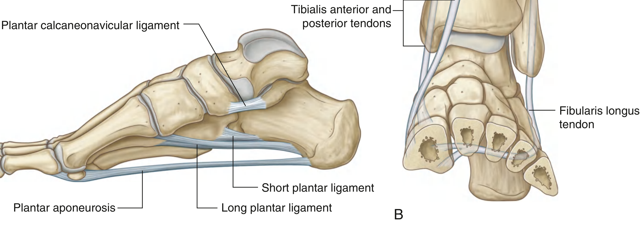
Fig: Support for arches of the foot. (A) Ligamentous support - medial view. (B) Cross-section showing tendinous supports. - Gray's Anatomy for Students
Muscular Support (Active / Dynamic)
- Tibialis posterior - crosses the medial ankle and inserts on the navicular; the primary dynamic stabilizer
- Flexor hallucis longus - runs beneath the sustentaculum tali; its tendon tightens the arch like the chord of an arc
- Flexor digitorum longus - supports the arch
- Flexor digitorum brevis - intrinsic support
- Abductor hallucis - medial support
- Tibialis anterior - pulls the arch up dynamically during walking
- THIEME Atlas, p. 488-489
2. LATERAL LONGITUDINAL ARCH
Bones (4 bones)
- Calcaneus (posterior pillar)
- Cuboid
- 4th and 5th metatarsals (anterior pillars)
Weight-bearing elements: calcaneus posteriorly and the heads of the 4th and 5th metatarsals anteriorly.
Characteristics
- Lower arch - it largely rests on the ground during standing (less curved)
- Provides stability rather than spring
- Forms the lateral ray of the tripod arrangement
Ligamentous Support
- Long plantar ligament - primary ligamentous support (runs from calcaneus to bases of 2nd-5th metatarsals)
- Short plantar ligament (plantar calcaneocuboid ligament) - secondary
- Plantar aponeurosis
Muscular Support
- Fibularis (Peroneus) longus - key stabilizer of the lateral arch as well as the transverse arch
- Flexor digitorum longus and brevis
- Abductor digiti minimi
- Quadratus plantae (flexor accessorius)
3. TRANSVERSE ARCH
Bones (9 bones)
- Cuboid
- 3 Cuneiform bones (medial, intermediate, lateral)
- Bases of 5 metatarsals
Characteristics
- The arch is highest in the coronal plane passing through the head of the talus and flattens toward the metatarsal heads
- The intermediate and lateral cuneiforms are wedge-shaped (narrower inferiorly), which is the primary osseous mechanism maintaining this arch
- Distally, the metatarsal heads are held together by the deep transverse metatarsal ligaments, preventing splaying
- The concavity faces plantar/inferiorly
Ligamentous and Muscular Support
- Interosseous ligaments (dorsal and plantar) between tarsal bones
- Deep transverse metatarsal ligaments at the metatarsal head level
- Fibularis (Peroneus) longus tendon - crosses from lateral to medial foot in the plantar groove of the cuboid and inserts on the medial cuneiform and base of 1st metatarsal, thereby acting as a tie-beam maintaining the transverse arch
- Gray's Anatomy for Students, p. 755; Imaging Anatomy Vol. 3, p. 929
4. PLANTAR APONEUROSIS
The plantar aponeurosis is a thickened layer of deep fascia anchored to the medial process of the calcaneal tuberosity posteriorly. It extends forward as a broad band of longitudinally arranged collagen fibers, divides into five digital bands at the level of the metatarsal heads, and attaches to the bones, ligaments, and dermis of the toes. Transverse interconnecting fibers form the superficial transverse metatarsal ligaments distally.
Functions:
- Supports the longitudinal arch of the foot
- Protects deeper structures in the sole
- The primary passive stabilizer of the medial longitudinal arch owing to its long lever arm
- Gray's Anatomy for Students, p. 755-756
5. THE WINDLASS MECHANISM
This is a key biomechanical concept for orthopaedic exams.
When the toes are dorsiflexed (as occurs during the toe-off phase of gait), the plantar fascia is pulled distally around the metatarsal head (acting as a windlass/capstan). This tightens the plantar aponeurosis, which:
- Raises the height of the medial longitudinal arch
- Converts the foot into a rigid lever for push-off
- Internally rotates the tibia and supinates the foot
This mechanism is fundamental to efficient toe-off during walking and explains why plantar fasciitis produces pain on first steps in the morning (the fascia is tight after overnight shortening) and why pes planus (flatfoot) disrupts the normal push-off mechanics.
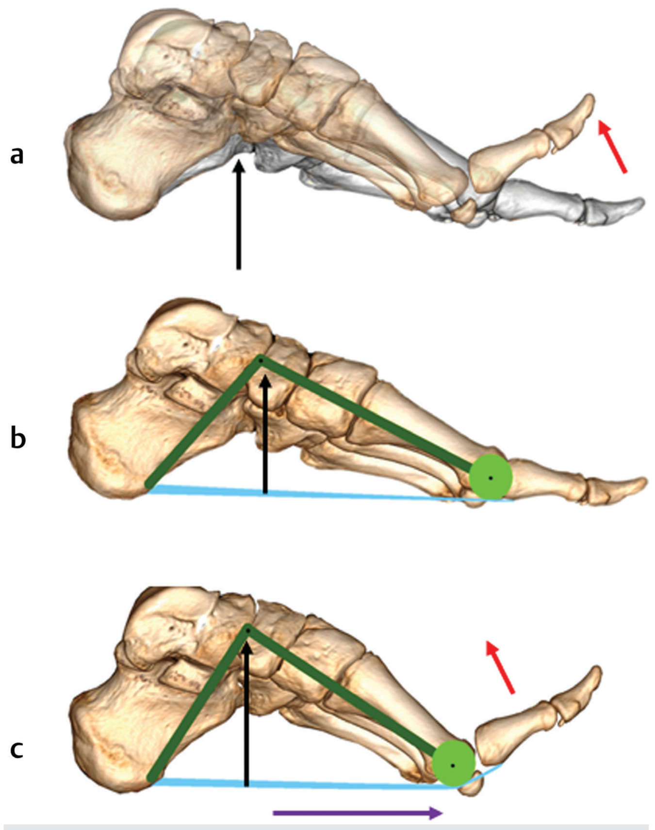
Fig: Windlass mechanism. As the toes dorsiflex (red arrow), the plantar fascia is pulled distally (purple arrow), leading to increase in height of the arch (black arrow). - Imaging Anatomy Vol. 3
6. BIOMECHANICS OF WEIGHT DISTRIBUTION
During stance, partial body weight at the talocrural joint is transmitted through the talus and distributed to:
- Forefoot (via medial and lateral rays to metatarsal heads)
- Hindfoot (via calcaneus to the ground)
The bony trabeculae of the foot are arranged parallel to the lines of compressive stress - the trabecular pattern mirrors the arch configuration, confirming the structural validity of the arch model. The cancellous trabeculae in the calcaneus and metatarsals run along stress lines, providing strength with minimum material.
The foot's tripod structure (heel + head of 1st metatarsal + head of 5th metatarsal) distributes forces across the sole. The footprint (podogram) is considerably larger than the bony triangle, as the plantar soft tissues cushion the intervening areas.
- THIEME Atlas, p. 487; Imaging Anatomy Vol. 3, p. 929
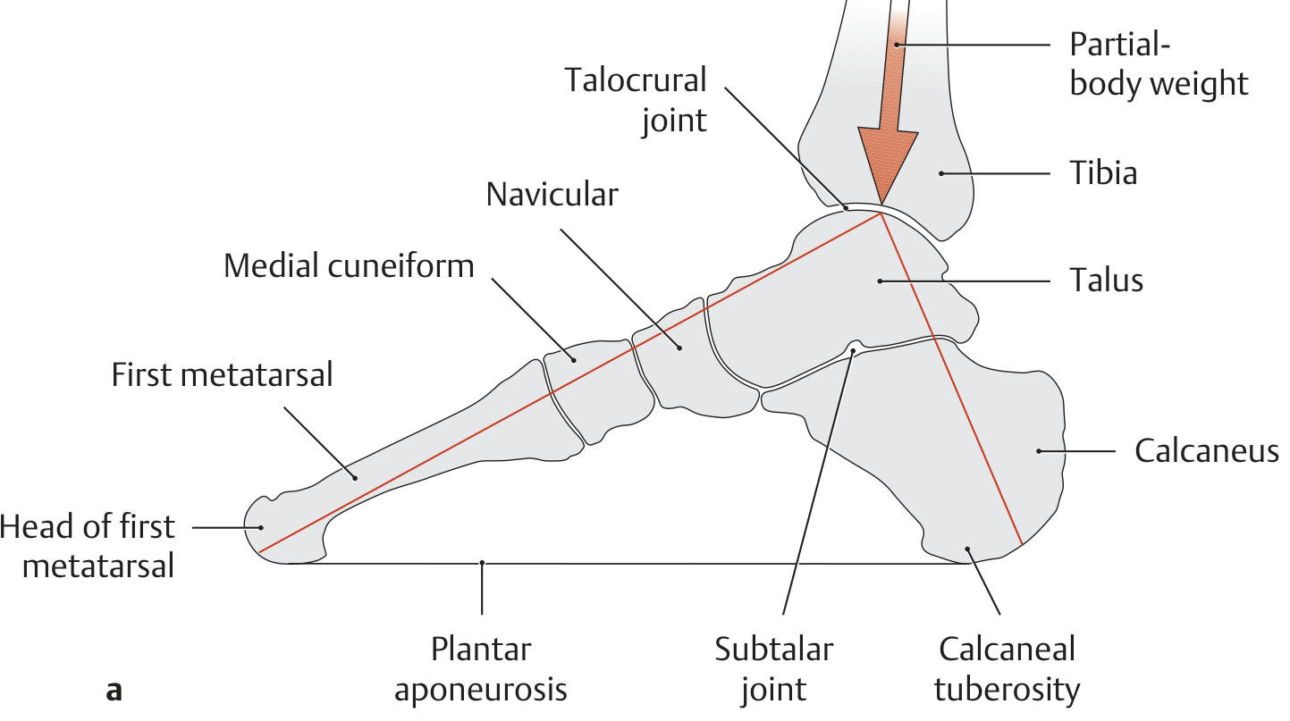
Fig: Transfer of compressive stresses. Body weight passes through the talocrural joint to the talus, then distributes to forefoot and hindfoot via medial longitudinal arch. - THIEME Atlas
7. CLINICAL CORRELATES
Pes Planus (Flat Foot)
Loss of the medial longitudinal arch. Occurs when the passive and active stabilizers fail (most commonly posterior tibial tendon insufficiency - PTTI). The calcaneus everts (hindfoot valgus), the talar head drops medially, and the forefoot abducts. On weight-bearing radiograph, the calcaneal pitch angle and talo-first metatarsal angle are abnormal. Failure of the spring ligament is a key contributing factor.
Pes Cavus (High Arch)
The medial longitudinal arch is elevated. Seen in neurological conditions such as Charcot-Marie-Tooth disease. Results from muscle imbalance (intrinsic foot muscle weakness), leading to plantarflexion of metatarsals, claw toes, and hindfoot varus. The Coleman block test is used to assess flexibility.
Plantar Fasciitis
Degeneration and micro-tears at the calcaneal origin of the plantar aponeurosis. The plantar fascia is the key passive stabilizer of the medial arch. Overload (obesity, prolonged standing, pes planus) leads to pain at the medial calcaneal tubercle.
Metatarsalgia / Splay Foot
Collapse of the transverse arch leading to spreading of the forefoot and pain under metatarsal heads.
- Gray's Anatomy for Students, p. 800; Imaging Anatomy Vol. 3, p. 929-930
SUMMARY TABLE
| Arch | Bones | Keystone | Weight-bearing Points | Key Ligaments | Key Muscles |
|---|---|---|---|---|---|
| Medial longitudinal | 9 (calcaneus, talus, navicular, 3 cuneiforms, medial 3 metatarsals) | Talus | Calcaneal tuberosity + medial 3 MT heads | Spring ligament, plantar aponeurosis | Tibialis posterior, FHL |
| Lateral longitudinal | 4 (calcaneus, cuboid, 4th + 5th metatarsals) | Cuboid | Calcaneus + 4th/5th MT heads | Long plantar, short plantar | Fibularis longus, abductor digiti minimi |
| Transverse | 9 (cuboid, 3 cuneiforms, 5 MT bases) | Intermediate cuneiform | None - spans side to side | Deep transverse MT ligament, interosseous ligaments | Fibularis longus (tie-beam) |
References:
- Gray's Anatomy for Students, 4th Ed - pp. 754-756, 800
- THIEME Atlas of General Anatomy and Musculoskeletal System - pp. 487-489
- Imaging Anatomy Vol. 3, Bones, Joints, Vessels and Nerves - pp. 929-931
This is a shared conversation. Sign in to Orris to start your own chat.