
Apoptosis
Reading File
Now let me search the medical library for apoptosis content in parallel.
Finding Sources
Finding Sources
The best sources are Robbins Pathology and Histology. Let me read those in parallel.
Reading File
Reading File
Reading File
Let me read more of Robbins Pathologic Basis (the most detailed source) on mechanisms, and also grab the histology images section.
Reading File
Reading File
Now let me fetch the key images and read more of Robbins (Pathologic Basis) on the mechanisms section.
Reading File
Loading Image
Excellent content gathered. Let me also fetch the Robbins apoptosis mechanism diagram and read the extrinsic pathway section.
Reading File
Loading Image
I now have comprehensive information. Let me also quickly fetch the Robbins mechanism diagram.
Finding Sources
Reading File
Loading Image
Loading Image
I now have all the key content and images. Let me compile a comprehensive, well-organized answer.
Apoptosis
Apoptosis is a tightly regulated, energy-dependent form of programmed cell death in which a cell activates its own internal "suicide" machinery. It is distinct from necrosis: the plasma membrane stays intact throughout, no inflammatory reaction is triggered, and cell fragments are efficiently cleared by phagocytes. The name comes from the Greek for "falling off" (as leaves fall from a tree).
1. Definition
"A pathway of cell death in which cells activate enzymes that degrade the cells' own nuclear DNA and nuclear and cytoplasmic proteins. Fragments of the apoptotic cells then break off... the plasma membrane of the apoptotic cell remains intact, but the membrane is altered in such a way that the fragments, called apoptotic bodies, are recognized and rapidly phagocytosed by macrophages."
- Robbins & Kumar Basic Pathology
2. Morphologic and Biochemical Features
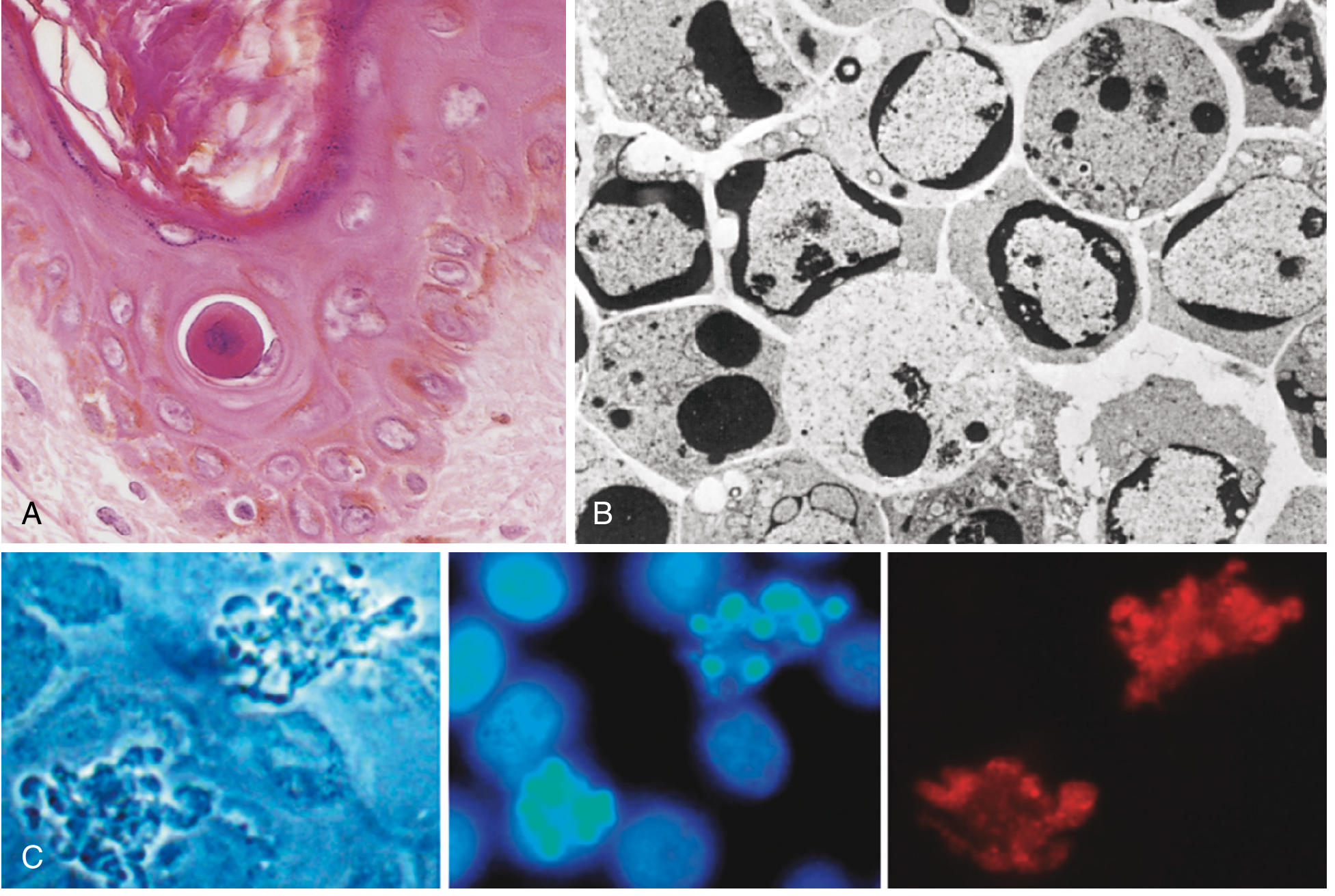
Light microscopy: The apoptotic cell appears as a round or oval mass of intensely eosinophilic cytoplasm with fragments of dense nuclear chromatin.
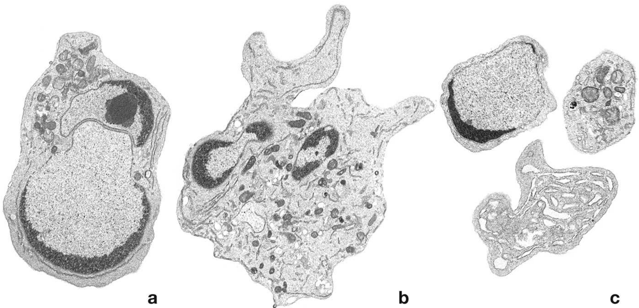
Four hallmark features (best seen on electron microscopy):
| Feature | Description |
|---|---|
| Cell shrinkage | Reduced size, dense eosinophilic cytoplasm; organelles packed tightly. Contrast with necrosis (cell swelling). |
| Chromatin condensation | Chromatin aggregates peripherally under the nuclear envelope into dense crescents/masses - the most characteristic feature |
| Membrane blebbing + apoptotic bodies | Surface blebs form, then fragment into membrane-bound apoptotic bodies (cytoplasm + organelles ± nuclear fragments) |
| Phagocytosis | Rapid ingestion by macrophages via "eat me" signals; no inflammatory response elicited |
Electron micrographs of the sequential stages - early nuclear fragmentation (a), advanced DNA/cytoplasmic fragmentation (b), and final apoptotic bodies (c):
3. Causes of Apoptosis
Physiologic
Apoptosis is essential for normal homeostasis - humans turn over ~1 million cells per second by this mechanism:
- Embryonic development - removal of supernumerary cells (e.g., web-space cells between digits); involution of primordial structures
- Hormone-dependent tissue involution - endometrial breakdown in menstrual cycle, ovarian follicular atresia in menopause, lactating breast regression after weaning
- Cell turnover in proliferating populations (e.g., intestinal crypt epithelium) to maintain constant cell numbers
- Lymphocyte selection - elimination of immature lymphocytes with non-functional antigen receptors in bone marrow/thymus; germinal center B cells that fail to produce high-affinity antibodies
- Self-tolerance - elimination of self-reactive lymphocytes to prevent autoimmunity
- End-of-response cleanup - neutrophils after acute inflammation; lymphocytes at end of immune response
Pathologic
- DNA damage - radiation and cytotoxic drugs damage DNA directly or via free radicals; if repair fails, p53 triggers intrinsic apoptosis, preventing malignant transformation
- Misfolded protein accumulation - ER stress response activates BH3-only proteins
- Viral infections - virus itself (e.g., adenovirus, HIV) or host CTL killing of infected cells
- Atrophy after duct obstruction - pancreas, parotid gland, kidney
4. Mechanisms of Apoptosis
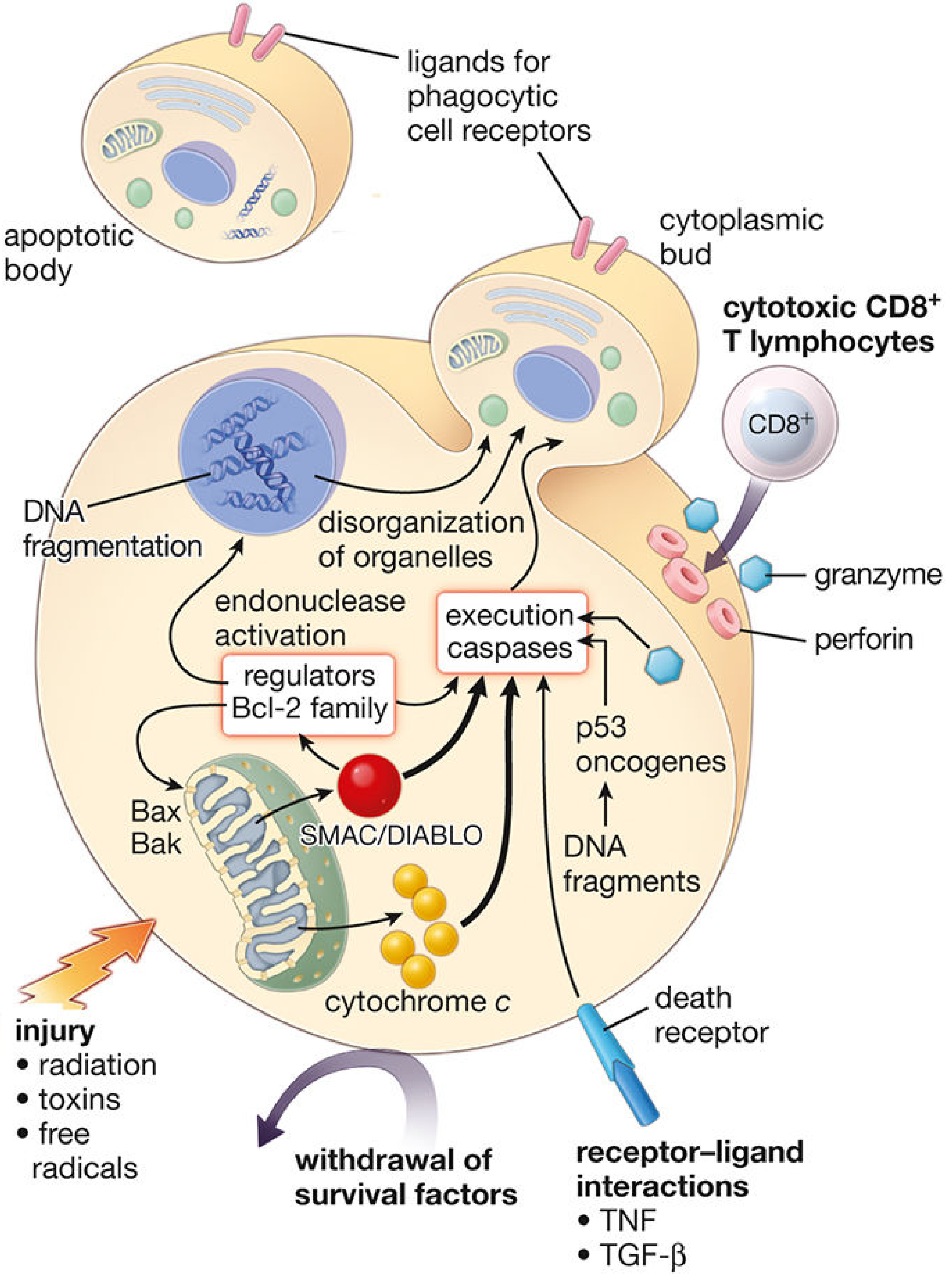
The central executioners are caspases - cysteine proteases that convey the apoptotic signal via a proteolytic cascade. Two major pathways converge on executioner caspases (caspase-3 and -6):
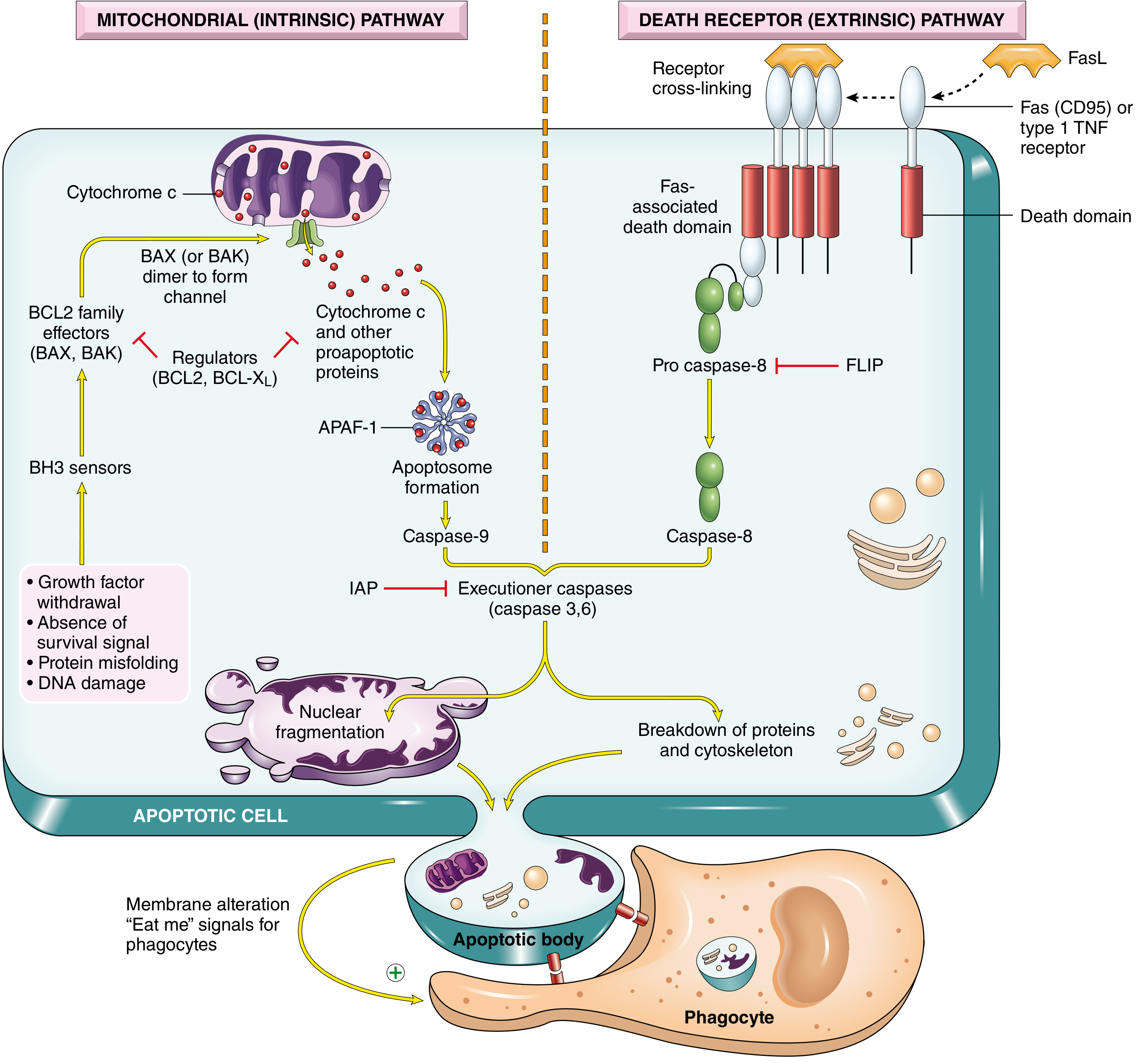
Pathway 1: Mitochondrial (Intrinsic) Pathway
Trigger: loss of survival signals, DNA damage, ER stress/misfolded proteins
The key is the BCL2 family of proteins, which controls permeability of the outer mitochondrial membrane:
| Group | Members | Function | BH domains |
|---|---|---|---|
| Anti-apoptotic | BCL2, BCL-XL, MCL1 | Keep outer membrane impermeable; block cytochrome c release | BH1-4 |
| Pro-apoptotic effectors | BAX, BAK | Oligomerize; form channels to permeabilize outer membrane | BH1-3 |
| BH3-only sensors | BAD, BIM, BID, PUMA, NOXA | Activated by cellular stress; activate BAX/BAK or block BCL2 | BH3 only |
Sequence of events:
- Cellular stress (DNA damage, ER stress, growth factor withdrawal) upregulates BH3-only proteins
- BH3-only proteins activate BAX/BAK and/or neutralize BCL2/BCL-XL
- BAX/BAK oligomers form channels in the outer mitochondrial membrane
- Cytochrome c (and SMAC/DIABLO) leaks from the intermembranous space into the cytosol
- Cytochrome c binds APAF-1 to form the apoptosome (multimeric wheel structure)
- Apoptosome recruits and activates caspase-9 (initiator caspase) via autocatalytic cleavage
- Active caspase-9 activates executioner caspases (3, 6, 7)
SMAC/DIABLO additionally neutralizes IAPs (inhibitor of apoptosis proteins), removing a brake on caspase activity.
Pathway 2: Death Receptor (Extrinsic) Pathway
Trigger: ligation of death receptors by specific ligands
- Death receptors: Fas (CD95) and TNF receptor type 1 (TNFR1); both are members of the TNF receptor superfamily
- Ligands: FasL (on cytotoxic T lymphocytes) and TNF (soluble or membrane-bound)
- Receptor cross-linking recruits adaptor proteins with a death domain (e.g., FADD - Fas-associated death domain)
- This complex, the DISC (death-inducing signaling complex), recruits and activates pro-caspase-8
- Active caspase-8 directly activates executioner caspases-3 and -6
- FLIP (FLICE inhibitory protein) is a cellular inhibitor that blocks caspase-8 activation - a key regulatory checkpoint
Physiologic role: elimination of self-reactive lymphocytes; CTL-mediated killing of virus-infected cells and tumor cells
CTL/Perforin-Granzyme Pathway
Cytotoxic CD8+ T lymphocytes insert perforin (pore-forming protein) into the target cell membrane and deliver granzymes (serine proteases) into the cytoplasm. Granzyme acts directly on caspase-3 and activates pro-apoptotic BCL2 family members, causing mitochondrial cytochrome c release.
5. Inhibitors of Apoptosis
| Inhibitor | Mechanism |
|---|---|
| BCL2, BCL-XL, MCL1 | Block BAX/BAK-mediated mitochondrial permeabilization |
| IAPs (e.g., XIAP) | Bind and inhibit active caspases-3, -7, -9 |
| FLIP | Blocks pro-caspase-8 activation at DISC |
| Survival factors (growth factors, estrogen, androgens, zinc, ECM interactions) | Stimulate BCL2 transcription; suppress BH3-only protein activity |
6. Clearance - Efferocytosis
Apoptotic cells display "eat me" signals that recruit phagocytes without triggering inflammation:
- Phosphatidylserine flip - normally on inner leaflet; flips to outer leaflet in apoptotic cells, recognized by macrophage receptors
- Soluble "find me" factors secreted by dying cells attract phagocytes
- Opsonization by C1q (complement) and natural antibodies
- Macrophages that engulf apoptotic cells produce fewer proinflammatory cytokines
This process (efferocytosis) is so efficient that apoptotic cells disappear within minutes. Since apoptosis occurs ~20x faster than mitosis, apoptotic cells are rarely seen in routine H&E preparations.
7. Apoptosis vs. Necrosis - Key Comparison
| Feature | Apoptosis | Necrosis |
|---|---|---|
| Trigger | Physiologic or controlled pathologic | Uncontrolled injury, ischemia |
| Cell size | Shrinks | Swells |
| Membrane integrity | Intact (until very late) | Lost early |
| Inflammation | None (anti-inflammatory) | Prominent |
| DNA fragmentation | Internucleosomal (laddering pattern) | Random |
| Caspases | Required | Not required |
| Energy | ATP-dependent | Passive |
| Apoptotic bodies | Yes | No |
8. Disease Relevance
| Condition | Role of Apoptosis |
|---|---|
| Cancer | Evasion of apoptosis is a hallmark of cancer; BCL2 overexpression in follicular lymphoma (t[14;18]); p53 loss prevents DNA-damage-induced apoptosis |
| Autoimmunity | Failure to delete self-reactive lymphocytes (Fas/FasL mutations cause lymphoproliferative disease - ALPS) |
| HIV/AIDS | Virus induces apoptosis of CD4+ T cells; immune evasion via viral anti-apoptotic proteins |
| Neurodegenerative diseases | Excessive neuronal apoptosis in Parkinson's, Alzheimer's, ALS |
| Ischemia-reperfusion injury | Apoptosis (caspase-mediated) contributes to cell loss after reperfusion |
| Chemotherapy | Most cytotoxic drugs kill cancer cells by inducing apoptosis |
| BH3 mimetics (e.g., venetoclax) | Drugs that mimic BH3-only proteins; block BCL2 to restore apoptosis in B cell malignancies |
9. Overview Diagram - Summary
Sources:
- Robbins, Cotran & Kumar - Pathologic Basis of Disease (9780443264528), Chapter 2: Cell Injury, Cell Death, and Adaptations
- Robbins & Kumar Basic Pathology (9780323790185), Chapter 1
- Histology: A Text and Atlas with Correlated Cell and Molecular Biology (9781975181512), Chapter 3
This is a shared conversation. Sign in to Orris to start your own chat.