
Note on Atherosclerosis.. discuss actiopathogenesis nd fate of thrombus
Reading File
Finding Sources
Finding Sources
Finding Sources
Finding Sources
Reading File
Finding Sources
Reading File
Reading File
Finding Sources
Finding Sources
Reading File
Reading File
Reading File
Reading File
Searching PubMed
atherosclerosis pathogenesis
Atherosclerosis: Aetiopathogenesis and Fate of Thrombus
1. Definition
Atherosclerosis is best understood as the vascular response to endothelial injury - a chronic inflammatory process of the arterial intima characterised by atheromas (atherosclerotic plaques): raised intimal lesions with a soft, lipid-rich necrotic core (cholesterol, cholesterol esters, debris) covered by a fibrous cap (Robbins & Kumar Basic Pathology, p. 313-314). It underlies coronary, cerebral, and peripheral vascular disease and causes roughly half of all deaths in Western countries.
2. Aetiopathogenesis - the "Response to Injury" Hypothesis
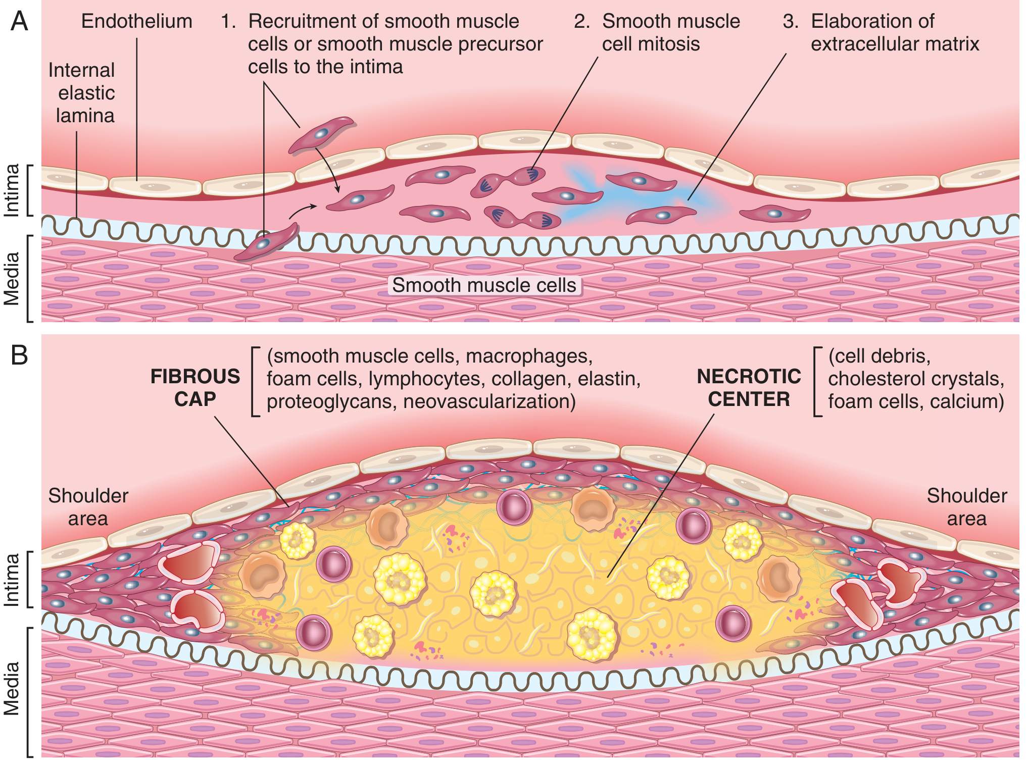
The currently accepted model views atherosclerosis as a chronic inflammatory and healing response of the arterial wall to endothelial injury, involving interaction of modified lipoproteins, monocyte-derived macrophages, T lymphocytes, and cells of the arterial wall. The sequence of events is:
- Endothelial injury/dysfunction - increased permeability, leukocyte adhesion, and a pro-thrombotic surface
- Accumulation of lipoproteins (mainly oxidised LDL and cholesterol crystals) within the intima
- Monocyte adhesion and migration into the intima, where they transform into macrophages and then lipid-laden foam cells
- Platelet adhesion to areas of injured/dysfunctional endothelium or exposed subendothelial matrix
- Growth factor and cytokine release from activated platelets, macrophages, and vessel-wall cells, recruiting smooth muscle cells (SMCs) from the media or circulating precursors
- Lipid accumulation both extracellularly and within macrophages/SMCs
- SMC proliferation, collagen/ECM production, and further recruitment of T cells, producing the fibrous cap over the lipid/necrotic core (Robbins & Kumar Basic Pathology, p. 316-317)
Key contributing factors:
- Endothelial dysfunction - triggered chiefly by hemodynamic disturbances (turbulent flow at branch points, ostia, posterior abdominal aorta) and hypercholesterolemia; also hypertension, cigarette smoke toxins, and inflammatory cytokines (e.g. TNF)
- Lipids - chronic hyperlipidemia (raised LDL, low HDL, raised Lp(a)) drives oxidative damage to endothelium; oxidised LDL is taken up by macrophage scavenger receptors, generating foam cells and further chemokine/growth-factor release
- Inflammation - cholesterol crystals and free fatty acids activate the inflammasome in macrophages, producing IL-1 and amplifying leukocyte recruitment; this drives both plaque progression and cap instability
- Risk factors - genetics/family history, age, gender (premenopausal women relatively protected), hypertension, smoking, diabetes; these act multiplicatively (two factors ~4x risk, three factors ~7x risk for MI) (Robbins & Kumar Basic Pathology, p. 314-315)
Morphological evolution: dysfunctional endothelium → fatty streak (intimal macrophage foam cells, Fig. 8.9) → fibrofatty atheroma with lipid core and fibrous cap → complicated/vulnerable plaque with erosion, calcification, neovascularisation, intraplaque haemorrhage, and rupture.
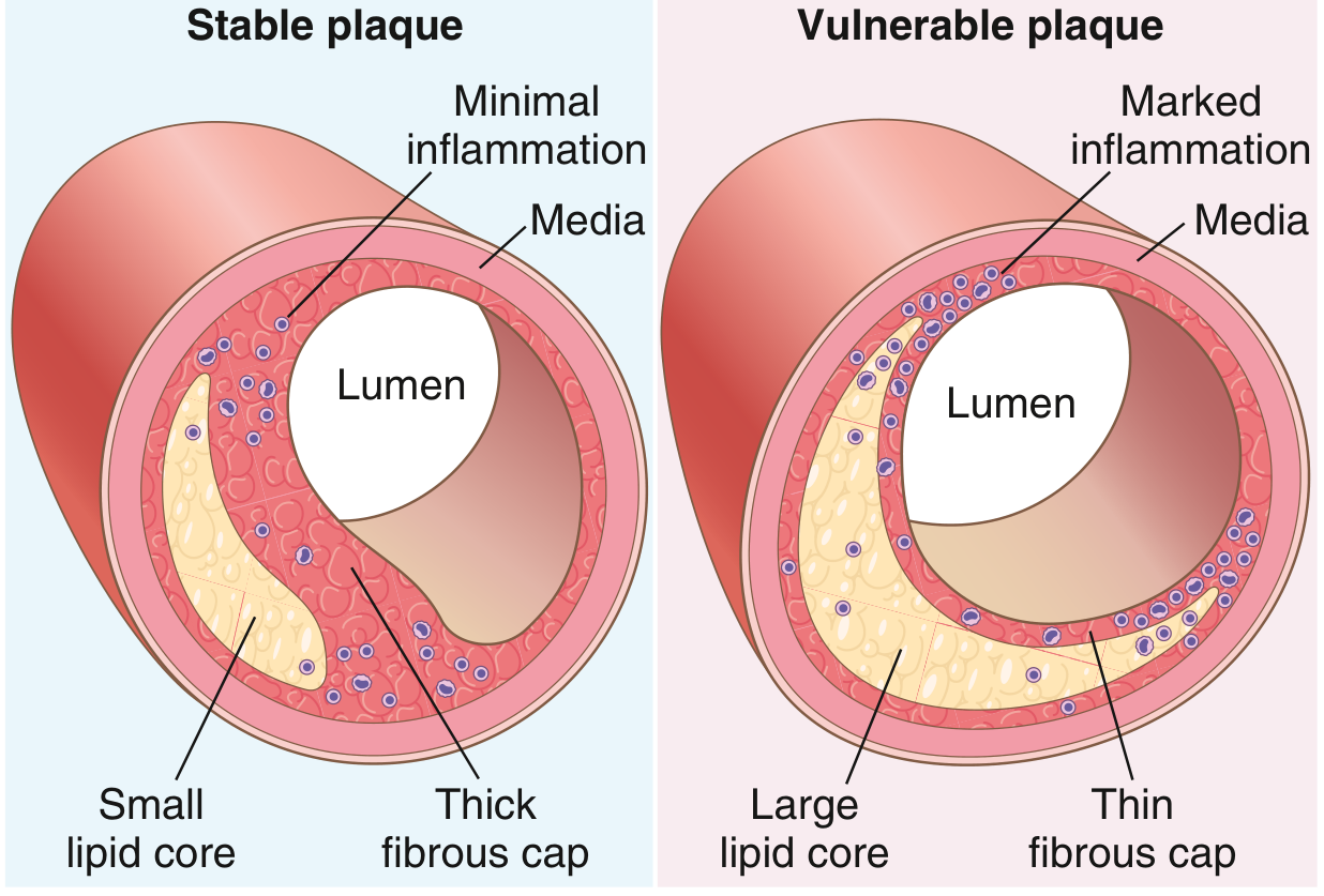
Plaque rupture and thrombosis: Plaques rich in foam cells, abundant extracellular lipid, a thin fibrous cap with few SMCs, and clusters of inflammatory cells are termed vulnerable plaques. Inflammation degrades collagen in the cap, weakening it against shear stress. Rupture or erosion exposes highly thrombogenic subendothelial collagen and necrotic core material to flowing blood, triggering acute thrombosis - the central event in acute coronary syndromes, most myocardial infarctions, and many ischaemic strokes. Disrupted plaques can also shower atheroemboli (cholesterol/plaque debris) distally.
3. Fate of the Thrombus
Once a thrombus forms (e.g. on a ruptured atherosclerotic plaque), if the patient survives the initial event it undergoes some combination of four outcomes over the following days to weeks (Robbins & Cotran, Pathologic Basis of Disease, p. 128-129):
-
Propagation - the thrombus accumulates additional platelets and fibrin, enlarging and increasing the risk of vascular occlusion. Arterial thrombi propagate retrograde, venous thrombi propagate toward the heart; the propagating tail is poorly attached and prone to fragmentation.
-
Embolization - part or all of the thrombus dislodges and travels through the bloodstream to lodge elsewhere (e.g. a coronary thrombus embolising distally, or a mural/aortic plaque thrombus producing atheroembolism), causing infarction at the new site.
-
Dissolution - fibrinolysis (via plasmin) causes rapid shrinkage and complete disappearance of recent thrombi. Older thrombi, with extensive fibrin cross-linking, are far more resistant to lysis - which is why therapeutic fibrinolytics (e.g. tPA) are only effective when given within the first few hours of an acute thrombotic event (e.g. acute coronary thrombosis).
-
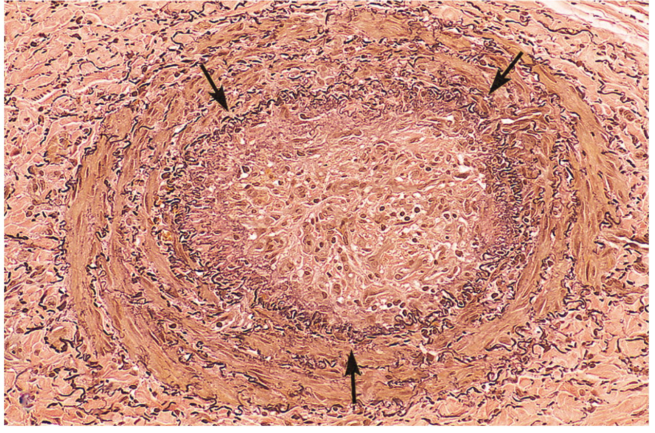
Organization and recanalization - older thrombi become organised by ingrowth of endothelial cells, smooth muscle cells, and fibroblasts. New capillary channels form that partially re-establish the continuity of the original lumen (recanalization). With continued remodeling, the thrombus may be reduced to a small fibrous mass incorporated into the vessel wall, so that eventually only a fibrous lump marks its original site.
Additional/occasional outcomes:
- Enzymatic digestion of the thrombus centre by lysosomal enzymes released from trapped leukocytes/platelets.
- Infection (septic thrombus) in the setting of bacteremia, producing an inflammatory mass that can erode and weaken the vessel wall, potentially leading to a mycotic aneurysm.
Clinical significance: Thrombi become clinically apparent when they obstruct the vessel lumen or embolise. Venous thrombi mainly threaten via pulmonary embolism; arterial thrombi (frequently superimposed on ruptured plaque) chiefly threaten by occluding critical vessels such as coronary or cerebral arteries, producing myocardial infarction or stroke - Robbins, Cotran & Kumar Pathologic Basis of Disease, p. 129.
Note on current literature: This account reflects the well-established "response to injury" model taught in current pathology texts. Recent systematic reviews (e.g. PMID 37676098 on Wnt signaling/sclerostin in vascular calcification, PMID 38977083 on intraplaque neovascularization) are refining understanding of specific molecular pathways (calcification signaling, plaque neovascularisation) but do not contradict this core framework - they add mechanistic detail rather than replacing it.
This is a shared conversation. Sign in to Orris to start your own chat.