
Malunion -cubitus varus and valgus for long answer question for orthopaedic MBBS exam
Reading File
Now let me search the medical library for cubitus varus and valgus content in parallel.
Finding Sources
Finding Sources
Excellent! Found great sources. Let me read the main sections in parallel.
Reading File
Reading File
Finding Sources
Good, reading more detailed content on complications and treatment now.
Reading File
Reading File
Reading File
Now let me get more content on the osteotomy techniques and tardy ulnar nerve palsy from cubitus valgus.
Reading File
Finding Sources
Excellent! Now let me get the tardy ulnar nerve palsy content and Miller's review summary, plus fetch key images.
Reading File
Reading File
Loading Image
I now have comprehensive content from multiple authoritative sources. Let me also fetch the carrying angle image for visual clarity.
Loading Image
Loading Image
I now have all the content needed for a comprehensive long-answer question response. Let me compile the full answer.
Malunion of the Elbow: Cubitus Varus and Cubitus Valgus
(Long Answer Question - Orthopaedics MBBS)
INTRODUCTION AND NORMAL CARRYING ANGLE
The carrying angle is the normal outward (valgus) deviation of the extended, supinated forearm from the axis of the arm. Normally it measures 10-15° (slightly more in females; 9-14° by some references, 2-3° greater in women). It is measured with the patient in the anatomical position - elbow fully extended and forearm supinated. The angle disappears on pronation or full flexion.
When the carrying angle is:
- Abnormally decreased or reversed - called Cubitus Varus
- Abnormally increased - called Cubitus Valgus
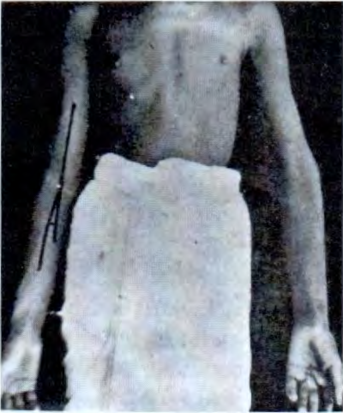
PART I: CUBITUS VARUS
Definition
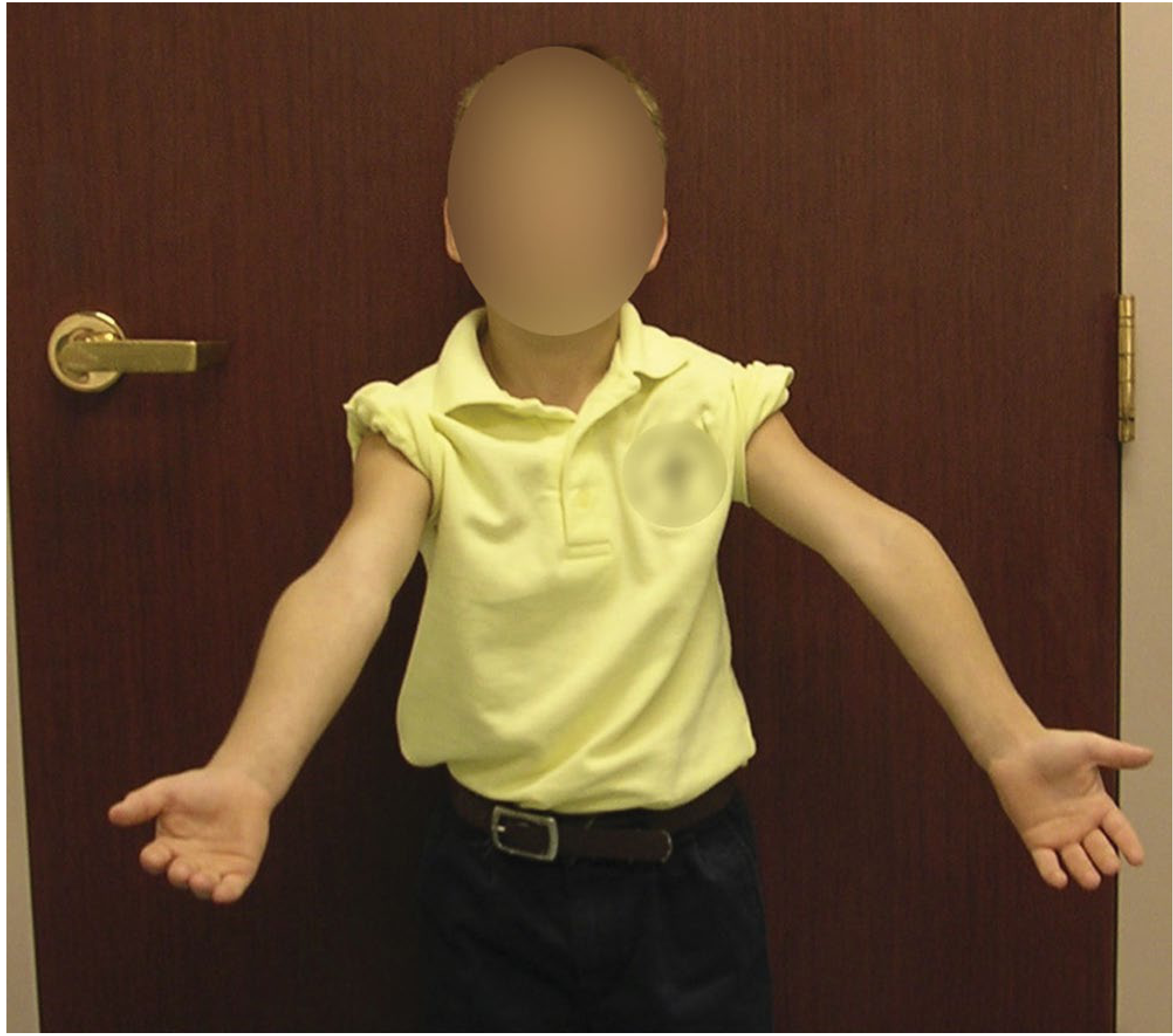
Cubitus varus is a deformity in which the carrying angle is reversed - the forearm deviates medially (inward) relative to the arm. It is also called the "gun-stock deformity" due to the characteristic appearance.
Aetiology / Cause
- Most common cause: Malunion of a supracondylar fracture of the humerus in children - it is the most common angular deformity resulting from supracondylar fractures
- Varus tilting of the distal fragment is the most important cause of the change in carrying angle
- Mechanisms of varus tilting (Fig. 38.19 - Campbell's):
- A. Medial impaction of the fracture
- B. Medial tilting of the distal fragment
- C. Horizontal rotation of the distal fragment
- Less common: medial column instability leading to collapse when treated with cast alone (without pinning) - even with apparently benign Type II fractures
- Rare cause: Osteonecrosis and delayed growth of the trochlea with relative overgrowth of the lateral side of the distal humeral epiphysis (progressive - cannot be prevented by stabilization)
Clinical Features
- Typically presents in a child with a history of a previous supracondylar fracture
- Obvious cosmetic deformity - the elbow has a gun-stock appearance
- Carrying angle is reduced or negative compared to the normal side
- Baumann's angle on X-ray is reduced (normal: 9-26°; reduced in cubitus varus)
- Function is generally preserved - historically considered a cosmetic problem
Complications (Why it is NOT just cosmetic)
More recent evidence has shown cubitus varus is associated with:
- Ulnar nerve dislocation and ulnar neuropathy
- Snapping of the medial head of the triceps over the medial epicondyle
- Secondary fractures - distal humeral or lateral condylar fractures (due to altered mechanics)
- Osteonecrosis of the distal humeral epiphysis
- Joint ganglia
- Osteoarthritis
- Posterolateral rotatory instability (PLRI) - occurring up to 51 years later
- The mechanical axis, olecranon, and triceps line of pull are all displaced medially
- This causes repetitive external rotation torque on the ulna, stretching the lateral collateral ligament complex
- Physical findings: positive posterolateral rotatory apprehension test, positive lateral pivot shift, posterolateral rotatory drawer signs
- Tardy ulnar nerve palsy can occur (though more classically associated with cubitus valgus)
Remodeling
- 100% remodeling has been shown in children younger than 5 years
- Only minimal remodeling occurs after age 8 years
- Malrotation of the distal humerus will not remodel but is compensated by shoulder motion
Treatment of Cubitus Varus
Conservative
- Observation is acceptable in mild deformity, especially in young children (<5 yrs) where remodeling may occur
Surgical (Corrective Osteotomy)
Indicated for significant deformity (>15°), functional impairment, or cosmetic concern.
Three basic types of osteotomy have been described:
| Osteotomy Type | Details |
|---|---|
| 1. Lateral Closing Wedge Osteotomy | Easiest, safest, most stable, and most commonly used. Corrects coronal plane; can correct sagittal plane if placed obliquely. Axial rotational deformity correctable with derotation. |
| 2. Medial Opening Wedge Osteotomy | With bone graft; less common |
| 3. Oblique Osteotomy with Derotation | Corrects rotational deformity |
| 4. Three-Dimensional Osteotomy (Uchida) | Corrects medial tilt, posterior tilt, and rotation simultaneously |
Lateral Closing Wedge Osteotomy - Key Steps (Campbell's Technique 38.5):
- Lateral approach to the elbow with tourniquet
- Under fluoroscopy, insert 2 K-wires into the lateral condyle before osteotomy
- Make a closing wedge osteotomy laterally, leaving the medial cortex intact; cuts can be angled in sagittal plane to correct flexion/extension deformity
- Weaken medial cortex with drill holes if needed; apply valgus stress in pronation to complete osteotomy
- Close osteotomy; advance K-wires or cannulated screws from lateral condyle into medial cortex of proximal fragment
- Stabilize with K-wires (younger patients) or plate-and-screw fixation (adolescents)
- Immobilize in long-arm cast at 60° flexion and full pronation
Postoperative care: K-wires removed at ~6 weeks; range-of-motion exercises started. Improved elbow flexion from 101° to 126° reported.
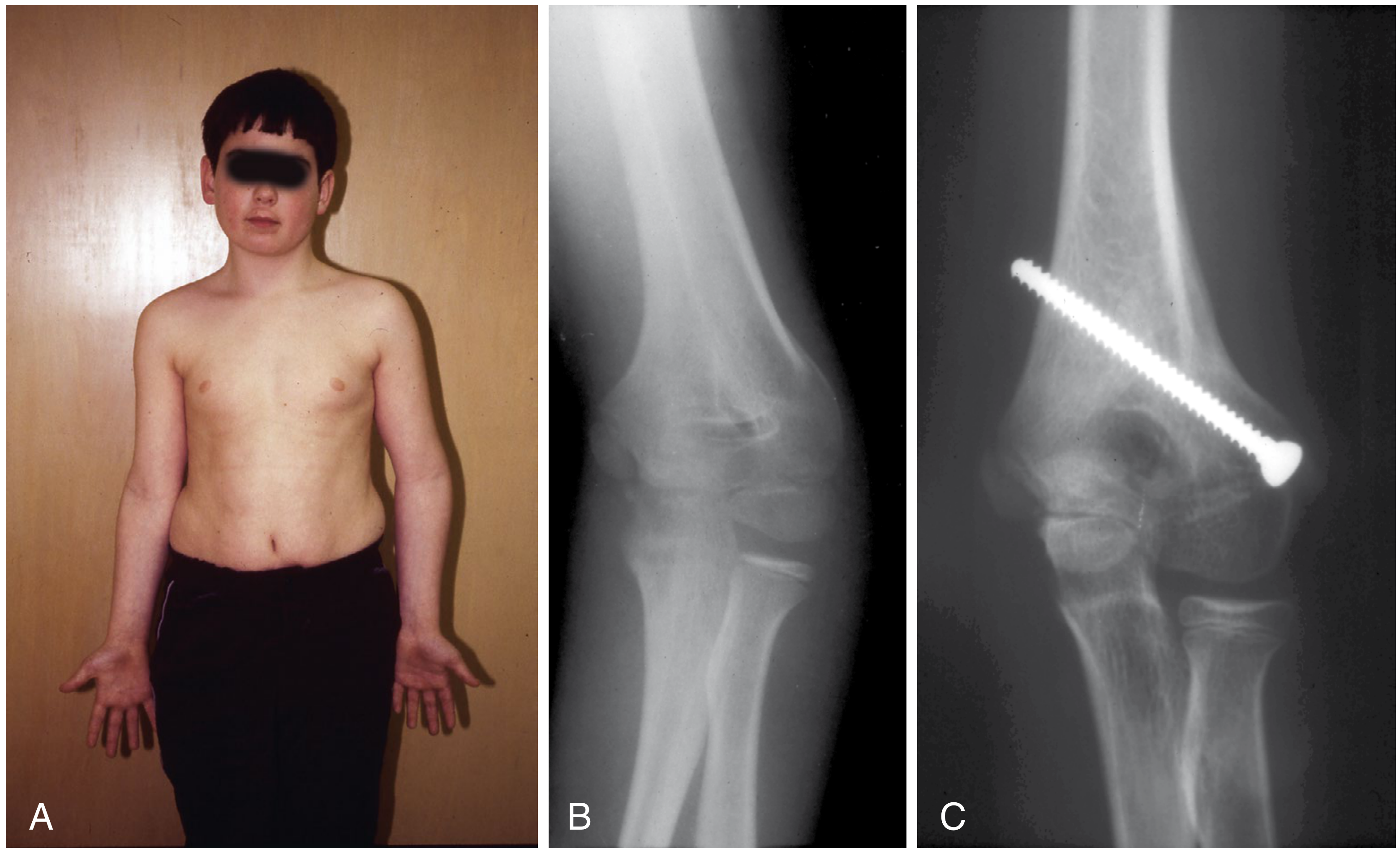
For associated instability: surgical treatment may include reconstruction of the lateral collateral ligament + osteotomy, ligament reconstruction alone, or total elbow arthroplasty.
PART II: CUBITUS VALGUS
Definition
Cubitus valgus is a deformity in which the carrying angle is increased beyond normal - the forearm deviates excessively laterally (outward). It is less common than cubitus varus.
Aetiology / Cause
- Most common cause: Nonunion of lateral condylar fractures - this is the most frequent cause of true cubitus valgus
- Malunion of a distal humeral fracture (including lateral condylar fractures in children)
- Lateral growth arrest following lateral condyle fractures (Milch classification)
- Note: Cubitus valgus after supracondylar fracture is mentioned in literature but rarely occurs (since the normal carrying angle increases from childhood to adulthood, an increase in valgus is less cosmetically noticeable)
Why Cubitus Valgus is Important
Cubitus valgus is clinically significant primarily because of its neurological complication:
Tardy Ulnar Nerve Palsy (TUNP)
- This is the most important complication of cubitus valgus
- "Tardy" = delayed (develops years to decades after the original injury)
- Mechanism: In cubitus valgus, the ulnar nerve is gradually stretched around the medial epicondyle as the valgus deformity increases the traction on the nerve over time
- This causes incomplete, progressive ulnar nerve paralysis - hence "tardy"
- The nerve is also susceptible to compression by callus or osteophytes at the malunited site
Features of Tardy Ulnar Nerve Palsy:
- Onset: Insidious, years after original fracture (average 10-20 years later, though can be much longer)
- Sensory: Numbness and tingling in the little finger and ring finger (ulnar 1.5 fingers)
- Motor:
- Wasting of hypothenar eminence and intrinsic muscles of the hand
- Claw hand (more pronounced in ring and little fingers - "ulnar claw")
- Weakness of grip
- Positive Froment's sign (weakness of adductor pollicis)
- Investigation: Nerve conduction studies confirm slowing at the elbow
Other Complications of Cubitus Valgus:
- Degenerative joint disease (osteoarthritis) of the elbow
- Restricted elbow movements
Treatment of Cubitus Valgus
For the Deformity:
- Corrective osteotomy (medial closing wedge or lateral opening wedge at the supracondylar level) - if the deformity is significant and causing functional problems
- This is less commonly needed than in cubitus varus, as valgus may be tolerated better
For Tardy Ulnar Nerve Palsy:
The nerve itself must be managed:
| Option | Details |
|---|---|
| Ulnar nerve decompression | Simple decompression in cubital tunnel (first-line for mild cases) |
| Anterior transposition of ulnar nerve | Subcutaneous, intramuscular, or submuscular - removes the nerve from its stretched/compressed position and places it anteriorly (most widely practiced) |
| Osteotomy | Corrects the underlying valgus deformity to relieve nerve tension |
| Combined | Osteotomy + nerve transposition for severe cases |
COMPARISON TABLE: CUBITUS VARUS vs CUBITUS VALGUS
| Feature | Cubitus Varus | Cubitus Valgus |
|---|---|---|
| Carrying angle | Decreased / reversed | Increased |
| Common name | Gun-stock deformity | - |
| Most common cause | Malunion of supracondylar fracture | Nonunion/malunion of lateral condyle fracture |
| Common in | Children (post-supracondylar) | Any age; often lateral condyle fractures |
| Cosmetic effect | Marked | Less noticeable (valgus increases naturally with age) |
| Primary nerve complication | Ulnar nerve dislocation, PLRI (late) | Tardy ulnar nerve palsy (classic) |
| Function affected | Usually preserved initially | Ulnar nerve deficits late |
| Remodeling potential | <5 yrs: good; >8 yrs: poor | Poor |
| Surgery of choice | Lateral closing wedge osteotomy | Nerve transposition ± corrective osteotomy |
IMPORTANT EXAM POINTS (High-yield)
- Cubitus varus = most common complication of supracondylar fracture in children
- Gun-stock deformity = synonym for cubitus varus
- Cubitus valgus = tardy ulnar nerve palsy - this association is the most tested fact
- Tardy UNP mechanism: Progressive stretching of ulnar nerve over the medial epicondyle due to valgus angulation
- Most common cause of cubitus valgus: Nonunion (not malunion) of lateral condylar fracture
- Surgery for cubitus varus: Lateral closing wedge osteotomy (supracondylar) - most common
- Baumann's angle on AP X-ray: reduced in cubitus varus (normal 9-26°; also described as angle between humeral shaft axis and lateral condylar physis/growth plate - normally 64-81° in some conventions - confirm the version used in your textbook)
- Cubitus varus - although historically "cosmetic only," now known to cause posterolateral rotatory instability and secondary fractures
- Carrying angle: 10-15° normally (slightly more in women)
- The carrying angle disappears on pronation or full flexion
Sources: Campbell's Operative Orthopaedics 15th Ed (2026); S Das - Manual on Clinical Surgery 13th Ed; Miller's Review of Orthopaedics 9th Ed; Bailey & Love's Short Practice of Surgery 28th Ed
This is a shared conversation. Sign in to Orris to start your own chat.