
So i have got a big boil under my groin. An area which touches down when i sit on a chair. It is too big and is painful and burning. What to do? I have been having these boils since a few days but they used to come and go away but this one has stayed and increased in size.
furuncle boil skin abscess groin perineal area
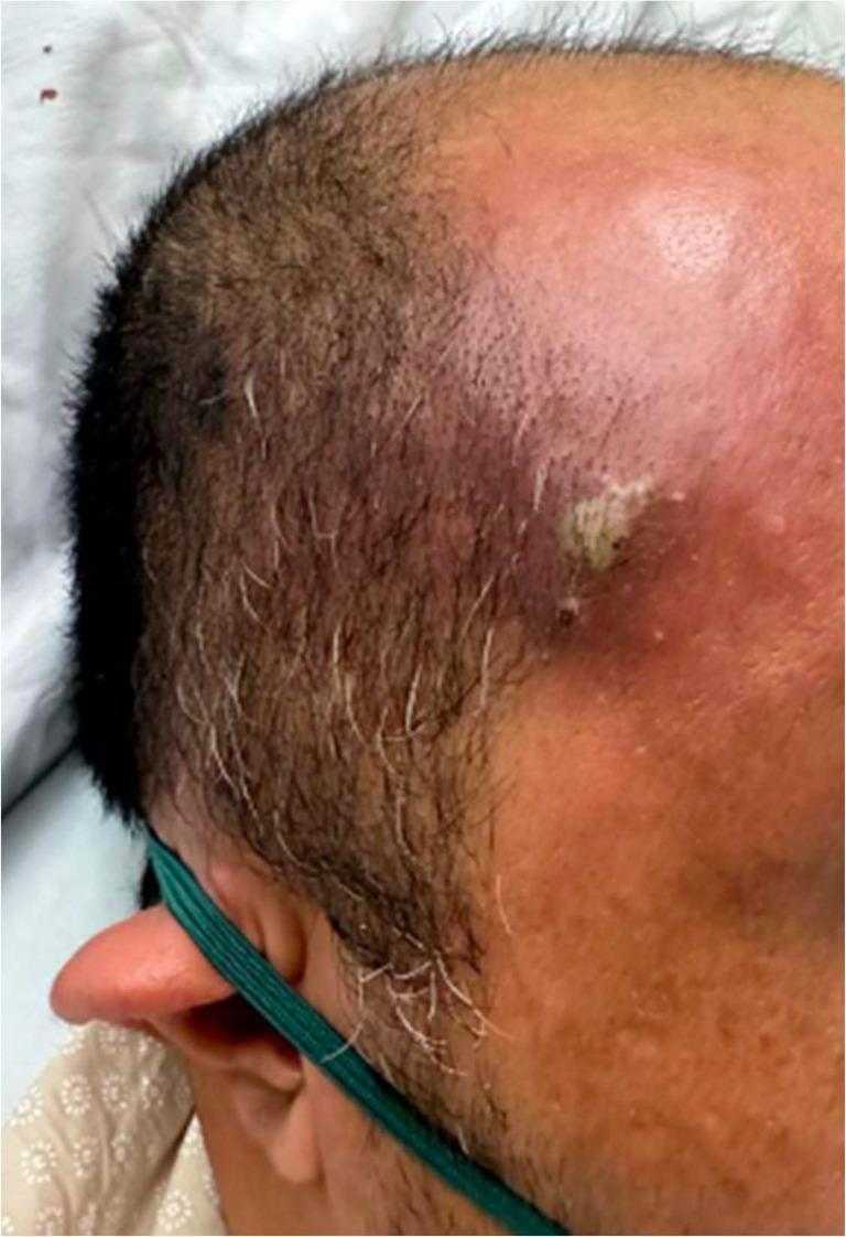
Clinical photograph showing a localized skin lesion, specifically a furuncle (boil), located on the right temporal/forehead region of a 44-year-old male. The lesion is characterized by an elevated, erythematous nodule approximately 1 cm in diameter, exhibiting central fluctuance and a small amount of yellowish-white purulent discharge (pus). There is significant surrounding perilesional erythema and soft tissue inflammation extending toward the scalp and forehead. The adjacent skin displays a slightly irregular texture with normal short hair distribution, including scattered graying. Clinically, such a lesion in a patient with poorly controlled diabetes mellitus can serve as a primary infectious nidus for hematogenous seeding, potentially leading to systemic complications such as septic pulmonary embolism (SPE). This image is used to demonstrate the morphology of a common cutaneous abscess and its potential role in the progression to sepsis.
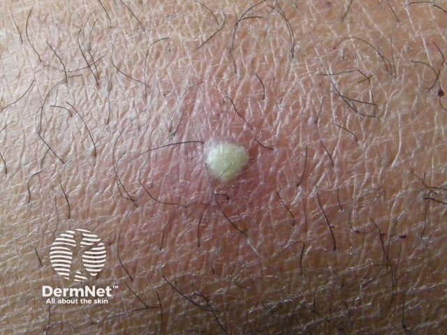
This dermatology clinical photograph documents a solitary boil (furuncle) on the cutaneous surface. Modality: Clinical photography; technique: macro close-up imaging of the skin with standard, noninvasive illumination. The lesion is a dome-shaped, erythematous nodule with a central yellowish-white pustular core representing purulent material within a hair follicle. Surrounding skin shows mild edema and perifollicular erythema. The lesion appears tender on palpation; no drainage is evident although the pus core is visible. The image captures typical signs of acute bacterial folliculitis progressing to furunculosis, most commonly caused by Staphylococcus aureus, with neutrophil-rich inflammatory infiltrate and abscess formation around the follicular unit. Clinically, this finding supports a diagnosis of furuncle rather than a simple acne lesion or epidermal cyst; differential diagnoses include carbuncle, abscess, or inflamed epidermoid cyst. Pathophysiology involves follicular occlusion, microbial colonization, and localized pus accumulation. Management implications include warm compresses, incision and drainage if fluctuance is present, and targeted antibiotics for extensive infection or cellulitis or systemic signs. This image is educational for medical trainees in recognizing cutaneous infections, dermatology case discussions, and demonstrations of localized skin abscesses and follicular infection. Useful for exam prep, clinical decision making, and patient education about boil treatment and outcomes globally.
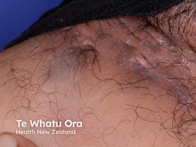
Clinical photograph of the groin region depicting hidradenitis suppurativa (acne inversa) in an intertriginous fold. Imaging modality: Clinical photography; Technique: macroscopic, color-corrected, frontal/anterior view of the inguinal area emphasizing skinfolds and surface lesions. Precise anatomical location: inguinal region with involvement of perineal skin and upper thigh junction; tissue involvement: dermis and superficial subcutaneous tissue with sinus tract formation. Visual features: inflamed, tender nodules and indurated plaques; draining sinus tracts with crusts and purulent exudate; surrounding erythema, edema, hyperpigmented scars, and tethering of the skin. Pathological correlation (clinical): chronic follicular occlusion precipitates abscess formation and sinus tract networks; histology would show follicular rupture with granulomatous inflammation around hair follicles and apocrine-bearing skin. Clinical significance: HS is a relapsing inflammatory dermatosis causing pain, malodor, scarring, and potential secondary infection; differential diagnoses include folliculitis, furunculosis, infected cysts, and cutaneous Crohn’s disease in some contexts. Diagnostic and educational value: helps stage disease (Hurley I–III), guides management including antibiotics (doxycycline, clindamycin–rifampin), biologics (adalimumab), hormonal therapy, and surgical planning. Use cases: documentation for baseline severity, progress monitoring, resident education, patient counseling, and research datasets; groin HS visualization supports dermatology, surgery, and primary care workflows. Images serve as illustrative teaching aids and research data.
What This Likely Is
1. Furuncle (Deep Boil) - Most likely for a single lesion
2. Hidradenitis Suppurativa (HS) - If this is truly recurrent
What To Do RIGHT NOW
1. See a Doctor - This is Urgent
- It is larger than 2 cm (roughly a coin-width)
- There is increasing redness spreading outward from the boil
- You have fever, chills, or feel unwell
- It has not started draining on its own after 5-7 days
- You feel increasing throbbing pain
2. Warm Compresses (While Waiting)
3. Antibiotics
- There is surrounding cellulitis (redness spreading outward)
- You have a fever
- The boil is not yet fluctuant (hasn't come to a soft head yet)
- You are diabetic or immunocompromised
4. Keep the Area Clean and Dry
- Wash gently with antibacterial soap (chlorhexidine or povidone-iodine)
- Wear loose-fitting, breathable underwear/clothing to reduce friction
- Do not share towels or clothing while the boil is active
- Wash your hands before and after touching the area
Warning Signs - Go to Emergency Immediately
- Fever above 38.5°C / 101.3°F with chills
- Red streaks spreading outward from the boil (sign of spreading infection/lymphangitis)
- Swelling rapidly increasing in size over hours
- Feeling very unwell, dizzy, or confused
- The boil is very close to or around the anus (may indicate a perianal/ischiorectal abscess, which is a surgical emergency)
If Boils Keep Coming Back - Address the Root Cause
- MRSA carrier state (bacteria living in your nose/skin) - your doctor may recommend mupirocin nasal ointment + chlorhexidine body washes to decolonize
- Hidradenitis suppurativa - requires a dermatologist referral for long-term management with antibiotics (clindamycin + rifampicin), biologics (adalimumab), or surgery
- Underlying diabetes or immunosuppression - a blood sugar check is worthwhile if you haven't had one
Disclaimer: This is medical information for educational purposes and is not a substitute for an in-person medical evaluation. Please see a qualified healthcare provider for diagnosis and treatment.
Would you be able to disgnose it better if i send you an image of it?
- Size and depth - whether it looks like a surface boil or a deeper abscess
- Fluctuance signs - whether it appears ready to drain (yellowish/white head visible)
- Surrounding redness - how far cellulitis has spread, which affects urgency
- Multiple lesions or sinus tracts - which would strongly point toward hidradenitis suppurativa rather than a simple furuncle
- Skin changes - scarring, pitting, or tunneling under the skin
- I can offer a better-informed opinion, but I cannot replace an in-person clinical exam - a doctor needs to physically feel whether the lump is fluctuant (ready to drain) and assess depth
- Image quality matters - good lighting and a clear, close-up shot will help
- If the area is too sensitive or private to photograph, that is completely fine - my previous assessment still stands and the advice to see a doctor today remains the same regardless


So these are 2 images of the thing that has happened. I don’t know whether it is actually a boil or something else. Kindly see and comment. I have marked it in red circles
What the Images Show
Image 1 (First boil - the main painful one)
- A single, dome-shaped, raised nodule approximately 1-1.5 cm in diameter
- Violaceous (purplish-dark red) color - this indicates deep-seated inflammation with blood pooling beneath the skin
- Two pale whitish-yellow "heads" (points) visible at the center - this is classic for a boil that has built up pus and is about to point/drain
- The overlying skin looks tense and shiny - a sign of pressure from pus underneath
- Mild surrounding redness
Image 2 (Second photo - two lesions marked)
- Upper lesion: An elongated, oval nodule with a pinkish-red shiny/pearlescent center, no active pus visible, minimal surrounding redness. This looks like it may be a partially resolving or previously drained boil, or a hypertrophic scar from a previous episode.
- Lower lesion: A smaller, darker purplish-pink, dome-shaped nodule. Intact surface, no discharge visible. Looks like a newer or earlier-stage boil, or a resolving one.
What This Pattern Tells Me
- Multiple lesions in the same intertriginous (skin-fold) area
- Lesions at different stages simultaneously (one active, one resolving, one early)
- Recurrent nature as you described ("they used to come and go")
- Location - groin/inner thigh is one of the classic HS zones (along with armpits and under breasts)
- Evidence of what appears to be scarring or fibrotic change in the second lesion (upper lesion, Image 2)
What To Do Based on the Images
Immediate (today/tomorrow)
Short-term (within 1-2 weeks)
Things that make HS worse (avoid these)
- Tight synthetic clothing rubbing against the area
- Shaving the area (try trimming instead)
- Obesity/excess weight (if relevant)
- Smoking (if applicable - it significantly worsens HS)
- Heat and sweating without prompt cleansing
Summary
| Lesion | What It Looks Like | Stage |
|---|---|---|
| Image 1 (main boil) | Furuncle with white heads - pus ready to drain | Active - needs drainage today |
| Image 2 upper | Shiny, pink, elongated nodule | Resolving / post-inflammatory scar |
| Image 2 lower | Small purple-pink dome | Early / healing boil |
Disclaimer: This visual assessment is educational and not a formal medical diagnosis. An in-person examination by a doctor is needed for definitive diagnosis and treatment.
Will boroline help?
What Boroline Is
- Zinc oxide (mild antiseptic/skin protectant)
- Boric acid (mild antiseptic)
- Lanolin (moisturizer/emollient)
Why It Won't Work Here
- The lanolin and occlusive base in Boroline can trap heat and moisture, which may actually encourage bacterial growth in an already infected area
- Applying a greasy cream over a pointing boil (with a pus head) can block natural drainage and worsen pressure/pain
- It has no meaningful antibacterial activity against Staphylococcus aureus, which is the bacteria causing this boil
What Actually Helps (Instead of Boroline)
| Situation | What to Use |
|---|---|
| While waiting to see a doctor | Warm moist compress 3-4 times/day for 15 min |
| If you want a topical antiseptic | Povidone-iodine (Betadine) dabbed gently on the surface only |
| If boil drains on its own | Keep clean with saline or dilute antiseptic, cover with gauze |
| Active boil with pus head (your situation) | See a doctor for incision & drainage - nothing topical fixes this |
| For recurrent HS | Topical clindamycin lotion is what dermatologists actually prescribe |