
Essay questions with neat labelled diagram and flowchart 1. Describe Ascending track 2. Explain corticospinal track 3. Functions and connections of thalamus 4. Structure connections and functions of cerebral cortex 5. Explain Visual pathway with the lesions 6. Organ of corti
Reading File
Now running all 6 searches in parallel:
Finding Sources
Finding Sources
Finding Sources
Finding Sources
Finding Sources
Finding Sources
Good - found Corticospinal tract and Organ of Corti. Now let me do broader searches for the other topics:
Finding Sources
Finding Sources
Finding Sources
Finding Sources
Reading File
I've used all my query_library_db calls. Now let me read the other textbook sections in parallel:
Reading File
Reading File
Reading File
Reading File
Reading File
Good. Now let me fetch images and read more relevant sections for ascending tracts, visual pathway lesions, and more thalamus/cortex content:
Loading Image
Reading File
Reading File
Reading File
Excellent - I have very good content now. Let me fetch the key visual pathway and organ of Corti images, plus read more about ascending tracts:
Loading Image
Loading Image
Loading Image
Reading File
Now let me fetch remaining images and get the thalamus functions table:
Loading Image
Loading Image
Reading File
I now have excellent content for all 6 topics. Let me read the thalamus connections table and fetch the Organ of Corti image:
Reading File
Loading Image
I now have all the key content and images needed. Let me compile the complete essay answers.Here are all six complete essay answers with labelled diagrams and flowcharts, compiled from authoritative medical textbooks.
ESSAY ANSWERS: NEUROANATOMY
1. ASCENDING TRACTS
Introduction
Ascending tracts (sensory tracts) carry sensory information from peripheral receptors upward through the spinal cord to higher brain centres. The two major systems are the Dorsal Column-Medial Lemniscal (DCML) pathway and the Spinothalamic tract (STT).
A. Dorsal Column-Medial Lemniscal Pathway
Modalities carried: Fine touch, vibration, proprioception (joint position sense), two-point discrimination
Three-neuron chain:
| Neuron | Location | Notes |
|---|---|---|
| 1st order | Peripheral receptor → Dorsal Root Ganglion (DRG) → Dorsal column | Ascends IPSILATERAL in fasciculus gracilis (legs) or fasciculus cuneatus (arms) |
| 2nd order | Nucleus gracilis / Nucleus cuneatus in medulla | Fibres CROSS (decussate) in medulla as internal arcuate fibres → form medial lemniscus → ascend CONTRALATERAL |
| 3rd order | VPL nucleus of thalamus | Projects to primary somatosensory cortex (postcentral gyrus, S1) |
Key features:
- Ipsilateral ascent in cord; crosses at medullary level
- Highly discriminative sensations
- Fasciculus gracilis = sacral, lumbar, lower thoracic (medial)
- Fasciculus cuneatus = upper thoracic, cervical (lateral)
B. Spinothalamic Tract
Modalities carried: Pain, temperature (lateral STT); crude touch, pressure (anterior STT)
Three-neuron chain:
| Neuron | Location | Notes |
|---|---|---|
| 1st order | Peripheral receptor → DRG | Enters dorsal horn |
| 2nd order | Dorsal horn (substantia gelatinosa, laminae I, IV-VI) | Crosses in ANTERIOR WHITE COMMISSURE of spinal cord → ascends CONTRALATERAL immediately |
| 3rd order | VPL nucleus of thalamus | Projects to S1 cortex |
Key features:
- Crosses at the level of entry (within 1-2 spinal segments)
- Ascending lateral to medial lemniscus in the brainstem
FLOWCHART: Ascending Pathways
DORSAL COLUMN-MEDIAL LEMNISCAL PATHWAY
Peripheral receptor (touch/vibration/proprioception)
        ↓
1st neuron: DRG → Dorsal column (IPSILATERAL)
   [Gracilis: legs | Cuneatus: arms]
        ↓
Synapse in Nucleus Gracilis / Cuneatus (medulla)
        ↓
2nd neuron: Internal arcuate fibres → DECUSSATE in medulla
        ↓
Medial lemniscus (CONTRALATERAL ascent)
        ↓
Synapse in VPL nucleus, Thalamus
        ↓
3rd neuron → Postcentral gyrus (S1 cortex)

──────────────────────────────────────────

SPINOTHALAMIC TRACT
Peripheral receptor (pain/temperature)
        ↓
1st neuron: DRG → Dorsal horn (Lamina I, II, IV-VI)
        ↓
2nd neuron: CROSSES in anterior white commissure (at same level)
        ↓
Contralateral lateral funiculus → ascends as STT
        ↓
Brainstem: lateral to medial lemniscus
        ↓
Synapse in VPL/VPM nucleus, Thalamus
        ↓
3rd neuron → Postcentral gyrus (S1 cortex)
LABELLED DIAGRAM: Ascending Pathways
                CORTEX (Postcentral gyrus S1)
                        ↑
               THALAMUS (VPL/VPM)
                  ↑           ↑
          Medial lemniscus   Spinothalamic
                  ↑           ↑
              MEDULLA
        [Nuc. Gracilis/Cuneatus]
              ↑  (crosses here: DCML)
    ┌─────────────────────────────────────┐
    │           SPINAL CORD               │
    │  Dorsal column (IPSI)  STT (CONTRA) │
    │  [Gracilis|Cuneatus]   [Pain/Temp]  │
    │         ↑               ↑           │
    │  DCML crosses           STT crosses │
    │  at medulla             at cord     │
    └─────────────────────────────────────┘
                        ↑
              Peripheral receptors
Clinical point: A hemisection of the spinal cord (Brown-Sequard syndrome) causes:
- Ipsilateral loss of touch/proprioception (DCML)
- Contralateral loss of pain/temperature (STT) - because STT already crossed below the lesion
(Source: Bradley and Daroff's Neurology in Clinical Practice)
2. CORTICOSPINAL TRACT (PYRAMIDAL TRACT)
Introduction
The corticospinal tract is the most important descending motor pathway, running from the cerebral cortex to spinal motor neurons. It is also called the pyramidal tract because it passes through the pyramids of the medulla.
Origin
The tract originates from three cortical areas:
- 30% - Primary motor cortex (precentral gyrus, area 4)
- 30% - Premotor and supplementary motor areas (area 6)
- 40% - Somatosensory cortex posterior to the central sulcus (areas 3, 1, 2)
Special cells of origin: Betz cells - giant pyramidal neurons (60 μm diameter) in primary motor cortex. Their axons conduct at ~70 m/sec. There are ~34,000 Betz cell fibres per tract, but the total corticospinal tract contains >1 million fibres (Betz cells = only ~3%).
Course (Descending Pathway)
FLOWCHART:
Motor Cortex (Primary + Premotor + Somatosensory)
        ↓
Corona radiata
        ↓
Posterior limb of Internal Capsule
(between caudate nucleus and putamen)
        ↓
Basis pedunculi / Crus cerebri (Midbrain)
        ↓
Longitudinal fascicles of Pons (scattered bundles)
        ↓
Medullary Pyramids
        ↓  ←— PYRAMIDAL DECUSSATION (lower medulla)
      ┌─────────────────┐
      ↓                 ↓
  ~85-90% cross       ~10-15% stay ipsilateral
(LATERAL cortico-     (VENTRAL cortico-
 spinal tract)         spinal tract)
      ↓                 ↓
Contralateral         Cross later at
dorsolateral          cervical/upper thoracic
funiculus of cord     spinal cord
      ↓
Synapse on interneurons (intermediate zone)
and directly on anterior horn motor neurons
        ↓
Lower Motor Neuron (alpha motor neuron)
        ↓
Skeletal muscle (voluntary movement)
LABELLED DIAGRAM
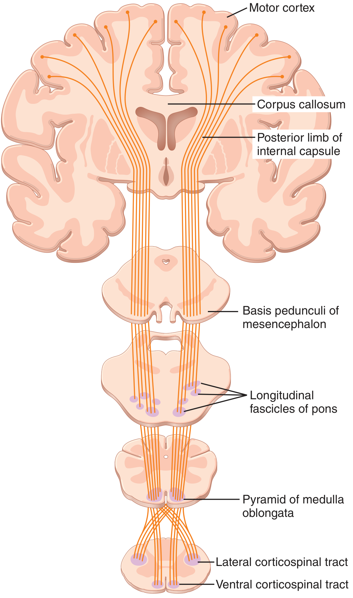
Corticospinal (pyramidal) tract. From Guyton & Hall Textbook of Medical Physiology.
Termination
Fibres terminate mainly on:
- Interneurons in the intermediate grey matter
- Sensory relay neurons in the dorsal horn
- A few directly on anterior motor neurons (direct excitation of muscle contraction)
Additional Fibre Pathways from Motor Cortex
- Short collaterals back to cortex from Betz cells - inhibit adjacent cortical areas ("sharpening" the excitatory signal)
- Fibres to caudate nucleus and putamen (basal ganglia) - postural muscle control
- Reticulospinal tract contributions
- Rubrospinal tract contributions (from red nucleus, midbrain)
Clinical Significance
- Damage to upper motor neurons (UMN) = spasticity, hyperreflexia, positive Babinski sign
- The lateral CST controls fine voluntary movements, especially distal limb muscles
- The ventral CST mainly controls bilateral postural movements
(Source: Guyton and Hall Textbook of Medical Physiology)
3. THALAMUS - FUNCTIONS AND CONNECTIONS
Introduction
The thalamus is the largest portion of the diencephalon and serves as the major synaptic relay station for all sensory information (except olfaction) reaching the cerebral cortex. It is located medial to the basal ganglia and lies on either side of the third ventricle.
Structural Organisation
The thalamus contains multiple nuclei separated by a Y-shaped sheet of myelinated fibres called the internal medullary lamina, which divides it into:
- Anterior nuclei
- Medial nuclei (Dorsomedial - DM)
- Lateral nuclei (dorsal tier: LD, LP, Pulvinar; ventral tier: VA, VL, VPL, VPM)
Additional nuclei:
- Reticular nucleus - thin inhibitory neuronal shell between external medullary lamina and internal capsule
- Intralaminar nuclei (Central Median - CM) - within the internal medullary lamina
- Midline nuclei - covers medial thalamic surface
- Geniculate bodies - LGB (visual), MGB (auditory)
LABELLED DIAGRAM: Thalamic Nuclei
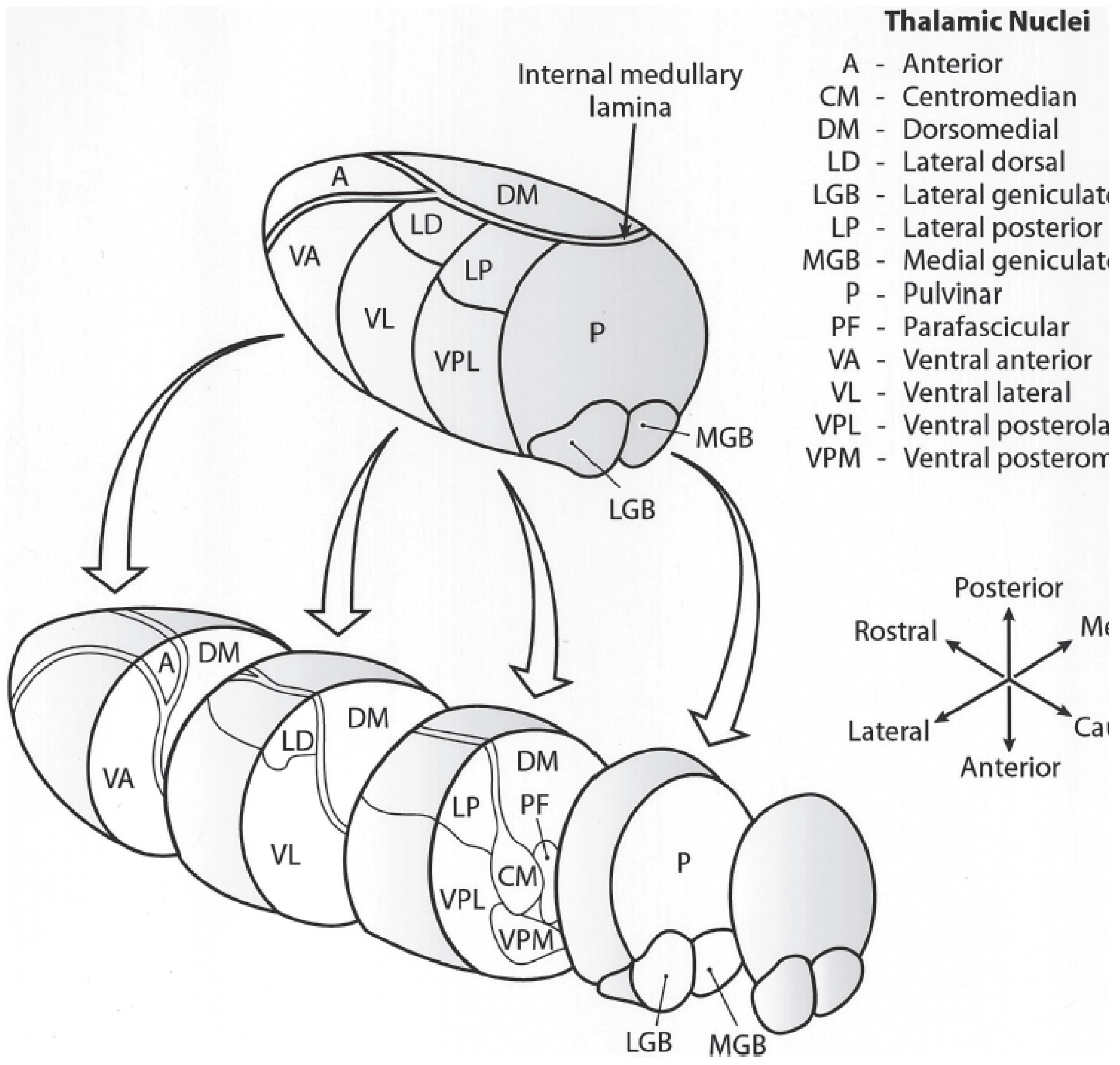
Thalamic nuclei organisation (Kaplan & Sadock's Comprehensive Textbook of Psychiatry)
Connections and Functions Table
Specific Relay Nuclei:
| Nucleus | Afferents (Input) | Efferents (Output) | Function |
|---|---|---|---|
| Anterior | Mammillary bodies, hippocampus | Cingulate cortex, parahippocampal gyrus | Emotion, learning, memory (Papez circuit) |
| Ventral Anterior (VA) | Globus pallidus, substantia nigra | Premotor cortex, frontal eye fields | Limb movement control, eye/face/head movements |
| Ventral Lateral (VL) | Dentate nucleus of cerebellum | Motor and premotor cortex | Head and limb movement control |
| VPL (Ventral Posterolateral) | Medial lemniscus + spinothalamic tract | Primary somatosensory cortex (S1) | Pain, temperature, touch, pressure, joint sense (body) |
| VPM (Ventral Posteromedial) | Sensory nuclei of trigeminal nerve | Primary somatosensory cortex (S1) | Pain, temperature, touch, pressure (face/head) |
| Medial Geniculate (MGB) | Inferior colliculus | Auditory cortex (temporal lobe) | Hearing |
| Lateral Geniculate (LGB) | Optic tract | Primary visual cortex (occipital lobe) | Vision |
Association Relay Nuclei:
| Nucleus | Afferents | Function |
|---|---|---|
| Lateral Dorsal (LD) | Pretectal area | Emotion (limbic connections) |
| Lateral Posterior (LP) | Superior colliculus, parietal cortex | Sensory integration |
| Pulvinar | Superior colliculus, temporal/parietal/occipital cortex | Integration of visual, auditory, somatosensory information |
| Medial Dorsal (MD) | Amygdala, hypothalamus, brainstem | Emotion, sensory integration; reciprocal with prefrontal cortex |
Diffuse-Projection Nuclei:
| Nucleus | Function |
|---|---|
| Midline nuclei | Modulation of cortical excitability |
| Intralaminar (CM) | Control of cortical activity; receives spinothalamic + reticular input |
| Reticular nucleus | Integrates thalamocortical activity; inhibitory (GABA) control of other thalamic nuclei |
FLOWCHART: Thalamic Circuitry
SENSORY PERIPHERY / SUBCORTICAL STRUCTURES
           ↓
  [Ascending sensory pathways]
           ↓
  SPECIFIC RELAY NUCLEI (VPL/VPM/LGB/MGB)
           ↓
  Primary Sensory Cortex (S1, V1, A1)
           ↓
  ←──── Feedback via Layer 6 pyramidal neurons ────→
           ↓
  ASSOCIATION RELAY NUCLEI (Pulvinar, MD, LD)
           ↓
  Association Cortex (prefrontal, parietal, temporal)
           ↑↓
  RETICULAR NUCLEUS
  (samples all corticothalamic traffic, modulates gain)
Functions Summary
- Relay station - All sensory modalities (except olfaction) synapse in thalamus before reaching cortex
- Motor integration - VA and VL relay cerebellar and basal ganglia output to motor cortex
- Consciousness and arousal - Intralaminar and midline nuclei regulate cortical excitability
- Emotion and memory - Anterior nucleus is part of Papez circuit
- Higher cortical integration - Pulvinar and MD link multiple cortical areas
- Pain modulation - Posterior nuclear group (nociceptive input)
- Cortico-thalamo-cortical relay - Higher-order relays (layer 5 → thalamus → another cortical area)
(Source: Kaplan & Sadock's Comprehensive Textbook of Psychiatry; Bradley and Daroff's Neurology)
4. CEREBRAL CORTEX - STRUCTURE, CONNECTIONS, AND FUNCTIONS
Introduction
The cerebral cortex is the outermost laminated sheet of neurons covering the cerebral hemispheres, several millimetres thick. It contains approximately 22 billion neurons communicating via ~165 trillion synapses, with ~12 million km of dendrites.
Structural Classification
By cell architecture (cytoarchitecture):
- Neocortex (Isocortex) - >90% of total cortical area; 6-layered structure (at some point in development); further divided into:
- Homotypical cortex (all 6 layers well-defined)
- Heterotypical cortex (some layers predominant - granular/agranular)
- Allocortex - remainder; includes:
- Paleocortex - base of telencephalon (olfactory areas)
- Archicortex - hippocampal formation
Six Layers of Neocortex (Outside to Inside)
| Layer | Name | Cell Types | Connections |
|---|---|---|---|
| I | Molecular layer | Few neurons, many dendrites and axons | Receives diffuse modulatory inputs |
| II | External granular | Small pyramidal + stellate cells | Corticocortical connections |
| III | External pyramidal | Medium pyramidal cells | Corticocortical (association/commissural) |
| IV | Internal granular | Dense stellate cells | Main INPUT layer from thalamus (thalamorecipient) |
| V | Internal pyramidal | Large pyramidal (including Betz cells) | Main OUTPUT to subcortical targets (brainstem, spinal cord) |
| VI | Multiform (fusiform) | Spindle-shaped cells | Corticothalamic projections |
Cell Types
Pyramidal neurons (~75% of neocortical neurons):
- Characteristic triangular soma
- Single apical dendrite ascending toward cortical surface
- Short basal dendrites spreading laterally
- Dendrites covered in dendritic spines (sites of excitatory synapses)
- Excitatory projection neurons using glutamate
- Found in layers II, III, V, VI
Stellate/Nonpyramidal neurons (~25%):
- Local circuit interneurons
- Use inhibitory neurotransmitter GABA
- Multiple subtypes distinguished by:
- Neuropeptides (VIP, NPY, somatostatin)
- Calcium-binding proteins (parvalbumin, calbindin)
- Morphology (chandelier cells, basket cells, bitufted cells)
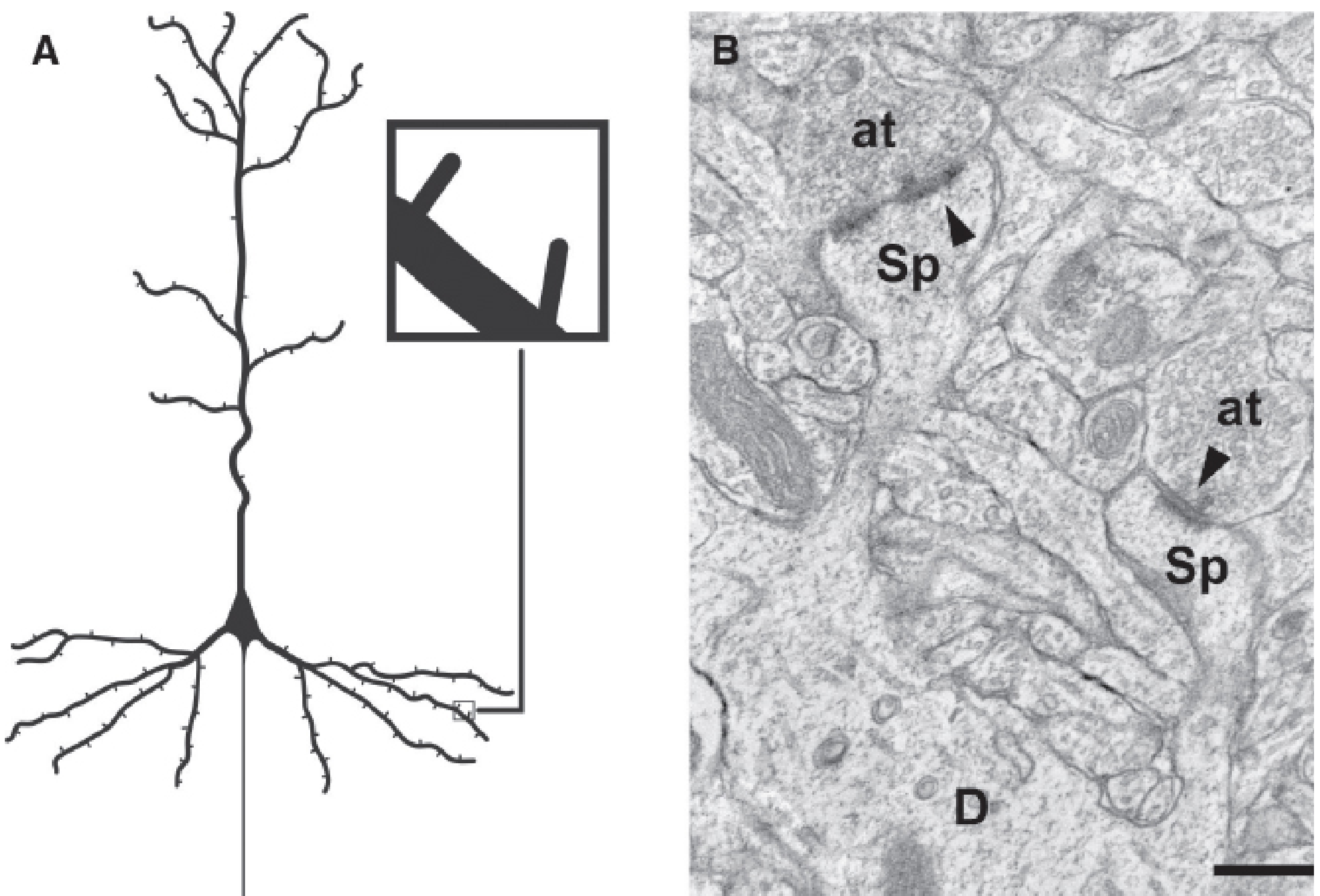
Dendritic spines in the cerebral cortex (Kaplan & Sadock's Comprehensive Textbook of Psychiatry)
Functional Areas (Brodmann Areas)
Primary Sensory Areas:
- Primary visual cortex (V1) - Occipital lobe (area 17), calcarine cortex
- Primary auditory cortex (A1) - Superior temporal gyrus (areas 41, 42)
- Primary somatosensory cortex (S1) - Postcentral gyrus (areas 3, 1, 2)
- Primary motor cortex (M1) - Precentral gyrus (area 4)
Association Cortices:
- Unimodal association cortex - Adjacent to primary areas; processes one modality in increasing complexity
- Heteromodal (polymodal) association cortex - Two principal sites:
- Posterior inferior parietal lobe (angular gyrus) - cross-modal integration
- Prefrontal cortex (DLPFC) - executive functions, working memory
Connections of the Cerebral Cortex
FLOWCHART: Types of Cortical Connections
CEREBRAL CORTEX
        │
        ├─── ASSOCIATION FIBRES (corticocortical, ipsilateral)
        │       ├── Short: connect adjacent gyri (U-fibres)
        │       └── Long: connect lobes (arcuate fasciculus, uncinate, etc.)
        │
        ├─── COMMISSURAL FIBRES (contralateral hemisphere)
        │       ├── Corpus callosum (main commissure)
        │       ├── Anterior commissure
        │       └── Posterior commissure
        │
        ├─── PROJECTION FIBRES (to/from subcortical structures)
        │       ├── Thalamocortical (afferent via internal capsule)
        │       ├── Corticospinal tract (to spinal cord)
        │       ├── Corticobulbar (to brainstem motor nuclei)
        │       ├── Corticopontine (to cerebellum via pons)
        │       └── Corticothalamic (feedback to thalamus)
        │
        └─── MODULATORY INPUTS (from subcortical systems)
                ├── Dopaminergic (from VTA/SNc)
                ├── Noradrenergic (from locus coeruleus)
                ├── Serotonergic (from raphe nuclei)
                └── Cholinergic (from basal forebrain)
Functional Hierarchy
PRIMARY CORTEX (raw sensation)
        ↓
UNIMODAL ASSOCIATION CORTEX (higher processing within one modality)
        ↓
HETEROMODAL ASSOCIATION CORTEX (cross-modal integration)
        ↓
PREFRONTAL CORTEX / LIMBIC CORTEX (executive, emotional, memory)
Functions Summary
- Voluntary motor control - Primary motor cortex (M1), corticospinal output
- Sensory perception - Somatosensory, visual, auditory primary areas
- Language - Broca's area (area 44/45, left IFG) = production; Wernicke's area (area 22, left STG) = comprehension
- Memory - Hippocampus + entorhinal cortex + association cortex
- Executive function and planning - Prefrontal cortex (DLPFC)
- Emotional processing - Limbic cortex, cingulate
- Visual object recognition - Ventral "what" stream (temporal)
- Spatial processing - Dorsal "where/how" stream (parietal)
(Source: Bradley and Daroff's Neurology; Kaplan & Sadock's Comprehensive Textbook of Psychiatry)
5. VISUAL PATHWAY WITH LESIONS
Introduction
The visual pathway transmits visual information from the retina to the primary visual cortex (V1). A precise knowledge of its anatomy allows localisation of lesions from specific visual field deficits.
Components of the Visual Pathway
- Retina (photoreceptors → bipolar cells → retinal ganglion cells)
- Optic nerve (CN II)
- Optic chiasm
- Optic tract
- Lateral Geniculate Nucleus (LGN) of thalamus
- Optic radiations (geniculocalcarine tract)
- Primary visual cortex (V1, area 17, calcarine cortex)
Detailed Pathway
FLOWCHART:
PHOTORECEPTORS (rods & cones) in retina
        ↓ synapse
BIPOLAR CELLS
        ↓ synapse
RETINAL GANGLION CELLS
        ↓
OPTIC NERVE (axons coalesce at optic disc → blindspot)
        ↓
OPTIC CHIASM (anterior to infundibular stalk)
   ┌────────────────────┐
   │  PARTIAL CROSSING: │
   │ Nasal retinal fibres│ → decussate → contralateral optic tract
   │ Temporal fibres    │ → stay ipsilateral
   └────────────────────┘
        ↓
OPTIC TRACT (courses around midbrain)
   ├── Most fibres → LGN of thalamus
   └── Small bundle → Pretectal area + Superior colliculus
                      (pupillary light reflex)
        ↓
LATERAL GENICULATE NUCLEUS (LGN), thalamus
        ↓
OPTIC RADIATIONS (Geniculocalcarine tract)
   ├── Upper fibres → through parietal lobe → upper calcarine cortex
   │                  (represent lower visual field)
   └── Lower fibres (MEYER'S LOOP) → through temporal lobe
                                → lower calcarine cortex
                                   (represent upper visual field)
        ↓
PRIMARY VISUAL CORTEX (V1)
Area 17, calcarine fissure, occipital lobe
[Macular fibres → posterior pole; peripheral fibres → anterior]
Retinal Representation Key Principle
- Right visual field → left half of each retina → left LGN → left V1
- Left visual field → right half of each retina → right LGN → right V1
- Nasal retina = contralateral visual field
- Temporal retina = ipsilateral visual field
LABELLED DIAGRAM: Visual Pathway and Optic Radiations
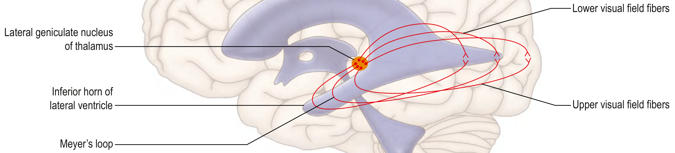
Optic radiations: Meyer's loop carries lower visual field fibres through the temporal lobe (Gray's Anatomy for Students)
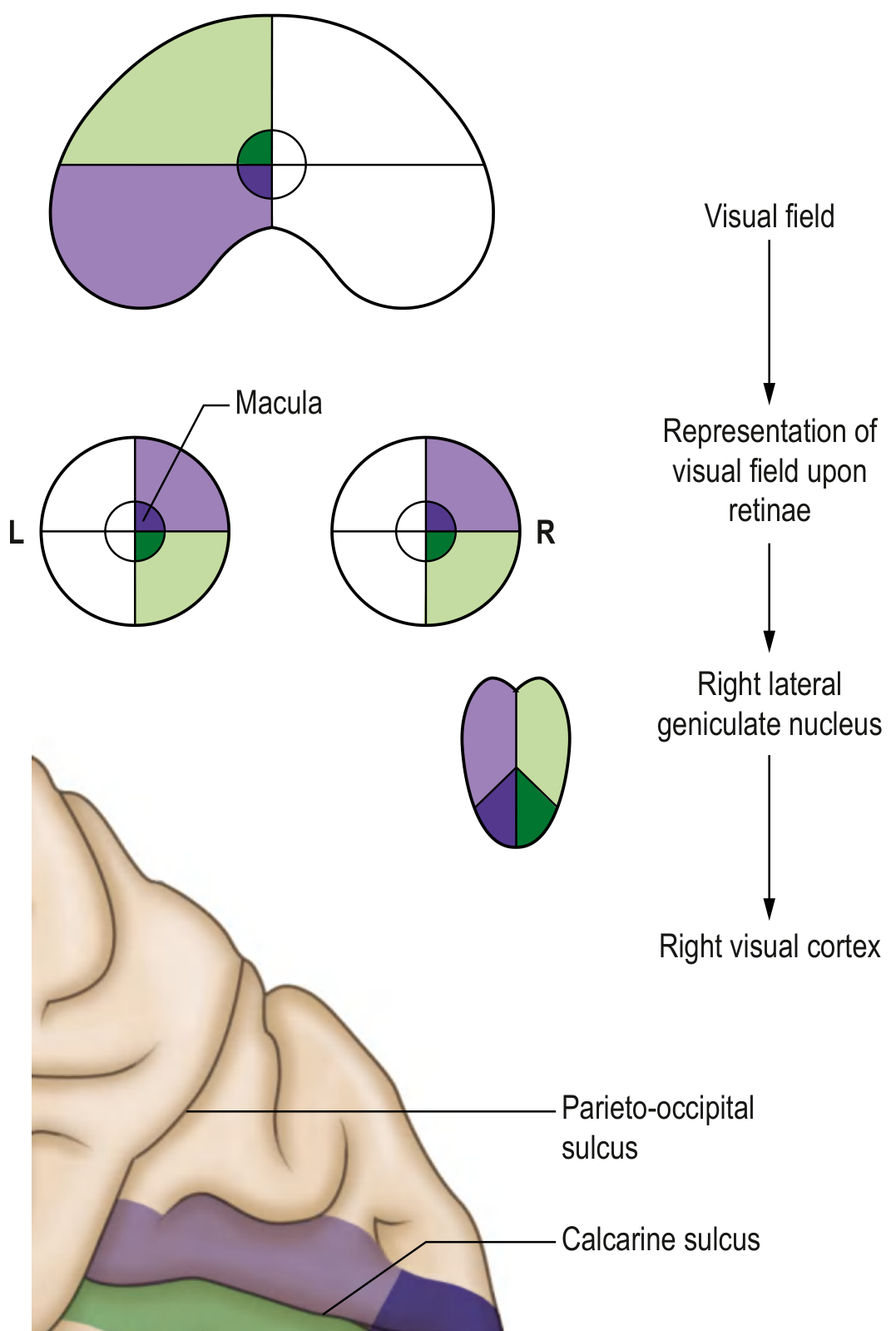
Representation of the left half of the visual field at various portions along the visual pathway (Gray's Anatomy for Students)
Visual Field Defects with Lesion Sites
| # | Lesion Site | Visual Field Defect | Cause / Notes |
|---|---|---|---|
| 1 | Optic nerve (one side) | Monocular blindness (ipsilateral eye) | Total loss of vision in one eye; e.g., optic neuritis, trauma |
| 2 | Optic chiasm - central | Bitemporal hemianopia ("tunnel vision") | Nasal fibres from both eyes decussate here; compressed by pituitary adenoma |
| 3 | Optic chiasm - lateral | Binasal hemianopia | Rare; bilateral lateral chiasm compression (aneurysm) |
| 4 | Optic tract (one side) | Contralateral homonymous hemianopia (incongruous) | Right optic tract lesion → left homonymous hemianopia |
| 5 | Meyer's loop (temporal lobe) | Contralateral upper quadrantanopia ("pie in the sky") | Temporal lobe surgery/tumour; affects lower visual field fibres |
| 6 | Parietal radiation (upper) | Contralateral lower quadrantanopia | Parietal lobe lesion |
| 7 | Complete optic radiation | Contralateral homonymous hemianopia (congruous) | With macular sparing possible if occipital pole spared |
| 8 | Occipital cortex (V1) | Contralateral homonymous hemianopia with MACULAR SPARING | Dual blood supply (MCA + PCA) preserves macular representation |
FLOWCHART: Lesion Localisation
Monocular visual loss
  → Lesion IPSILATERAL to blind eye
  → Optic nerve / retina

Bitemporal hemianopia
  → Optic CHIASM (central)

Homonymous hemianopia (both eyes same side)
  → Lesion CONTRALATERAL to visual field loss
  → Post-chiasmal: tract → radiation → cortex

Upper quadrantanopia
  → Meyer's loop (temporal lobe)

Lower quadrantanopia
  → Parietal optic radiation

Homonymous hemianopia + macular sparing
  → Occipital cortex (posterior cerebral artery territory)
(Source: Gray's Anatomy for Students; Scott-Brown's Otorhinolaryngology; Bradley and Daroff's Neurology)
6. ORGAN OF CORTI
Introduction
The organ of Corti is the sensory end-organ of hearing, located within the cochlea of the inner ear. It lies on the basilar membrane within the scala media (cochlear duct) and is covered by the tectorial membrane.
Location in the Cochlea
COCHLEA (cross-section)
        │
   ┌────┴─────────────┐
   │  SCALA VESTIBULI  │ (perilymph, above)
   │  ───────────────  │
   │  SCALA MEDIA      │ ← Organ of Corti lies here (on basilar membrane)
   │  (endolymph)      │   Reissner's membrane above; Basilar membrane below
   │  ───────────────  │
   │  SCALA TYMPANI    │ (perilymph, below)
   └───────────────────┘
Structure of the Organ of Corti
Components:
| Structure | Description |
|---|---|
| Basilar membrane | Foundation; ~35 mm long in humans; width and mass increase from base to apex; determines frequency tuning (tonotopy) |
| Inner hair cells (IHCs) | Single row, ~3,500 cells, ~12 μm diameter; primary transducers (90-95% of auditory nerve fibres innervate IHCs) |
| Outer hair cells (OHCs) | 3-4 rows, ~12,000 cells, ~8 μm diameter; electromotile - amplify cochlear response ("tuning"); receive retrograde (efferent) innervation |
| Stereocilia (hair bundles) | Project upward from hair cells; tips embedded in tectorial membrane (IHCs) or free-standing (OHCs) |
| Tectorial membrane | Overlies hair cells; gelatinous; stereocilia deflect against it during basilar membrane vibration |
| Rods of Corti (pillar cells) | Triangular supporting structures; form tunnel of Corti |
| Tunnel of Corti | Between IHC and OHC regions; filled with perilymph |
| Spaces of Nuel | Around OHCs; filled with perilymph |
| Reticular lamina | Rigid plate at apical surface of hair cells; outer ends of hair cells fixed here |
| Spiral ganglion (of Corti) | In modiolus (central axis of cochlea); contains ~30,000 bipolar neurons; sends axons as cochlear nerve to brainstem |
LABELLED DIAGRAM (Schematic)
          TECTORIAL MEMBRANE (overlying)
                    │
         ┌──────────┴──────────────────┐
   Scala │  IHC  Tunnel  OHC OHC OHC  │
   media │   │   of Corti  │   │   │  │
         │   │←Rods of Corti→│   │  │  │
         │   └───────────────────────┘  │
         │        RETICULAR LAMINA       │
         │                               │
         └────────BASILAR MEMBRANE───────┘
                    │
             SPIRAL LIGAMENT
                    │
              SCALA TYMPANI
KEY:
IHC = Inner Hair Cell (single row, ~3,500)
OHC = Outer Hair Cell (3-4 rows, ~12,000)
Spaces of Nuel surround OHCs
Mechanism of Transduction
FLOWCHART:
Sound wave → Vibration of tympanic membrane
        ↓
Ossicles (malleus → incus → stapes)
        ↓
Oval window → fluid waves in perilymph (scala vestibuli)
        ↓
Reissner's membrane → scala media
        ↓
BASILAR MEMBRANE VIBRATES
[High freq. = base; Low freq. = apex → Tonotopy]
        ↓
Reticular lamina + rods of Corti move as RIGID UNIT
        ↓
Hair stereocilia SHEAR against tectorial membrane
        ↓
Bending TOWARD tallest stereocilium → DEPOLARISATION
(K+ enters via mechanosensitive ion channels from endolymph)
Bending AWAY from tallest stereocilium → HYPERPOLARISATION
        ↓
Receptor potential in IHC
        ↓
Glutamate release at base → excitation of spiral ganglion
        ↓
Cochlear nerve (CN VIII, ~30,000 fibres)
        ↓
Cochlear nucleus (medulla)
        ↓
Superior olivary nucleus → bilateral projections
        ↓
Lateral lemniscus
        ↓
Inferior colliculus (midbrain)
        ↓
Medial geniculate nucleus (thalamus)
        ↓
Primary auditory cortex (Heschl's gyrus, area 41/42)
Tonotopy
The basilar membrane is mechanically tuned:
- Base (narrow, stiff) → responds to high frequencies
- Apex (wide, compliant) → responds to low frequencies
- This tonotopic map is preserved all the way up to the auditory cortex
Role of Outer Hair Cells
OHCs are electromotile - they change length in response to voltage changes, actively amplifying basilar membrane motion by up to 1000-fold. They act as a "cochlear amplifier" and fine-tune frequency selectivity. Efferent (olivocochlear) fibres from the brainstem modulate OHC stiffness/length, enabling the brain to modify its own hearing sensitivity.
Clinical Points
- Sensorineural hearing loss - damage to hair cells or spiral ganglion (noise trauma, ototoxic drugs, ageing)
- OHCs damaged preferentially by noise and ototoxins (aminoglycosides, cisplatin)
- Loss of OHCs reduces sensitivity and frequency selectivity but IHCs remain functional
- Otoacoustic emissions (OAEs) are sounds produced by OHC electromotility - used in neonatal hearing screening
(Sources: Guyton & Hall Textbook of Medical Physiology; Scott-Brown's Otorhinolaryngology Head & Neck Surgery; Costanzo Physiology 7th Edition)
Note: All content sourced from authoritative medical textbooks including Guyton & Hall Medical Physiology, Bradley & Daroff's Neurology, Gray's Anatomy for Students, Kaplan & Sadock's Comprehensive Textbook of Psychiatry, and Scott-Brown's Otorhinolaryngology. These essays cover the standard exam requirements with detailed anatomy, mechanisms, clinical correlations, labelled diagrams, and flowcharts.
This is a shared conversation. Sign in to Orris to start your own chat.