
Acute onset SOB and pedal edema with Dilated RA Moderate PAH severe TR with Chest X ray showing unilateral white out lung? Probable diagnosis, evaluation and management
Reading File
Finding Sources
Finding Sources
Finding Sources
Searching PubMed
"pleural effusion" AND "pulmonary embolism"
Searching the Web
unilateral opaque hemithorax acute dyspnea diagnostic approach massive pleural effusion guidelines
Reading File
Finding Sources
Finding Sources
Reading Web Page
https://pmc.ncbi.nlm.nih.gov/articles/PMC4753987/
Searching PubMed
"acute pulmonary embolism" AND "practice guideline"
This presentation requires urgent hospital assessment, ideally in an emergency department or high-dependency setting.
Most likely working diagnosis
Massive unilateral pleural effusion causing compressive atelectasis, producing the unilateral “white-out” on chest X-ray, with decompensated right-sided heart failure from pulmonary hypertension and severe functional tricuspid regurgitation.
However, acute pulmonary embolism (PE) on a background of chronic pulmonary hypertension must be actively excluded. Acute PE can abruptly worsen dyspnea and right-heart pressure overload, while chronic pulmonary hypertension produces right atrial enlargement, right ventricular dilatation, and moderate-to-severe TR. Textbook of Clinical Echocardiography describes this chronic progression, while noting that acute PE can cause abrupt RV pressure overload (block 2, lines 4417-4419).
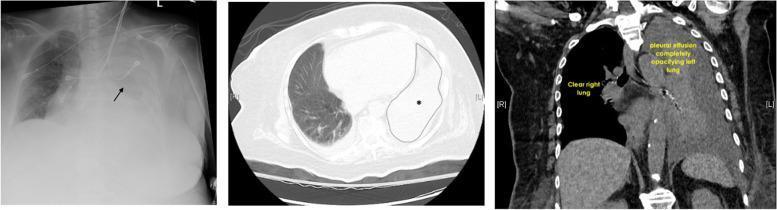
Important differential diagnosis of a unilateral white-out hemithorax
The direction of mediastinal shift is very helpful:
| X-ray finding | Leading possibilities |
|---|---|
| Shift away from the opacity | Massive pleural effusion, tension hydrothorax, occasionally massive intrathoracic mass |
| Shift toward the opacity | Whole-lung collapse from a main bronchial obstruction, mucus plug, or endobronchial tumor |
| No shift | Extensive consolidation, combined collapse plus effusion, fixed mediastinum from malignancy/fibrosis |
A massive effusion usually shifts the mediastinum to the opposite side. If it does not, consider an ipsilateral main-stem bronchial obstruction with collapse, malignant pleural disease, or mediastinal fixation. Review of pleural-effusion imaging supports this distinction.
Etiology of the large effusion to consider
Do not assume it is solely due to heart failure, especially when it is unilateral and massive.
- Heart failure-related transudative effusion, possibly worsened by severe TR and pulmonary hypertension. Usually bilateral or right-sided, but can be unilateral.
- Malignancy, a common cause of massive unilateral effusion.
- Parapneumonic effusion or empyema, especially with fever, leukocytosis, pleuritic pain, or elevated inflammatory markers.
- Tuberculous pleuritis, depending on epidemiology and subacute constitutional symptoms.
- Hemothorax, if trauma, anticoagulation, procedure, or a falling hemoglobin level.
- Pulmonary embolism, which may produce pleural effusion and acute RV strain, though a complete white-out would be less typical.
- Whole-lung collapse, if ultrasound does not confirm a large fluid collection.
Immediate evaluation
1. Stabilize first
- ABC assessment, continuous pulse oximetry and cardiac monitoring.
- Sit upright.
- Give supplemental oxygen, targeting adequate saturation. Use ventilatory support if fatigued, hypercapnic, or severely hypoxemic.
- Two IV lines, blood pressure monitoring, urine output.
- Look immediately for shock: hypotension, altered sensorium, cool extremities, rising lactate, oliguria.
If hypotension with suspected massive PE or tension hydrothorax is present, involve critical care, cardiology, respiratory medicine, and interventional radiology/thoracic surgery immediately.
2. Bedside tests
- POCUS of chest: confirm fluid versus consolidation/collapse; assess volume, septations, echogenicity, and a safe drainage site.
- Focused cardiac ultrasound/urgent echocardiography:
- RV size and systolic function
- LV systolic/diastolic function
- TR severity and estimated pulmonary artery systolic pressure
- IVC size/collapsibility
- Pericardial effusion/tamponade
- Right-heart thrombus, if present
- ECG for tachyarrhythmia, ischemia, or right-heart strain.
- ABG/VBG with lactate in a sick patient.
3. Blood tests
- CBC, renal function, electrolytes, liver profile, glucose.
- Troponin and BNP/NT-proBNP.
- CRP/procalcitonin and blood cultures before antibiotics if infection is plausible.
- PT/INR, aPTT, platelet count and blood group/crossmatch before drainage.
- D-dimer only if pretest probability for PE is low/intermediate and it will change imaging decisions. Do not use it to delay imaging in a high-probability case.
- Hemoglobin trend if hemothorax is possible.
4. Definitive imaging
- Repeat portable chest X-ray if needed, paying close attention to mediastinal shift.
- Contrast CT chest after initial stabilization to define fluid, lung collapse, mass, pleura, infection, and lymphadenopathy.
- CT pulmonary angiography if PE is clinically suspected and renal function/hemodynamics permit.
- If too unstable for CT, bedside echo plus lower-limb compression ultrasound can help guide decisions.
Pleural-fluid evaluation
Unless this is clearly a small, typical bilateral heart-failure effusion that resolves promptly with diuresis, perform ultrasound-guided diagnostic thoracentesis.
Send pleural fluid for:
- Appearance and odor
- Cell count and differential
- Protein and LDH, with simultaneous serum protein and LDH for Light's criteria
- pH and glucose, particularly if infection is possible
- Gram stain and bacterial culture
- Cytology
- AFB smear/culture and ADA when tuberculosis is clinically plausible
- Triglycerides if chylothorax is suspected
- Hematocrit if hemorrhagic fluid is obtained
Chest ultrasound can distinguish fluid from a solid or consolidated lung and can guide a safer thoracentesis. This review summarizes its role.
Management
A. If this is a large symptomatic pleural effusion
- Ultrasound-guided therapeutic thoracentesis for dyspnea/hypoxemia.
- Drain in a controlled fashion and stop for chest pain, persistent cough, vasovagal symptoms, or clinical deterioration. Avoid rapid, excessive drainage because re-expansion pulmonary edema can occur.
- In emergency medicine guidance, therapeutic drainage of roughly 1.0-1.5 L is suggested for dyspnea at rest, with caution regarding larger acute volumes. Tintinalli's Emergency Medicine, treatment section (block 5, lines 3923-3948).
- If pus, positive Gram stain/culture, loculations, or a complicated parapneumonic effusion: intercostal chest-tube drainage plus IV antibiotics, with specialist review for intrapleural therapy or surgery.
B. Treat decompensated right-sided heart failure, but cautiously
- IV loop diuretic if clinically volume overloaded, with close monitoring of blood pressure, renal function, potassium, and urine output.
- Salt and fluid restriction where appropriate.
- Treat triggers: infection, arrhythmia, anemia, renal failure, pulmonary embolism, medication nonadherence, or left-sided cardiac disease.
- Avoid indiscriminate large fluid boluses in a congested, failing right ventricle.
C. If PE is suspected
- Assess hemodynamic risk immediately.
- If no major contraindication and imaging is delayed but clinical probability is high, clinicians may begin therapeutic anticoagulation after considering bleeding risk and the possibility of hemothorax/procedural drainage.
- Hypotension or shock with confirmed/highly likely PE requires urgent PE-response or critical-care input for reperfusion options such as systemic thrombolysis, catheter-based therapy, or embolectomy as appropriate.
- A current multidisciplinary US guideline for acute PE is indexed as the 2026 AHA/ACC/ACCP/ACEP/CHEST guideline.
Bottom line
The immediate label should be:
Acute hypoxemic dyspnea due to a large unilateral pleural effusion with compressive atelectasis, in a patient with chronic pulmonary hypertension, severe TR, and right-sided heart failure. Rule out acute PE, malignancy, infection/empyema, tuberculosis, and whole-lung collapse from bronchial obstruction.
The urgent next actions are bedside chest ultrasound, echocardiography, oxygen/supportive stabilization, diagnostic plus symptom-relieving ultrasound-guided thoracentesis, and CT/CTPA when stable.
This is a shared conversation. Sign in to Orris to start your own chat.