
Tell me about renal ccell carcinoma renal oncytoma nephroblastoma urothelisl carcinoma of bladder complete concept with every single detail in paragraph
Renal Cell Carcinoma, Renal Oncocytoma, Nephroblastoma, and Urothelial Carcinoma of the Bladder
1. RENAL CELL CARCINOMA (RCC)
Epidemiology and Overview
Familial Syndromes
Classification and Pathogenesis
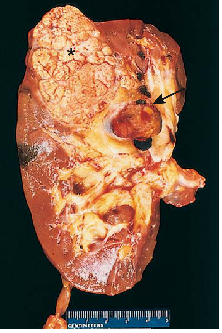
Gross Morphology
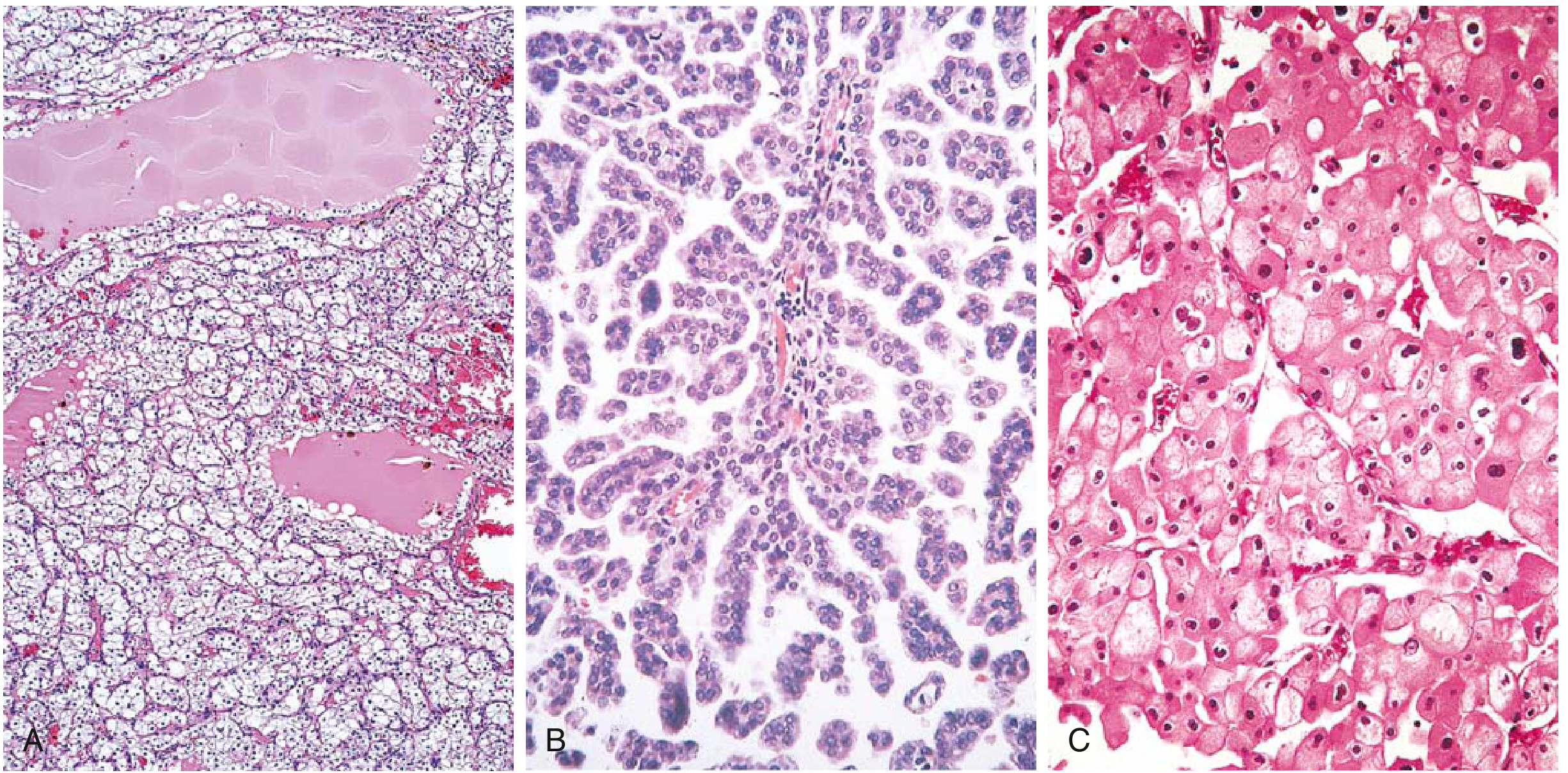
Histology
Clinical Features
Imaging
Treatment and Prognosis
2. RENAL ONCOCYTOMA
Definition and Cell of Origin
Pathogenesis and Molecular Features
Gross Morphology
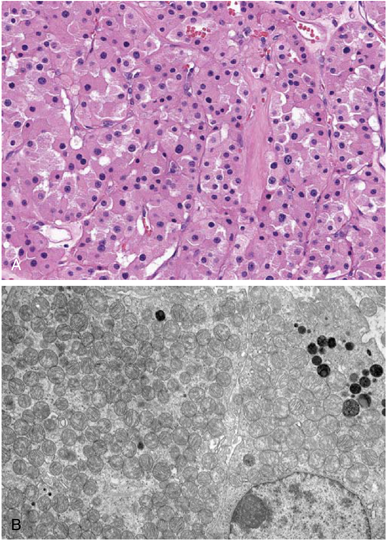
Histology
Diagnostic Challenge
3. NEPHROBLASTOMA (WILMS TUMOR)
Epidemiology and Overview
Associated Syndromes
- WAGR syndrome (Wilms tumor, Aniridia, Genitourinary malformations, and intellectual disability/mental Retardation): caused by deletions at chromosome 11p13 that encompass the WT1 gene.
- Beckwith-Wiedemann syndrome: an overgrowth syndrome characterized by macroglossia, visceromegaly, and hemihypertrophy, associated with alterations in the IGF1, H19, and p57 genes (linked to chromosome 11p15 imprinting).
- Isolated hemihypertrophy: associated with increased risk even without other features of Beckwith-Wiedemann.
- Denys-Drash syndrome: WT1 mutation causing gonadal dysgenesis, nephropathy, and Wilms tumor.
- Genitourinary abnormalities (hypospadias, cryptorchidism, renal fusion) are found in 4.5-7.5% of patients with unilateral Wilms tumor and up to 13.4% of those with bilateral disease.
Pathogenesis and Genetics
Precursor Lesions: Nephrogenic Rests
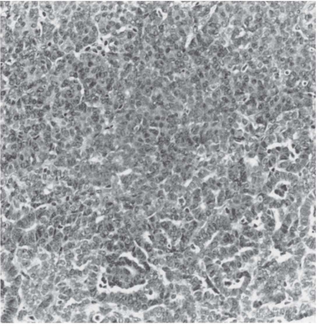
Pathology and Histology
Histologic Prognosis: Favorable vs. Unfavorable
Staging (NWTS System)
- Stage I: Tumor limited to the kidney and completely excised; no capsule penetration or renal sinus vessel involvement.
- Stage II: Tumor extends beyond the kidney but is completely removed; may involve capsule penetration, renal sinus vessel invasion, local biopsy, or local spillage.
- Stage III: Residual non-hematogenous tumor confined to the abdomen (e.g., positive lymph nodes, peritoneal contamination, incomplete resection, or tumor spillage not confined to the flank).
- Stage IV: Hematogenous metastases (lung, liver, bone, brain) or lymph node metastases beyond the abdomino-pelvic region.
- Stage V: Bilateral renal involvement at diagnosis.
Clinical Presentation
Treatment
4. UROTHELIAL CARCINOMA OF THE BLADDER
Epidemiology and Overview
Classification: Non-Muscle-Invasive vs. Muscle-Invasive
Molecular Pathogenesis
Histologic Grading
Histologic Variants
Gross and Pathologic Features
TNM Staging
- Ta: Non-invasive papillary carcinoma
- Tis: Carcinoma in situ (flat)
- T1: Invasion into subepithelial connective tissue (lamina propria)
- T2a/T2b: Invasion into superficial/deep muscularis propria
- T3a/T3b: Microscopic/macroscopic invasion into perivesical fat
- T4a: Invasion into prostate stroma, uterus, or vagina
- T4b: Invasion into pelvic or abdominal wall
- N1-3: Regional lymph node involvement
- M1: Distant metastasis
Clinical Presentation
Diagnosis
Treatment
Summary Comparison Table
| Feature | RCC (Clear Cell) | Renal Oncocytoma | Nephroblastoma | Urothelial Ca (Bladder) |
|---|---|---|---|---|
| Cell of origin | Proximal tubule | Intercalated cells (collecting duct) | Metanephric blastema | Transitional epithelium |
| Age | 6th-7th decade | Any adult | Peak ~3 years | 6th-8th decade |
| Gender | M:F = 2:1 | Slight male predominance | Equal | M:F = 3:1 |
| Nature | Malignant | Benign | Malignant | Malignant |
| Key gene | VHL (3p25) | Complex I genes; chr 1/Y loss | WT1 (11p13) | p53, Rb, FGFR3 |
| Gross | Yellow, necrotic, vascular | Tan/mahogany, central scar | Large, multilobulated, gray | Papillary or flat/indurated |
| Classic symptom | Hematuria + flank pain + mass | Incidental | Abdominal mass | Painless hematuria |
| Spread | Renal vein, IVC | None (benign) | Renal vein, lungs, liver | Lymphatics, hematogenous |
| Key treatment | Partial/radical nephrectomy ± targeted therapy | Partial nephrectomy | Nephrectomy + chemo +/- RT | TURBT + BCG / Cystectomy + neoadjuvant chemo |
Tell me more in detail i'm not understanding
Which topic(s) are you finding hard to understand? · What part is confusing you most? · What is your level / purpose?
Complete Student Guide: Renal & Bladder Tumors - Explained From Scratch
HOW TO READ THIS GUIDE
- Where does it come from? (Cell of origin)
- Why does it happen? (Pathogenesis/molecular mechanism - step by step)
- What does it look like? (Gross + histology)
- How does the patient present? (Clinical features)
- How do we treat it? (Management)
TOPIC 1: RENAL CELL CARCINOMA (RCC)
Step 1 - Where Does It Come From?
Step 2 - Why Does It Happen? (Pathogenesis, explained simply)
A) CLEAR CELL RCC (most common - 65-80%)
- Both copies of the VHL gene are lost/mutated (two-hit model: chromosome 3p deletion knocks out one copy; somatic mutation/hypermethylation knocks out the second copy).
- With no VHL "garbage collector" functioning, HIF-1 is never destroyed.
- HIF-1 accumulates even with normal oxygen → it acts as if the cell is hypoxic all the time.
- This causes constant overexpression of VEGF (→ rich blood vessel formation in the tumor, explaining its high vascularity) and IGF-1 (→ cell proliferation).
- HIF-1 also teams up with MYC to rewire cell metabolism to favor growth.
- Additionally, mutations in histone-methylation genes alter the epigenome, contributing further.
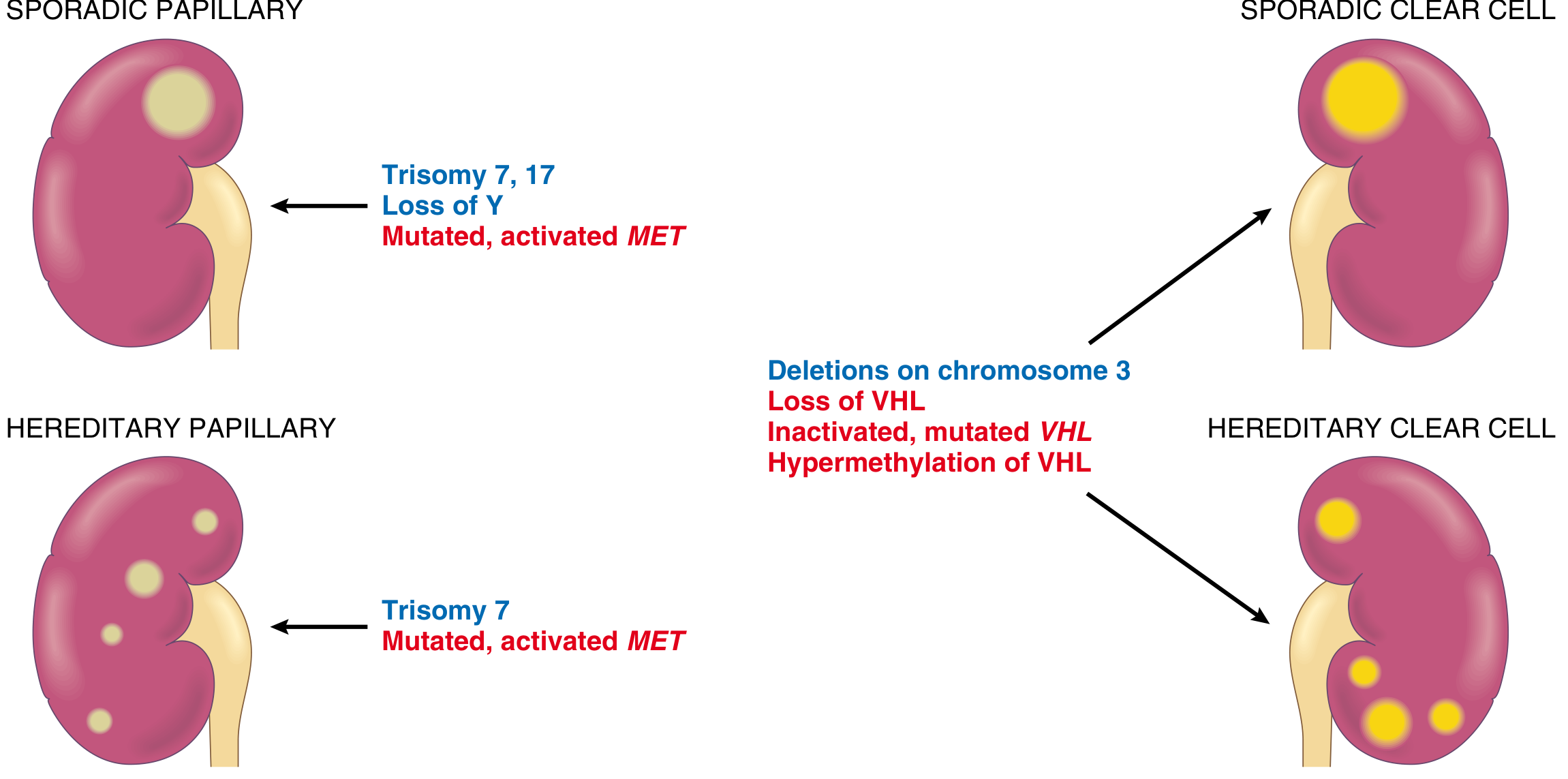
B) PAPILLARY RCC (10-15%)
- Gain-of-function mutations or extra copies of MET (from trisomies of chromosome 7 and 17) cause the MET receptor to be permanently overactive - it fires growth signals non-stop, even without HGF.
- This drives abnormal proliferation of proximal/distal tubular cells.
- Since the mutation can be in every kidney cell (germline/hereditary form) or in many cells (sporadic, from trisomy 7), these tumors are often multifocal and bilateral.
C) CHROMOPHOBE RCC (5%)
Step 3 - What Does It Look Like?
Gross Appearance (what you see with naked eye)
Histology (what you see under microscope)
Step 4 - How Does the Patient Present?
- Polycythemia (high RBC count) - tumor secretes erythropoietin - seen in 5-10%
- Hypercalcemia - tumor secretes PTHrP
- Hypertension - tumor secretes renin
- Cushing syndrome - ectopic ACTH
- Hepatic dysfunction without liver metastasis (Stauffer syndrome) - rare
- Fever, weight loss, fatigue (constitutional symptoms)
Step 5 - How Do We Treat It?
- Partial nephrectomy (remove only the tumor, preserve rest of kidney) - preferred for tumors <4 cm (T1a) and when feasible for larger tumors.
- Radical nephrectomy (remove entire kidney + fat + adrenal) for larger/complex tumors.
- Even with renal vein/IVC thrombus - surgery can still be curative!
- Anti-VEGF/anti-angiogenic drugs (sunitinib, pazopanib, axitinib) - block the VEGF pathway driven by HIF overactivation.
- mTOR inhibitors (everolimus, temsirolimus)
- Immune checkpoint inhibitors (nivolumab + ipilimumab combination, pembrolizumab) - now first-line for many metastatic patients.
- No metastases → ~100% 5-year survival
- Renal vein invasion or perinephric fat involvement → ~60% 5-year survival
- Overall average → ~70% 5-year survival
TOPIC 2: RENAL ONCOCYTOMA
The Big Picture First
Cell of Origin
Why Does It Happen? (Pathogenesis)
- Cytoplasm is full of mitochondria → appears bright pink/eosinophilic (granular) under the microscope (mitochondria take up eosin stain).
- The massive mitochondrial content makes the tumor brownish-tan/mahogany in color to the naked eye (mitochondria give it that color).
- On electron microscopy, you can literally see the cytoplasm packed densely with mitochondria.
Gross Appearance
- Tan or mahogany brown color (not yellow like clear cell RCC)
- Well-circumscribed, with a fibrous capsule
- Central stellate scar - a characteristic central scar in a star/spoke shape - present in about 1/3 of cases. This scar can sometimes be seen on CT/MRI and suggests oncocytoma, but is NOT diagnostic.
- Can grow very large - up to 12 cm.
Histology
Clinical Features and Why It's Tricky
Treatment
TOPIC 3: NEPHROBLASTOMA (WILMS TUMOR)
The Big Picture First
Cell of Origin and Concept
Pathogenesis - The Genetics (Step by Step)
The Two-Hit Hypothesis (Knudson, 1972)
- Sporadic form: Both mutations happen after birth, in the same kidney cell (post-zygotic). These patients typically present later, with unilateral tumor.
- Familial/hereditary form: The first mutation is present in every cell from birth (inherited germline mutation). Only ONE more somatic mutation is needed in any kidney cell to start a tumor. This explains why familial cases present EARLIER in life and are more likely to be BILATERAL and MULTIFOCAL.
Key Gene: WT1 (Chromosome 11p13)
Associated Syndromes (Very High-Yield for Exams)
| Syndrome | Features | Gene |
|---|---|---|
| WAGR | Wilms + Aniridia + Genitourinary malformations + Retardation | Deletion 11p13 (includes WT1 and PAX6) |
| Beckwith-Wiedemann | Macroglossia + macrosomia + organomegaly + hemihypertrophy + ear creases | 11p15 (IGF-1, H19, p57 imprinting) |
| Denys-Drash | Gonadal dysgenesis + nephropathy + Wilms tumor | WT1 point mutation |
Gross Appearance
Histology - The Triphasic Pattern (The Key to Recognizing Wilms on Biopsy)
-
Blastema - Primitive small round blue cells, tightly packed, with hyperchromatic nuclei and scant cytoplasm. This represents the undifferentiated embryonic kidney cells that failed to mature. This is the most cellular-looking and "blue" component.
-
Epithelium - Tubular and glomeruloid structures attempting to form kidney-like units. You see primitive tubules and structures resembling early glomeruli. This is the differentiating part.
-
Stroma - Loose spindle cells, often with smooth or skeletal muscle differentiation. This is the supportive/connective tissue component.
Histologic Grading: Favorable vs. Unfavorable
Clinical Features
- Classic presentation: A parent notices a smooth, large, non-tender abdominal mass in a 3-year-old child while bathing them. This is the textbook scenario.
- Hypertension from renin secretion by the tumor.
- Hematuria (microscopic or gross).
- Fever.
- Abdominal pain is less common.
NWTS Staging
- Stage I: Tumor entirely within kidney, completely removed.
- Stage II: Beyond kidney but completely removed (capsule breach, local spillage).
- Stage III: Residual disease in abdomen (positive nodes, peritoneal spill, incomplete resection).
- Stage IV: Hematogenous metastases (lung, liver, bone, brain).
- Stage V: Bilateral kidney involvement.
Treatment (Multimodality - Surgery + Chemo + RT)
- Stages I-III favorable histology: Actinomycin D + Vincristine
- Stage IV or unfavorable histology: Actinomycin D + Vincristine + Doxorubicin
TOPIC 4: UROTHELIAL CARCINOMA OF THE BLADDER
The Big Picture First
Epidemiology
- 4th most common cancer in men, 9th in women (in Western countries).
- Male:Female = 3:1
- Peak: 6th-8th decade.
- #1 risk factor: Cigarette smoking (responsible for ~50% of cases). Carcinogens in tobacco are excreted in urine, bathing the urothelium.
- Occupational exposure to arylamines (2-naphthylamine, benzidine) - dye workers, rubber workers, leather workers. These chemicals are also excreted in urine and directly contact the urothelium.
- Cyclophosphamide (immunosuppressant/chemotherapy) - its metabolite acrolein is directly toxic to the urothelium.
- Pelvic radiation.
- Schistosoma haematobium infection (leads more to squamous cell carcinoma, not urothelial).
Why Does It Happen? Two Molecular Pathways
Classification: The Most Important Clinical Distinction
- Ta: Papillary tumor confined to the urothelium only (hasn't even invaded the lamina propria)
- Tis (CIS): Flat, high-grade carcinoma in situ - cells look malignant but haven't invaded yet
- T1: Invades into the lamina propria (the layer below the urothelium) but not the muscle
- T2: Into the muscle (T2a = superficial muscle, T2b = deep muscle)
- T3: Through the muscle into perivesical fat
- T4: Into adjacent organs (prostate, uterus, vagina, pelvic wall)
Gross Appearance
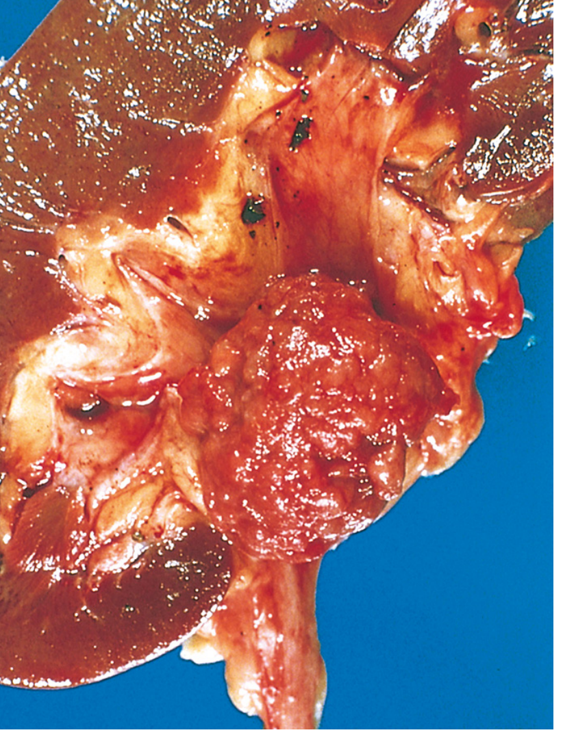
Histology
Clinical Features
Diagnosis
- Cystoscopy + TURBT (Transurethral Resection of Bladder Tumor): Gold standard. The urologist passes a camera into the bladder, visualizes the tumor, and removes it endoscopically. This is both diagnostic and therapeutic for NMIBC.
- Urine cytology: Sensitive for high-grade disease and CIS (malignant-looking cells shed into urine). Less useful for low-grade tumors.
- CT urography: To evaluate the entire upper urinary tract (ureters, renal pelvis) and detect extravesical spread, lymph node involvement.
Treatment
Non-Muscle-Invasive Disease (NMIBC)
- Step 1: TURBT - Remove all visible tumor endoscopically.
- Step 2: Immediate post-operative intravesical mitomycin C (a single instillation into the bladder right after TURBT) - for low-risk patients to reduce recurrence.
- Step 3: BCG (Bacillus Calmette-Guerin) intravesical therapy - for intermediate/high-risk NMIBC (T1 tumors, CIS, high-grade Ta). BCG is a bacterial immunotherapy instilled directly into the bladder; it triggers an immune response that kills residual tumor cells and prevents recurrence. This is the most effective intravesical treatment. Maintenance BCG is given for 1-3 years.
- Re-TURBT: A second TURBT 6 weeks later is done for T1 tumors - because 30-50% of these have residual tumor left behind after the first resection.
- Patients who fail BCG: Should undergo radical cystectomy to prevent progression to muscle-invasive disease.
Muscle-Invasive Disease (MIBC)
- Neoadjuvant cisplatin-based chemotherapy FIRST - then surgery. This is the standard of care. The regimen is MVAC (Methotrexate + Vinblastine + doxorubicin/Adriamycin + Cisplatin) or gemcitabine + cisplatin. Given before surgery to eliminate micrometastatic disease and shrink the primary tumor. Complete pathologic response in 20-40% of patients. Provides a survival advantage over surgery alone.
- Radical cystectomy: Remove the entire bladder + perivesical fat + regional lymph nodes. In men, also remove prostate and seminal vesicles. In women, also remove uterus, cervix, and anterior vaginal wall. Then urinary diversion is needed (ileal conduit/neobladder).
- Important: Carboplatin is NOT a substitute for cisplatin in bladder cancer even if the patient cannot tolerate cisplatin - it is significantly less effective. Patients who cannot receive cisplatin should go straight to cystectomy without neoadjuvant chemotherapy.
- Bladder-preserving trimodality therapy (for those who refuse/cannot have cystectomy): Maximum TURBT + concurrent chemoradiation (cisplatin + radiation). Approximately 30-50% can achieve long-term disease-free survival with intact bladder. Contraindicated if CIS, multifocal disease, or hydronephrosis is present.
Metastatic Disease
- First-line: MVAC or gemcitabine + cisplatin
- Second-line: Immune checkpoint inhibitors (pembrolizumab, atezolizumab)
- Novel agents: Enfortumab vedotin (antibody-drug conjugate) + pembrolizumab combination is now a major first-line option in metastatic disease.
- FGFR3 mutations: Erdafitinib (FGFR inhibitor) is approved for FGFR2/3-altered tumors.
MASTER COMPARISON - All 4 Tumors at a Glance
| RCC | Oncocytoma | Nephroblastoma | Urothelial Ca | |
|---|---|---|---|---|
| Nature | Malignant | Benign | Malignant | Malignant |
| Who gets it | Adults, 60s, M>F | Adults | Children, age 3, M=F | Adults, 60-70s, M>F 3:1 |
| Cell of origin | Proximal tubule epithelium | Intercalated cells of collecting duct | Metanephric blastema | Urothelial epithelium |
| Location | Renal cortex | Renal cortex | Anywhere in kidney | Bladder mucosa |
| Key gene / molecular change | VHL loss (clear cell), MET gain (papillary), Chr losses (chromophobe) | Complex I loss → mitochondrial proliferation | WT1 (11p13) loss | p53/Rb loss (invasive), FGFR3/RAS gain (papillary) |
| Gross color | Yellow-orange (lipid-rich) | Tan/mahogany brown (mitochondria-rich) | Grey-tan with hemorrhage/necrosis | Papillary/pink OR ulcerated |
| Classic histology | Nests of clear cells + delicate vasculature | Uniform pink granular cells + central scar | TRIPHASIC: blastema + epithelium + stroma | Papillary fronds OR flat CIS |
| #1 symptom | Painless hematuria | Incidental finding | Abdominal mass (parent finds it) | Painless intermittent gross hematuria |
| Famous spread | Renal vein → IVC → right heart | Does NOT spread (benign) | Lungs (85-95%), then liver | Superficial recurrence; → muscle invasion → metastasis |
| Treatment | Partial/radical nephrectomy ± TKI/immunotherapy | Partial nephrectomy (curative) | Nephrectomy + actinomycin D/vincristine ± RT | TURBT + BCG (NMIBC) / Neoadjuvant chemo + cystectomy (MIBC) |
| Prognosis | ~70% 5-yr overall | Excellent (benign) | >85% 5-yr overall | Variable: NMIBC good; MIBC ~50% 5-yr |
Can you explain uworld qid 19002
- Paste the question text here (copy the stem, answer choices, and the explanation if you have it), or
- Take a screenshot of the question and share the image
A 38-year-old man comes to the office with headaches, loss of libido, and erectile dysfunction for the last 3 months. The patient has no chronic medical conditions and takes no prescription or over-the-counter medications. He does not use tobacco, alcohol, or illicit drugs. Vital signs are normal. The examination is notable for peripheral vision loss, bilateral gynecomastia, and soft testes without masses. Laboratory evaluation shows normal serum prolactin, normal β-hCG, low serum LH, and low serum testosterone. Which of the following is the most likely mechanism for this patient's gynecomastia? A. Decreased systemic clearance of estrogen [7%] B. Defective androgen receptor function [10%] C. Enzymatic defect in the testosterone biosynthetic pathway [17%] D. Excessive estrogen production in the testes [13%] E. Persistent aromatization of adrenal estrogen precursors [51%] Incorrect. Correct answer is E 51% answered correctly Explanation: This patient's headaches and peripheral vision loss (ie, bitemporal hemianopsia) suggest a pituitary mass with compression of the optic chiasm. In addition, he has features of hypogonadism, including decreased libido, erectile dysfunction, testicular atrophy, and a low circulating testosterone level; his low LH level confirms secondary (central) hypogonadism. Secondary hypogonadism is common in patients with pituitary masses. Potential etiologies include the following: High prolactin levels from a prolactin-secreting adenoma (prolactinoma) can suppress secretion of GnRH, leading to decreased LH secretion and lower testosterone production Non–prolactin-secreting tumors can disrupt the inhibitory dopaminergic regulation of prolactin secretion, leading to hyperprolactinemia and (usually mild) hypogonadism Direct compression of gonadotrope cells with loss of LH secretion (independent of prolactin effect), which is likely in this patient with a normal prolactin level Gynecomastia is the abnormal growth of male breast tissue, typically due to an increased estrogen/testosterone ratio. This patient has low testosterone, but production of estrogen precursors (eg, androstenedione, dehydroepiandrosterone [DHEA], DHEA sulfate) continues uninterrupted in the zona reticularis of the adrenal cortex. These steroid hormones are subsequently aromatized to estrogens in peripheral tissues. When testosterone levels are normal, the inhibitory effect of testosterone prevents breast growth, but when testosterone is low, the adrenal-derived estrogens can produce significant gynecomastia. (Choice A) Estrogen is cleared primarily in the liver. Patients with cirrhosis commonly develop gynecomastia due to decreased clearance of estrogen along with increased production and aromatization of estrogen precursors. However, this patient has no risk factors for cirrhosis (eg, alcohol use), and his bitemporal hemianopsia suggests a pituitary etiology. (Choice B) Androgen receptor defects typically present at birth with a female phenotype or ambiguous genitalia (with 46,XY karyotype), although some variants (partial androgen insensitivity syndrome) may present in adulthood with gynecomastia and undervirilization (eg, decreased facial and pubic hair). However, androgen production is unaffected, and testosterone levels would be normal to high. (Choice C) Testosterone biosynthetic defects generally present at birth or during development with abnormal genitalia and/or undervirilization in males. Biochemical testing shows primary hypogonadism (ie, low testosterone, high LH). (Choice D) Estrogen is synthesized in the testes by the aromatization of androgens, producing high local concentrations. However, this patient has decreased testicular testosterone production, so subsequent estrogen synthesis would also be low. Educational objective:
UWorld QID 19002 - Full Breakdown
First, Build the Diagnosis Step by Step
| Clue in Stem | What It Tells You |
|---|---|
| Headaches + peripheral (bitemporal) vision loss | Mass compressing the optic chiasm from below = pituitary tumor |
| Low libido + erectile dysfunction | Low testosterone (hypogonadism) |
| Soft testes (atrophy) | Testes not being stimulated = hypogonadism |
| Low LH | The problem is ABOVE the testes (central/secondary), not in the testes themselves |
| Low testosterone | Confirms hypogonadism |
| Normal prolactin | NOT a prolactinoma; instead the tumor is directly compressing gonadotrope cells |
| Normal β-hCG | Rules out an hCG-secreting germ cell tumor |
| Bilateral gynecomastia | Abnormal estrogen/androgen ratio |
The Core Concept You Must Understand: The Estrogen/Androgen Ratio
- Testosterone pulls the rope toward "no breast growth"
- Estrogen pulls toward "breast growth"
Where Does Estrogen Come From in Men? (This is the Key)
Source 1: The Testes
Source 2: The Adrenal Cortex (Zona Reticularis) → Peripheral Aromatization
- Testosterone ↓↓↓ (because LH is low, testes aren't stimulated)
- Adrenal androgen precursors → NORMAL (ACTH is fine)
- Peripheral estrogen from aromatization → RELATIVELY PRESERVED
Why Answer E is Correct
- Pituitary tumor → compresses gonadotropes → ↓LH
- ↓LH → testes not stimulated → ↓testosterone
- Adrenal zona reticularis → continues making androstenedione + DHEA (ACTH is intact)
- Peripheral aromatase → converts these precursors to estrogen
- With low testosterone to oppose estrogen → breast tissue grows = gynecomastia
Why Each Wrong Answer is Wrong (Detailed)
A. Decreased systemic clearance of estrogen (7% chose this)
B. Defective androgen receptor function (10% chose this)
- In AIS, testosterone levels are normal or HIGH (because without feedback, the pituitary keeps pumping out LH trying to get a response). This patient has low LH AND low testosterone - the opposite pattern.
- Complete AIS presents at birth with female phenotype (46,XY female). Partial AIS can present in adulthood with undervirilization and gynecomastia, but this does NOT cause low LH.
C. Enzymatic defect in testosterone biosynthetic pathway (17% chose this)
- Low testosterone ✓ (same as this patient)
- HIGH LH ✗ (this patient has LOW LH)
D. Excessive estrogen production in the testes (13% chose this - very tempting!)
The Full Pathophysiology Summary in One Diagram
Pituitary Macroadenoma
↓
Compresses gonadotrope cells
↓
↓ LH secretion (FSH also low)
↓
Testes not stimulated
↓
↓ Testosterone production Adrenal zona reticularis
↓ (ACTH still intact)
Less testosterone to oppose ↓
estrogen action in breast tissue → Androstenedione, DHEA
still secreted normally
↓
Peripheral aromatase
(fat, skin, muscle)
↓
Estrone + Estradiol
(relatively preserved)
↓
ESTROGEN/ANDROGEN RATIO ↑↑↑ → GYNECOMASTIA
Additional Layers to Lock This In
One-Line Summary for the Exam
A pituitary mass suppresses LH → low testosterone from testes, but adrenal continues making androgen precursors (androstenedione, DHEA) under ACTH control → these are aromatized peripherally to estrogen → with low testosterone to oppose it, the estrogen/androgen ratio rises → gynecomastia. Answer = E.
Explain chiari malformations I and 2 and dandy walker malformations and syringomyelia
Chiari I, Chiari II, Dandy-Walker, and Syringomyelia - Complete Concept
ANATOMY FIRST: Understand the Posterior Fossa
- The cerebellum (balance and coordination)
- The brainstem (pons and medulla)
- The fourth ventricle (CSF channel between the brainstem and cerebellum)
TOPIC 1: CHIARI MALFORMATION TYPE I (CM-I)
What Is It? (The Fundamental Defect)
- The herniated tonsils compress the cervicomedullary junction (where the medulla meets the spinal cord)
- They obstruct the normal flow of CSF through the foramen magnum
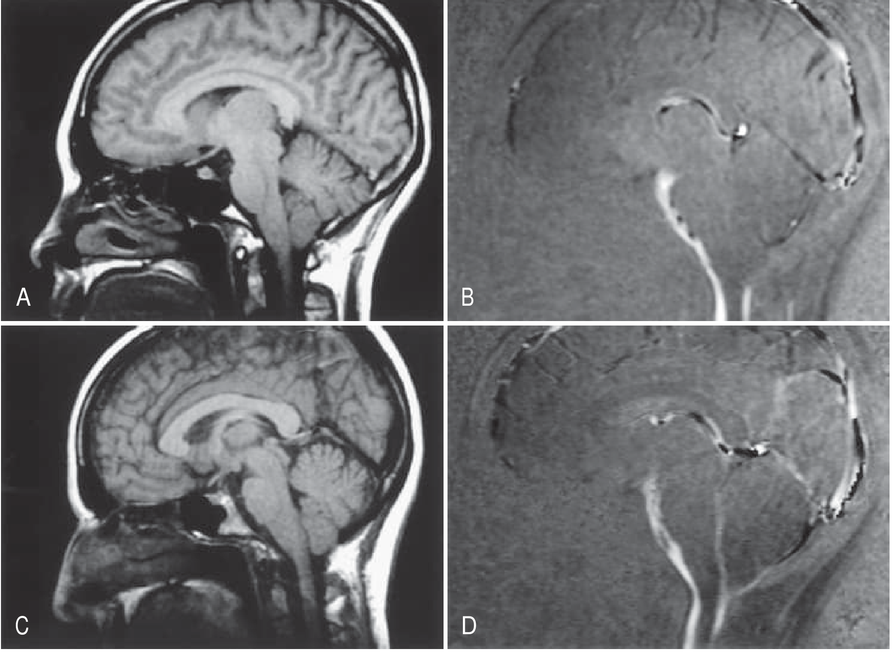
Why Does the Posterior Fossa Become Too Small?
| Mechanism | Cause | What Happens |
|---|---|---|
| "Squeeze down" (too small box) | Classic CM-I, craniosynostosis, achondroplasia | Small posterior fossa forces tonsils down |
| "Pull down" (tethered cord) | Tethered cord syndrome, CM-II | Spinal cord pulled down, brainstem follows |
| "Push down" (raised ICP) | Hydrocephalus, tumors, hematoma | Increased cranial pressure pushes brain caudally |
| "Suck down" (low spinal pressure) | CSF leaks, prolonged lumboperitoneal shunt | Pressure gradient draws tonsils down |
| "Shake down" (cranial settling) | Ehlers-Danlos, occipitocervical instability | Skull sinks down over unstable joints |
Who Gets It and When?
- Prevalence: 0.1-0.5% of the population
- Women are affected 3 times more often than men
- Most patients have no symptoms and are discovered incidentally on MRI
- When symptoms do appear, they often start in adolescence or early adulthood
- Interestingly, at least a quarter of patients first develop symptoms after a relatively minor head or neck injury
- There is a hereditary tendency, with transmissibility approaching 12%
Normal Values for Tonsillar Position by Age (Exam High-Yield)
| Decade | Upper Limit of Normal (mm below foramen magnum) |
|---|---|
| 1st decade (0-9 years) | 6 mm |
| 2nd-3rd decade | 5 mm |
| 4th-8th decade | 4 mm |
| 9th decade | 3 mm |
Clinical Features (How the Patient Presents)
- Ocular disturbances (78%) - blurring, floaters, diplopia, photophobia
- Acoustic and vestibular complaints (74%) - dizziness, dysequilibrium, tinnitus
- Dysesthesias (59%) - numbness, tingling, burning sensations in the arms and/or hands (often due to associated syringomyelia)
- Chronic fatigue (58%)
- Bulbar dysfunction (52%) - difficulty swallowing (dysphagia), hoarseness, sleep apnea
- Myelopathy - weakness, spasticity, sensory loss - from compression at the cervicomedullary junction or from syringomyelia
- Nystagmus - often downbeat nystagmus, a classic sign of craniocervical junction pathology
Diagnosis
- Cerebellar tonsils displaced below the foramen magnum (>5 mm in adults)
- Tonsillar morphology: instead of rounded, the tonsils appear peglike/pointed (this is more specific than just position)
- CSF flow studies (phase-contrast MRI) demonstrate reduced or absent CSF pulsation at the foramen magnum level - this correlates with symptoms better than the degree of descent alone
Treatment
TOPIC 2: CHIARI MALFORMATION TYPE II (CM-II / Arnold-Chiari)
The Key Difference From CM-I
What Is the Defect?
- The caudal vermis AND lower medulla are displaced downward (not just the tonsils)
- The medulla itself is distorted and kinked ("cervicomedullary kink")
- The fourth ventricle is elongated and displaced into the cervical spinal canal
- The foramen magnum is actually enlarged (opposite of CM-I) because the spinal cord pulls the neural structures down
- The brainstem and upper cervical cord are compressed and distorted
- Hydrocephalus develops in nearly all cases (from obstruction of CSF flow)
- Additional brain anomalies: beaking of the tectum (the top of the midbrain looks like a beak), abnormal cerebral gyration (stenogyria), hypoplastic cerebellar hemispheres, dysgenesis of the corpus callosum
CM-I vs CM-II: The Key Differences (High-Yield)
| Feature | Chiari I | Chiari II |
|---|---|---|
| What herniates | Cerebellar tonsils only | Cerebellar vermis + lower medulla |
| Posterior fossa | Small (too small box) | Small |
| Foramen magnum | Small/stenosed | Large (enlarged) |
| Neural tube defect | ABSENT | ALWAYS present (myelomeningocele) |
| Hydrocephalus | Uncommon | Almost universal |
| Timing of presentation | Adolescence/adulthood | Birth (or prenatal) |
| Brainstem anomalies | Absent/mild | Severe, multiple |
| Spinal cord defect | Often syringomyelia | Myelomeningocele + often syrinx |
| Associated with | None specifically | Spina bifida, hydrocephalus, tethered cord |
Clinical Features of CM-II
- Lower limb paralysis and sensory loss from the spinal cord defect (level depends on where the myelomeningocele is)
- Bladder and bowel dysfunction (neurogenic bladder)
- Hydrocephalus - most patients require a ventriculoperitoneal (VP) shunt
- Brainstem dysfunction - stridor, apnea (life-threatening in infants), swallowing difficulty, vocal cord paralysis
- Syringomyelia may coexist
- Long-term survivors have intellectual disabilities to varying degrees, though patients with lumbar (lower) myelomeningocele fare better cognitively than those with thoracic levels
Treatment
TOPIC 3: DANDY-WALKER MALFORMATION
The Fundamental Defect
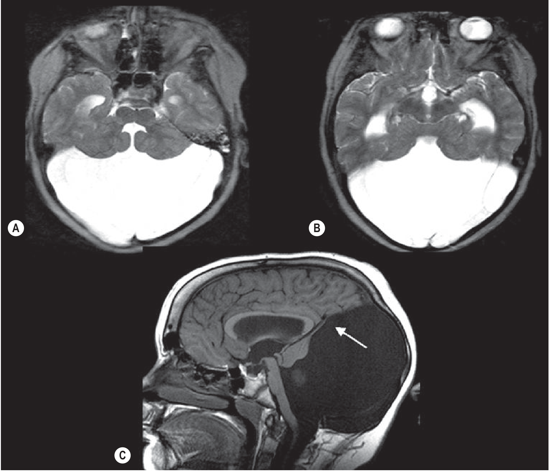
- Cystic dilatation of the fourth ventricle - the fourth ventricle balloons into a huge posterior fossa cyst
- Aplasia or severe hypoplasia of the cerebellar vermis - the midline cerebellum is absent or severely underdeveloped, and the remnant is rotated upward
- Enlarged posterior fossa with upward displacement of the tentorium, torcula (confluence of venous sinuses), and straight sinus above the lambdoid suture
Pathogenesis: Why Does It Happen?
- Foramen of Magendie (midline, posterior)
- Two Foramina of Luschka (lateral)
Dandy-Walker Spectrum (Important for Exams)
| Entity | 4th Ventricle | Vermis | Posterior Fossa | Clinical Significance |
|---|---|---|---|---|
| Classic Dandy-Walker | Massively dilated, opens into cyst | Aplastic/severely hypoplastic, rotated | Enlarged, torcula elevated | Severe; usually hydrocephalus |
| Dandy-Walker variant | Mildly enlarged | Partially formed (mild-moderate hypoplasia) | Mildly enlarged | Variable |
| Mega cisterna magna | Normal | Normal | Normal to slightly enlarged | Incidental finding, benign |
| Blake's pouch cyst | Variable | Normal | Normal | Obstructive hydrocephalus possible |
Associated Anomalies
- Hydrocephalus (almost universal in classic form) - from obstruction of CSF outflow
- Agenesis or partial agenesis of the corpus callosum - a major commissure between hemispheres
- Cortical dysplasia - abnormal gyration (pachygyria), heterotopias
- Occipital bone bulging (seen on plain films and CT) - the enlarged cyst pushes the occipital bone outward
- Elevation of the torcula above the lambdoid suture - visible on imaging, sometimes on plain skull X-ray
Clinical Features
- Most patients present in infancy with progressive head enlargement (macrocephaly from hydrocephalus)
- Developmental delay is common (though severity varies)
- Seizures
- Cerebellar signs - ataxia, nystagmus, truncal instability
- Signs of raised intracranial pressure: irritability, bulging fontanelle, vomiting, sunset sign (eyes deviated downward)
- If hydrocephalus is treated promptly, prognosis can be acceptable - the neurological deficits correlate more with associated brain malformations (cortical dysplasia, corpus callosum agenesis) than with the hydrocephalus itself
- Incidentally discovered Dandy-Walker variant in an older child or adult may be completely asymptomatic
Treatment
- VP shunting for hydrocephalus (most common intervention)
- Some surgeons prefer to shunt the posterior fossa cyst directly (cystoperitoneal shunt)
- Posterior fossa decompression may be needed
- Associated malformations are managed as needed
TOPIC 4: SYRINGOMYELIA
What Is It?
Location and Anatomy of the Syrinx
- The cervical cord is most commonly affected (most cases start here)
- The syrinx typically runs along many segments - sometimes the entire cervical and thoracic cord
- It usually begins in the central cord, near the central canal
- The spinal cord is enlarged in about 80% of cases (you can see this on MRI as a swollen cord with a dark fluid-filled center on T2-weighted images)
- The size of the cord and syrinx on MRI does NOT reliably predict clinical severity - this is a common misconception
Pathogenesis: Why Does the Syrinx Form?
- Chiari I tonsils herniate and plug the foramen magnum, partially or completely
- Normal CSF pulsations (driven by the cardiac cycle) cannot pass freely between the cranial and spinal compartments
- Each heartbeat creates a pressure wave that normally dissipates through the foramen magnum. When it's blocked, the pressure wave is transmitted instead into the central canal of the spinal cord (through the obex at the base of the fourth ventricle)
- Repeated pressure pulsations distend the central canal and eventually rupture it, creating a cavity that propagates within the cord substance
- Once formed, the cavity propagates further by hydrodynamic forces acting on normal cord tissue
- Post-traumatic syringomyelia - after spinal cord injury, scar tissue can obstruct CSF flow
- Spinal cord tumors (intramedullary) - ependymoma and astrocytoma can have an associated syrinx ("tumor cyst")
- Adhesive arachnoiditis - inflammation/scarring around the spinal cord obstructs CSF flow
- Tethered cord syndrome
- Idiopathic - no cause found in 10-20% of cases
The Classic Clinical Syndrome: "Cape Distribution" Sensory Loss
- Wasting of intrinsic hand muscles (classic sign - the small muscles of the hand atrophy, giving a "claw-like" appearance)
- Fasciculations
- Loss of deep tendon reflexes in the arms (LMN pattern)
- Spastic paraparesis (UMN pattern, legs)
- Hyperreflexia and extensor plantar responses (Babinski) in the legs
- Bladder dysfunction
Additional Association: Scoliosis
- Left-sided curves (idiopathic is typically right-sided)
- Thoracic kyphosis >40° instead of the hypokyphosis of idiopathic scoliosis
- Lack of rotation through the apex
- Loss of superficial abdominal reflexes (an important clinical sign - feel for this on exam)
Diagnosis
- The syrinx appears as a well-circumscribed cavity within the cord
- T1: hypointense (dark) - same signal as CSF
- T2: hyperintense (bright) - same signal as CSF
- The cord may be expanded around the syrinx
- CSF flow studies (phase-contrast MRI) assess pulsation dynamics at the foramen magnum
- The entire craniovertebral junction must be imaged to look for a Chiari malformation
Treatment
MASTER COMPARISON TABLE
| Chiari I | Chiari II | Dandy-Walker | Syringomyelia | |
|---|---|---|---|---|
| What herniates/forms | Cerebellar tonsils herniate DOWN | Cerebellar vermis + medulla herniate DOWN | 4th ventricle BALLOONS + vermis fails to form | Fluid cavity FORMS INSIDE spinal cord |
| Posterior fossa | Too SMALL | Small | Too LARGE (cyst) | Normal |
| Associated spinal defect | Syringomyelia (65-70%) | Myelomeningocele (100%) | None specifically | IS the spinal lesion |
| Hydrocephalus | Uncommon | Near universal | Universal (classic form) | Not directly |
| Age of onset | Adolescence/adulthood | Birth (prenatal) | Infancy | Any age (often young adult) |
| #1 symptom | Cough/exertional occipital headache | Symptoms of spinal defect + hydrocephalus | Macrocephaly, developmental delay | Cape dissociated sensory loss + hand wasting |
| Classic MRI finding | Pointed tonsils >5mm below FM | Medullary kink, beaked tectum, hydrocephalus | Massive 4th ventricle cyst, elevated torcula | Fluid-filled cavity inside cord (dark T1, bright T2) |
| Key imaging | Foramen magnum SMALL | Foramen magnum LARGE | Posterior fossa LARGE, elevated torcula | Expanded cord with central CSF signal |
| Treatment | Posterior fossa decompression | Myelomeningocele closure + VP shunt | VP/cystoperitoneal shunt | Foramen magnum decompression (if Chiari) or syrinx shunting |
The Chiari-Syringomyelia Connection (The Most Important Link)
Underdeveloped posterior fossa (CM-I)
↓
Cerebellar tonsils herniate into foramen magnum
↓
CSF flow obstructed at foramen magnum
↓
Pressure waves transmitted into central canal
↓
Syrinx develops in cervical cord
↓
Dissociated sensory loss + hand wasting + UMN legs
↓
In children: atypical LEFT-sided scoliosis
↓
Treatment: Decompress the posterior fossa
→ CSF flow restored → Syrinx collapses
Tell me
Continue explaining Chiari/Dandy-Walker/syringomyelia with more depth
Deep Continuation: Chiari, Dandy-Walker, and Syringomyelia
PART 1: GOING DEEPER INTO CHIARI II - WHAT ACTUALLY HAPPENS TO THE BRAIN
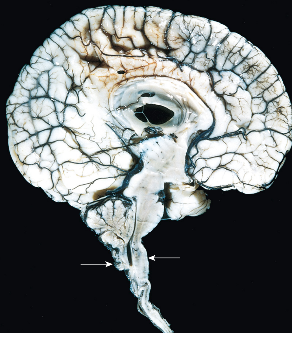
Every Structure Displaced and Deformed in CM-II
- Stenogyria - abnormally small, tightly packed gyri, especially over the medial surfaces
- Gray matter heterotopias - clumps of neurons stranded in wrong locations during migration
- Aqueductal stenosis - narrowing of the cerebral aqueduct between the third and fourth ventricles, contributing to or worsening hydrocephalus
- Corpus callosum dysgenesis - the major white matter bridge between hemispheres may be partially or completely absent
PART 2: THE NEURAL TUBE DEFECT - WHY MYELOMENINGOCELE AND CM-II ALWAYS GO TOGETHER
The Hydrodynamic Theory
PART 3: HYDROMYELIA vs SYRINGOMYELIA - The Distinction You Need
- Hydromyelia: ependymal lining (real canal dilated)
- Syringomyelia: glial lining (new cavity in cord substance)
PART 4: SYRINGOMYELIA - THE COMPLETE NEUROLOGICAL SYNDROME LEVEL BY LEVEL
The Spinal Cord Cross-Section (What's Where)
- Center: Central canal (where the syrinx begins)
- Inner gray matter: Anterior commissure (crossing pain/temperature fibers)
- Anterior horns: Lower motor neurons to muscles
- Lateral horns (cervical only): Sympathetic preganglionic neurons
- Posterior horns: Sensory relay neurons
- Lateral funiculi: Corticospinal tracts (going down) + Spinothalamic tracts (going up)
- Posterior funiculi: Dorsal columns - vibration and proprioception
Stage 1: Anterior Commissure Destruction - The Signature Finding
- Pain and temperature sensation is lost bilaterally at the levels of the syrinx
- Vibration and proprioception (carried in dorsal columns, which are untouched) remain perfectly preserved
- This combination = dissociated sensory loss (selectively losing pain/temp while preserving light touch/vibration)
- The distribution is bilateral and dermatomal - if the syrinx occupies C4-T1, you lose pain/temp in both arms and hands but can still feel vibration and joint position in those areas
- The classic term is "suspended sensory level" - the deficit is suspended in the middle of the body (arms and trunk) rather than extending to the legs (which haven't been affected yet because the lower cord is still fine)
Stage 2: Lateral Horn Destruction - Horner Syndrome
- Ptosis (drooping eyelid - from loss of superior tarsal muscle innervation)
- Miosis (small pupil - from loss of pupil dilator innervation)
- Anhidrosis of the ipsilateral face and neck (from loss of sudomotor innervation)
- = Ipsilateral Horner syndrome
Stage 3: Anterior Horn Destruction - Lower Motor Neuron Signs in Arms
- Wasting and atrophy of intrinsic hand muscles (thenar, hypothenar, interossei) - this is the most visible sign, giving the hand a "skeleton hand" appearance
- Wasting of forearm, arm, and shoulder muscles depending on level
- Fasciculations (visible muscle twitching from denervated motor neurons)
- Absent deep tendon reflexes in the arms (LMN pattern: biceps reflex, triceps reflex lost)
Stage 4: Lateral Funiculus Destruction - Upper Motor Neuron Signs in Legs
- Spastic paraparesis (stiff, weak legs - UMN pattern)
- Hyperreflexia in the legs (exaggerated knee and ankle jerks)
- Extensor plantar responses (Babinski sign)
- Bladder dysfunction - urgency, hesitancy, incontinence
- Bowel dysfunction
Stage 5: Brainstem Extension - Syringobulbia
- Palatal and vocal cord paralysis - hoarseness, nasal voice, dysphagia (cranial nerves IX, X)
- Tongue weakness and atrophy (CN XII involvement)
- Dysarthria (slurred speech)
- Horizontal or vertical nystagmus
- Dizziness or vertigo (CN VIII)
- Facial numbness - the descending trigeminal tract runs down to C2 level, so a high syrinx or syringobulbia disrupts CN V sensation on the face
- Respiratory irregularity - because the respiratory center in the medulla may be involved
Complete Syndrome Summary in One Flow
SYRINX CENTER (starts here)
↓
Anterior commissure → Dissociated sensory loss (cape distribution)
↓
Lateral horn (C8-T1) → Horner syndrome
↓
Anterior horn (cervical) → Wasted hands, absent arm reflexes, fasciculations
↓
Lateral funiculi → Spastic legs, hyperreflexia, Babinski, bladder dysfunction
↓
Extension to medulla → Syringobulbia (cranial nerve palsies, nystagmus)
MRI of Syringomyelia - What You Actually See
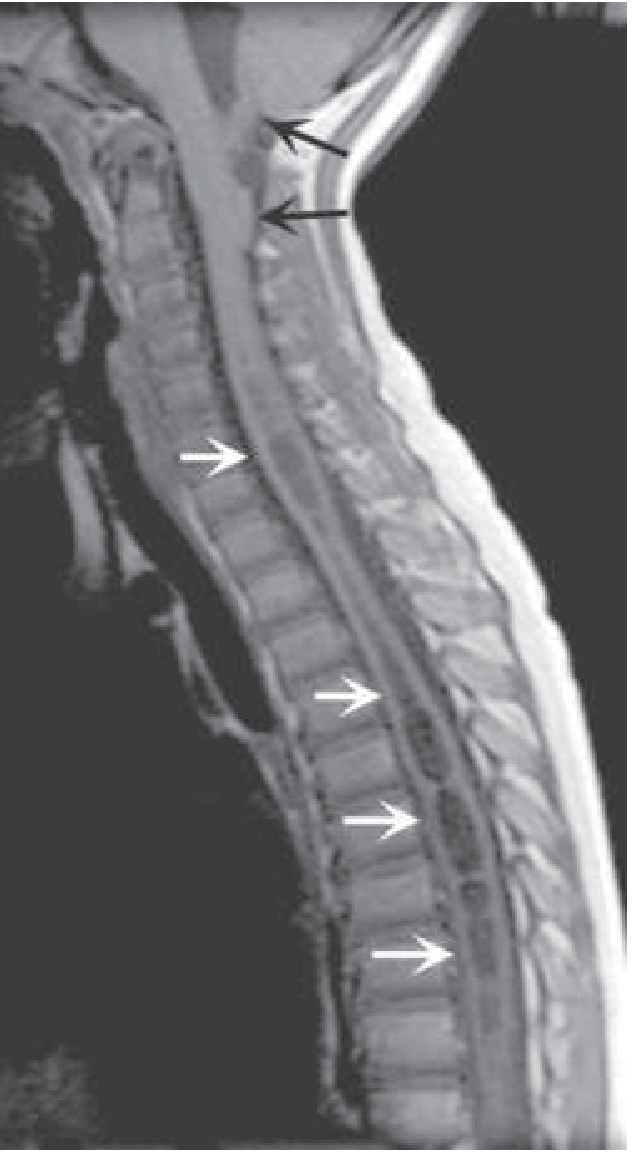
- The cerebellar tonsils are clearly below the base of the skull (the foramen magnum) - that is the Chiari I
- Running down the center of the spinal cord is a long, continuous, dark (hypointense on T1) fluid-filled cavity - the syrinx
- The cord is visibly enlarged around the syrinx
- The syrinx runs from the cervical region all the way into the thoracic cord - this is a large syrinx
- Sagittal T1 and T2 of the ENTIRE spine (to map the full extent of the syrinx)
- Sagittal T1 of the BRAIN (to assess the posterior fossa and look for Chiari)
- Axial cuts through the cord at multiple levels
- CSF flow studies at the foramen magnum (to assess whether CSF pulsation is obstructed)
- Assessment for hydrocephalus (dilated ventricles)
PART 5: DANDY-WALKER - THE PATHOLOGY IN DEPTH
What the Cerebellar Vermis Is Supposed to Do
- Truncal ataxia (wobbling, inability to sit or stand without support)
- Gait ataxia (wide-based, unsteady walking)
- Titubation (rhythmic tremor of the head and trunk)
The Cyst in Detail
- The cyst is lined by ependymal cells on its inner surface (because it is derived from the ventricular system)
- The outer surface is continuous with the leptomeninges (pia and arachnoid)
- The roof of the fourth ventricle (area membranacea posterior) normally forms the choroid plexus and the foramina of Magendie and Luschka; in Dandy-Walker, this roof fails to open and persists as the cyst wall
- Dysplasias of brainstem nuclei are commonly found alongside - the inferior olivary nuclei, dentate nucleus, and other brainstem structures may be abnormally formed (this contributes to the neurological deficits beyond just the vermian absence)
Torcula/Venous Sinus Displacement - Clinical and Imaging Significance
- Plain skull X-ray: the lambdoid suture line is lower than the torcula (normally they should be level)
- CT scan: the tentorium is tent-shaped and elevated; the posterior fossa is massively enlarged
- MRI sagittal: the straight sinus curves upward above the lambdoid - a pathognomonic sign
Hydrocephalus Mechanism
Blake's Pouch Cyst vs Dandy-Walker vs Mega Cisterna Magna
| What failed to open | Result | What you see |
|---|---|---|
| The entire roof never develops normally + vermis fails to form | Classic Dandy-Walker | No vermis, massive cyst, enlarged posterior fossa, elevated torcula, hydrocephalus |
| Only the foramen of Magendie fails to open (vermis is NORMAL) | Blake's Pouch Cyst | Normal vermis, 4th ventricle communicates with CSF collection, obstructive hydrocephalus possible |
| The subarachnoid space behind the cerebellum is simply larger than normal | Mega Cisterna Magna | Normal vermis, normal 4th ventricle, large CSF space behind cerebellum - BENIGN, no treatment |
- Vermis absent/hypoplastic/rotated = Dandy-Walker
- Vermis normal = Blake's Pouch Cyst or Mega Cisterna Magna
PART 6: THE THREE-WAY CONNECTION - CHIARI, DANDY-WALKER, AND SYRINGOMYELIA
POSTERIOR FOSSA TOO SMALL
(Chiari I: tonsils herniate)
↓
CSF obstruction at foramen magnum
↓
SYRINGOMYELIA forms
(70-90% of syringes)
↓
Treat cause (Chiari) → syrinx resolves
POSTERIOR FOSSA TOO LARGE
(Dandy-Walker: cyst expands)
↓
4th ventricle outlets blocked
↓
HYDROCEPHALUS forms
(ventricular dilation)
↓
Treat with VP shunting
NEURAL TUBE FAILS TO CLOSE
(Myelomeningocele)
↓
CSF leaks → posterior fossa
never properly inflates
↓
CHIARI II forms
(vermis + medulla herniate down)
↓
Hydrocephalus (nearly universal)
+ Syringomyelia may also develop
PART 7: TREATMENT DECISIONS IN DETAIL
When to Operate on Chiari I
- Patient positioned prone (face down)
- Skin incision over the back of the skull/upper neck
- Remove a small section of the occipital bone (suboccipital craniectomy)
- Remove the posterior arch of C1 (cervical laminectomy)
- Open the dura (the fibrous covering of the brain) - duraplasty: the dura is opened in a Y shape and a patch is sewn in to enlarge the space
- Sometimes the tonsils themselves are shrunk (using cautery) or the arachnoid scar tissue around the foramen magnum is removed
- Result: more space, restored CSF pulsation through the foramen magnum
Treatment of Dandy-Walker
- Shunt the lateral ventricles (most common) - drains the supratentorial CSF buildup
- Shunt the posterior fossa cyst directly (cystoperitoneal shunt) - some surgeons prefer this as it directly addresses the problematic cyst
- Sometimes both shunts are needed (combined approach)
- Associated brain anomalies - the corpus callosum agenesis, cortical dysplasias, and heterotopias present in many Dandy-Walker patients are far more responsible for intellectual disability than the hydrocephalus itself. If the brain is otherwise structurally normal (isolated Dandy-Walker), shunting can result in normal or near-normal development.
- Chromosomal abnormalities - Dandy-Walker associated with trisomy 18 or 13 carries a very poor prognosis regardless of treatment. Chromosomal microarray is routinely performed.
PART 8: SYRINGOHYDROMYELIA - HYDROMYELIA vs SYRINGOMYELIA vs SYRINGOBULBIA
| Term | Definition | Location | Lining | Cause |
|---|---|---|---|---|
| Hydromyelia | Dilation of existing central canal | Within ependymal canal | Ependymal cells | Obstructed CSF flow (Chiari, hydrocephalus) |
| Syringomyelia | New fluid cavity in cord parenchyma | Within cord tissue outside canal | Glial cells | Chiari, trauma, tumor, arachnoiditis |
| Syringohydromyelia | Combined - both central canal + parenchymal | Both | Mixed | Same as above |
| Syringobulbia | Extension into brainstem (medulla/pons) | Within medulla/pons | Glial cells | Severe syringomyelia extending rostrally |
PART 9: HIGH-YIELD EXAM SUMMARY TABLES
Chiari I vs II - Every Key Difference
| Feature | Chiari I | Chiari II |
|---|---|---|
| What herniates | Cerebellar tonsils only (pointy/peg-shaped) | Cerebellar vermis + lower medulla + 4th ventricle |
| Posterior fossa size | Small (box too small for tonsils) | Small |
| Foramen magnum size | Small/stenosed (transversely constricted) | Large/enlarged |
| Spinal cord defect | NONE | Myelomeningocele (100% association) |
| Hydrocephalus | Uncommon | Nearly universal |
| Tectal beaking | Absent | Present (beaked midbrain) |
| Medullary kink | Absent | Present (cervicomedullary kink) |
| Age of onset | Adolescence/adulthood (often incidental) | Birth/prenatal |
| Sex ratio | Women 3x more | Equal |
| Syringomyelia | Yes (65-70%) | Yes (frequently) |
| Key associated feature | Syrinx → cape sensory loss | Spina bifida + hydrocephalus + paraplegia |
| Corpus callosum | Usually normal | Often dysgenetic |
| Treatment | Posterior fossa decompression | MMC closure + VP shunting |
| In utero surgery | Not applicable | Fetal MMC repair reduces CM-II severity |
Syringomyelia - Deficits by Level Destroyed
| Structure Destroyed | Deficit |
|---|---|
| Anterior commissure (crossing fibers) | Bilateral dissociated sensory loss (cape distribution) - HALLMARK |
| Lateral horn (C8-T1) | Ipsilateral Horner syndrome |
| Anterior horn (C5-T1) | Wasted hand/arm muscles, fasciculations, absent arm reflexes (LMN) |
| Lateral funiculus (corticospinal) | Spastic paraparesis, hyperreflexia in legs, Babinski (UMN) |
| Medullary extension (syringobulbia) | Palatal/vocal cord palsy, tongue atrophy, nystagmus, dysphagia, facial numbness |
Dandy-Walker Spectrum
| Entity | 4th Ventricle | Vermis | Posterior Fossa | Torcula | Clinical |
|---|---|---|---|---|---|
| Classic Dandy-Walker | Massively cystic | Absent/severely hypoplastic | Enlarged | Elevated above lambdoid | Severe, hydrocephalus, developmental delay |
| DW variant | Moderately enlarged | Partially formed | Mildly enlarged | Normal or mildly elevated | Variable |
| Blake's Pouch Cyst | Connected to CSF collection | Normal | Normal | Normal | Obstructive hydrocephalus possible |
| Mega Cisterna Magna | Normal | Normal | Normal/slightly enlarged | Normal | Incidental - benign, no treatment |
THE COMPLETE CLINICAL SCENARIO (For Exam Pattern Recognition)
Explain only these 4 topics chiari 1 2 syringomyelia dandy walker malformations
What specifically is still unclear or missing from the previous explanations? · How would you like it presented this time?
Chiari I, Chiari II, Syringomyelia, Dandy-Walker - Fresh Start, Simple Language
BEFORE ANYTHING - ONE PICTURE YOU MUST HAVE IN YOUR HEAD
- Upper floor = cerebral hemispheres (thinking brain)
- Lower floor (posterior fossa) = cerebellum + brainstem
1. CHIARI TYPE I MALFORMATION
What is the problem, in one sentence?
Why does the box become too small?
What does this cause?
How does the patient feel?
Who gets it and when?
How is it diagnosed?
How is it treated?
2. CHIARI TYPE II MALFORMATION (Arnold-Chiari)
What is the problem, in one sentence?
The absolutely critical difference from Chiari I
Why does Chiari II happen? (Understanding the mechanism)
What exactly herniates and what is deformed?
How is the patient found?
- "Lemon sign" - the frontal bones of the skull are indented inward (like the ends of a lemon) because of the low intracranial pressure from CSF leaking out
- "Banana sign" - the cerebellum is curved and banana-shaped because it is being pulled downward
Treatment
Prognosis
3. SYRINGOMYELIA
What is it, in one sentence?
Why does the cavity form?
What does the syrinx destroy, and what symptoms does that cause?
Diagnosis
Treatment
4. DANDY-WALKER MALFORMATION
What is it, in one sentence?
Notice the opposite relationship to Chiari
- In Chiari I and II, the posterior fossa is too small, so things get pushed downward out of it.
- In Dandy-Walker, the posterior fossa is too large, because a big CSF-filled cyst has expanded it from within.
Why does it happen?
What does it look like?
Associated brain abnormalities
How does the baby/child present?
What is the difference between Dandy-Walker and mega cisterna magna?
Treatment
THE SIMPLEST COMPARISON TO LOCK IT ALL IN
Tell me about renal tumors bladder every single detail every concept in paragraph
Renal Tumors and Bladder Tumors - Complete Concept in Paragraphs
PART ONE: RENAL TUMORS
Understanding the Kidney First
RENAL CELL CARCINOMA (RCC)
What it is and who gets it
Risk factors
Familial syndromes
Classification of RCC subtypes
Gross pathology
Histology
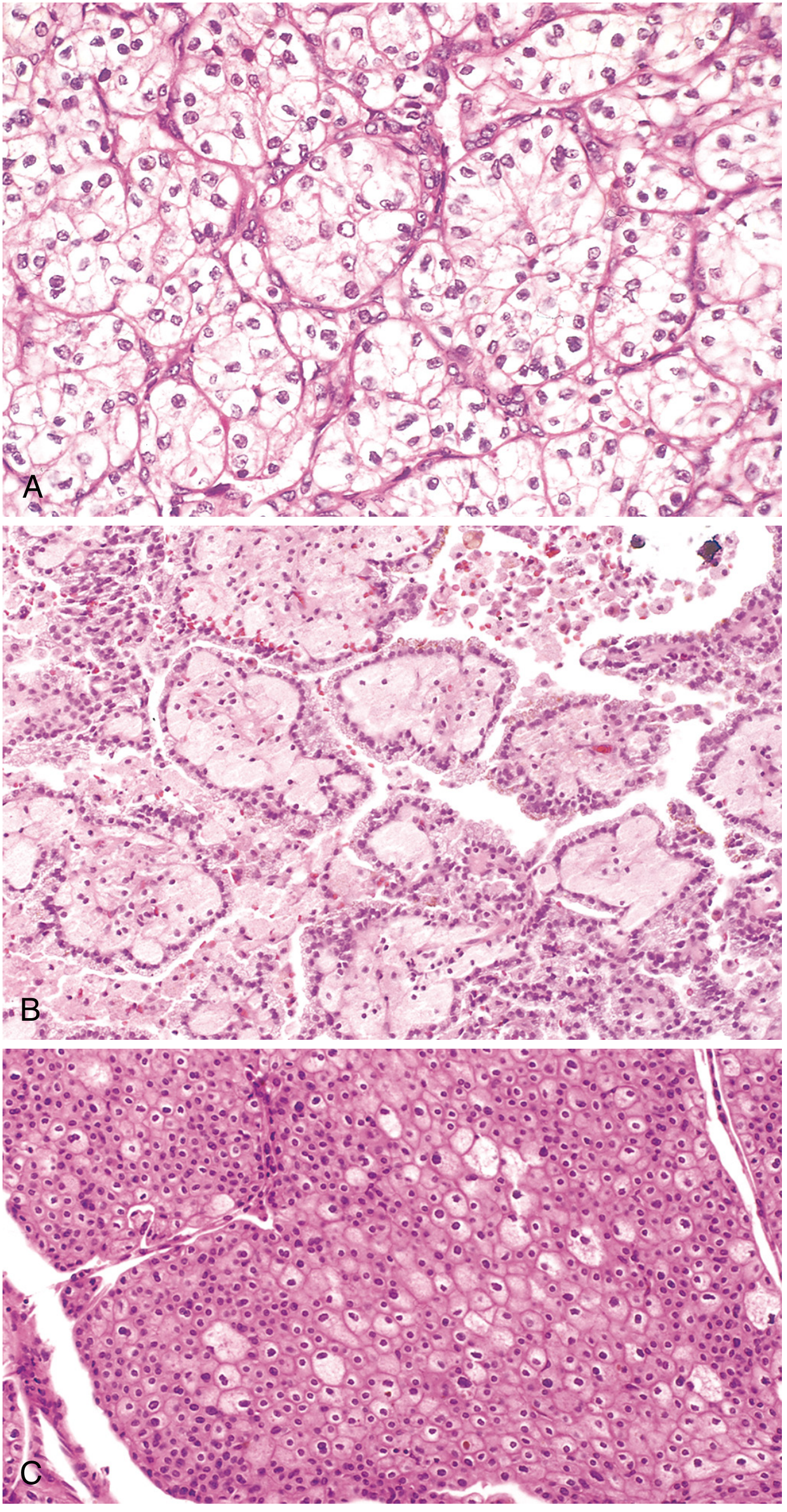
Clinical features - "The Great Mimic"
Staging, treatment and prognosis
RENAL ONCOCYTOMA
NEPHROBLASTOMA (WILMS TUMOR)
Overview and epidemiology
Pathogenesis and genetics
Pathology
Histological prognostic classification
NWTS Staging
Clinical features and treatment
UROTHELIAL CARCINOMA OF THE RENAL PELVIS
PART TWO: BLADDER TUMORS
Understanding the Bladder First
UROTHELIAL (TRANSITIONAL CELL) CARCINOMA OF THE BLADDER
Epidemiology
Risk factors
Molecular pathogenesis - Two Distinct Pathways
WHO grading system
TNM Staging - The Most Important Clinical Classification
Gross and histological features
Field cancerization - why bladder cancer is multifocal
Clinical features
Diagnosis
Treatment - Non-Muscle-Invasive Disease (NMIBC)
Treatment - Muscle-Invasive Disease (MIBC)
Treatment - Metastatic Disease
Prognosis
NON-UROTHELIAL BLADDER TUMORS
FINAL SUMMARY: All Renal and Bladder Tumors at a Glance
A 67-year-old man comes to the office due to generalized weakness, easy fatigability, anorexia, and intermittent nausea for the past several months. He also says that he is "itching and scratching a lot." Physical examination shows bilateral lower extremity pitting edema and skin excoriations. Laboratory results show a serum creatinine level of 3.4 mg/dL and a blood urea nitrogen level of 48 mg/dL. A renal biopsy is performed. Light microscopy of the tissue sample shows widespread narrowing of the renal arterioles with deposition of homogeneous, glassy material in the vessel walls that stains pink with periodic acid-Schiff (PAS) stain. This patient most likely has which of the following underlying conditions? A. Atheroembolic renal disease [9%] B. Diabetes mellitus [37%] C. Malignant hypertension [24%] D. Multiple myeloma [17%] E. Rapidly progressive glomerulonephritis [10%] Incorrect. Correct answer is B 37% answered correctly Explanation: This patient's symptoms (eg, fatigue, weakness, itching) are most likely due to accumulation of uremia toxins secondary to progressive chronic kidney disease. His renal biopsy shows deposition of eosinophilic hyaline material in the intima and media of small arteries and arterioles, which is characteristic of hyaline arteriolosclerosis. It is typically seen in patients with untreated or poorly controlled hypertension (HTN) or diabetes mellitus. Chronic/repetitive endothelial injury caused by hemodynamic stress or hyperglycemia causes leakage of plasma constituents across the vascular endothelium and stimulates smooth muscle cell (SMC) proliferation and excessive extracellular matrix production. (Choice A) Atheroembolic renal disease typically occurs after manipulation of the aorta (eg, abdominal aortic aneurysm repair) in adults with widespread atherosclerosis. Atheroemboli with cholesterol clefts would be seen within the arterial lumen. (Choice C) Malignant hypertension (extreme or rapidly developing hypertension) causes fibrinoid necrosis and hyperplastic arteriolosclerosis. Fibrinoid necrosis is characterized by localized destruction of the vascular wall with a circumferential ring of pink, amorphous material surrounding the lumen. Hyperplastic arteriolosclerosis consists of onion-like, concentric thickening of the walls of arterioles due to laminated layers of SMCs with intervening basement membrane reduplication (onion skinning). This patient's lack of concentric SMC thickening and absence of vascular necrosis are more suggestive of hyaline arteriolosclerosis. (Choice D) Nephropathy in multiple myeloma is most often due to excess excretion of free light chains (Bence Jones proteins) that precipitate with Tamm-Horsfall protein to form obstructing tubular casts (cast nephropathy). These casts are seen as amorphous hyaline material in the tubular lumen. (Choice E) Rapidly progressive glomerulonephritis (RPGN) is characterized by the formation of glomerular crescents composed of proliferating parietal cells, lymphocytes, macrophages, and fibrin. RPGN may occur in the absence of a systemic vasculitic syndrome; therefore, renal arteriolar lesions are not a defining feature. Educational objective: Homogeneous deposition of eosinophilic hyaline material in the intima and media of small arteries and arterioles characterizes hyaline arteriolosclerosis. This is typically produced by untreated or poorly controlled hypertension and/or diabetes.
Explain this mcqs in simple
Explain mcq that I have aend