
Intensive care and Obstetric tactics
Reading File
Reading File
This is a broad area. It can mean ICU care in pregnancy, obstetric emergencies, or ICU operational tactics. I can focus it properly if you select the intended scope.
Asking for Preferences
Which topic do you want?
This is a broad area. It can mean ICU care in pregnancy, obstetric emergencies, or ICU operational tactics. I can focus it properly if you select the intended scope.“Intensive care and obstetric tactics” is a broad topic. It usually includes:
- Critical care in pregnancy - physiological changes, shock, sepsis, respiratory failure, cardiac disease, and ICU monitoring.
- Obstetric emergencies - postpartum hemorrhage, eclampsia, sepsis, uterine rupture, embolism, and massive transfusion.
- Tactical obstetric decision-making - when to stabilize the mother first, timing and mode of delivery, fetal monitoring, multidisciplinary escalation, and transfer to a higher-level center.
- Special procedures - airway management, ventilation, vasopressors, resuscitation, blood products, anesthesia, and perimortem cesarean delivery.
- ICU organization - communication between obstetricians, anesthetists, intensivists, neonatologists, blood bank, and operating theatre.
Core rule: maternal resuscitation and stabilization take priority, because improving maternal oxygenation, circulation, and perfusion is usually the best initial fetal treatment. Delivery is considered when it is required for definitive maternal treatment or there is a primary obstetric indication.
Tell me whether you want exam notes, obstetric emergency algorithms, or ICU management in pregnancy, and I will focus on that one.
Quick answer
“Intensive care and obstetric tactics” is too broad to answer as one defined topic. It usually refers to critical care management of pregnant or postpartum patients, alongside rapid management of obstetric emergencies.
Key areas include:
-
Maternal resuscitation
- Use standard ABCDE assessment.
- Give high-flow oxygen when indicated, obtain IV/IO access, monitor continuously.
- In cardiac arrest after about 20 weeks, manually displace the uterus to the left to relieve aortocaval compression.
- Consider resuscitative cesarean delivery if return of spontaneous circulation is not achieved promptly.
-
Obstetric hemorrhage
- Recognize and treat postpartum hemorrhage immediately.
- Uterine massage, uterotonics, tranexamic acid early, blood-product resuscitation, correction of hypothermia/acidosis/coagulopathy.
- Escalate to balloon tamponade, compression sutures, arterial embolization, or hysterectomy when needed.
-
Hypertensive emergency and eclampsia
- Stabilize airway and prevent injury during seizures.
- Magnesium sulfate is used for seizure prevention/treatment.
- Treat severe hypertension urgently with appropriate antihypertensives.
- Definitive management is delivery once the mother is stabilized.
-
Sepsis in pregnancy/postpartum
- Obtain cultures when feasible but do not delay antibiotics.
- Start broad-spectrum antimicrobials, fluids, source control, and vasopressors if shock persists.
- Consider obstetric sources such as chorioamnionitis, septic abortion, pyelonephritis, endometritis, and retained products.
-
Respiratory failure
- Pregnancy increases oxygen consumption and reduces functional residual capacity, so hypoxemia can develop rapidly.
- Escalate early from oxygen to ventilatory support when needed.
- In late pregnancy, airway management may be difficult and aspiration risk is increased.
-
Venous thromboembolism and pulmonary embolism
- Pregnancy and the puerperium are hypercoagulable states.
- Suspect PE in acute dyspnea, chest pain, syncope, tachycardia, or unexplained hypoxemia.
- Stabilize first, then investigate and anticoagulate when indicated.
-
Timing and mode of delivery
- Delivery is not automatically required solely because a patient is in ICU.
- It may be therapeutic or life-saving in conditions such as uncontrolled hemorrhage, eclampsia/HELLP syndrome, severe maternal deterioration, fetal compromise, or when uterine size interferes with maternal resuscitation or ventilation.
- Decisions require joint input from obstetrics, anesthesia, ICU, neonatology, and relevant specialists.
-
Team tactics
- Activate an obstetric emergency/major hemorrhage protocol early.
- Assign clear roles: airway, circulation, medications, documentation, blood bank liaison, obstetric procedure lead, neonatal team.
- Use closed-loop communication and frequent reassessment.
- Arrange early transfer to a higher-level maternal critical-care center if needed.
If you meant a specific topic, such as ICU care in eclampsia, postpartum hemorrhage protocol, maternal cardiac arrest, or obstetric shock, name it and I can give a focused, exam-style outline.
Explain in detail Intensive care and Obstetric tactics
Reading File
Quick answer
Intensive Care and Obstetric Tactics
This topic concerns the management of a critically ill woman during pregnancy, labor, or the postpartum period. It combines intensive care principles with obstetric decisions, especially the question: Should pregnancy be continued, or should delivery be undertaken to improve maternal or fetal outcome?
The central principle is:
Resuscitate and stabilize the mother first.
Maternal oxygenation, circulation, and treatment of the underlying cause are the best initial treatment for the fetus.
Care should involve a multidisciplinary team: obstetrician, intensivist, anesthetist, neonatologist, hematologist or blood bank, and relevant medical or surgical specialists.
1. Goals of obstetric intensive care
- Identify life-threatening maternal illness early.
- Support airway, breathing, circulation, renal function, and neurologic status.
- Diagnose and treat the underlying cause rapidly.
- Monitor fetal well-being when the fetus is viable and monitoring will alter management.
- Decide the safest timing, place, and method of delivery.
- Prevent complications such as hemorrhage, thromboembolism, aspiration, renal failure, disseminated intravascular coagulation, and multiorgan dysfunction.
2. Why pregnancy changes intensive-care management
Pregnancy produces normal physiological changes that can mask severe disease or cause deterioration to occur quickly.
Cardiovascular changes
- Blood volume and cardiac output increase.
- Heart rate rises modestly.
- Systemic vascular resistance falls, so blood pressure may be lower than before pregnancy.
- A pregnant woman may lose a substantial blood volume before hypotension becomes obvious.
Clinical implication: Do not be reassured by an apparently normal blood pressure in a woman with suspected bleeding. Tachycardia, pallor, altered mental state, oliguria, rising lactate, and cool peripheries may indicate shock earlier.
Respiratory changes
- Oxygen consumption rises.
- Functional residual capacity decreases because the uterus elevates the diaphragm.
- Pregnancy-related progesterone causes mild chronic respiratory alkalosis.
Clinical implication: Hypoxemia develops rapidly during apnea, sedation, pulmonary edema, pneumonia, or difficult intubation. Airway edema and aspiration risk also make intubation more difficult.
Hematologic changes
- Plasma volume increases more than red-cell mass, causing physiological anemia.
- Pregnancy is a hypercoagulable state.
- Fibrinogen is physiologically elevated.
Clinical implication: A “normal” fibrinogen in a bleeding obstetric patient can be concerning, because it may represent a marked fall from the pregnancy baseline. The risk of deep-vein thrombosis and pulmonary embolism is increased.
Aortocaval compression
After approximately 20 weeks, the gravid uterus can compress the inferior vena cava and aorta in the supine position.
Clinical implication: Position the woman with left uterine displacement or a left lateral tilt during resuscitation, anesthesia, and cardiac arrest management.
3. Indications for ICU or high-dependency care
A pregnant or postpartum patient should be considered for ICU/HDU care with:
- Respiratory failure, severe hypoxemia, need for noninvasive or invasive ventilation
- Septic shock or persistent hypotension
- Massive obstetric hemorrhage or major transfusion requirement
- Severe pre-eclampsia, eclampsia, HELLP syndrome, pulmonary edema, or stroke
- Acute kidney injury with oliguria, electrolyte disturbance, or need for renal replacement therapy
- Cardiac disease with heart failure, arrhythmia, cardiomyopathy, pulmonary hypertension, or ischemia
- Major trauma, burns, poisoning, or postoperative complications
- Severe diabetic ketoacidosis
- Acute liver failure or acute fatty liver of pregnancy
- Thromboembolism or suspected massive pulmonary embolism
- Altered consciousness, seizures, coma, or intracranial hemorrhage
- Multiorgan dysfunction.
4. Initial approach: ABCDE with obstetric modifications
A. Airway
Assess whether the airway is patent and whether the woman can protect it.
Consider early airway control if there is:
- Reduced consciousness
- Recurrent seizures
- Severe respiratory distress
- Massive bleeding with aspiration risk
- Severe agitation or inability to cooperate with treatment
- Need for emergency surgery.
Obstetric airway tactics
- Anticipate difficult intubation because of airway edema, weight gain, enlarged breasts, and reduced oxygen reserve.
- Pre-oxygenate thoroughly.
- Use appropriately sized equipment, often a smaller endotracheal tube may be required.
- Have a difficult-airway plan and senior anesthesia support.
- Treat every emergency general anesthetic in pregnancy as an aspiration-risk situation.
B. Breathing
Assess respiratory rate, oxygen saturation, work of breathing, chest signs, arterial blood gases, and chest imaging when necessary.
Management may include:
- Supplemental oxygen
- Noninvasive respiratory support in carefully selected patients
- Mechanical ventilation if respiratory failure occurs
- Treatment of pneumonia, asthma, pulmonary edema, pulmonary embolism, aspiration, or acute respiratory distress syndrome.
Ventilation principle
Avoid maternal hypoxemia. Maternal oxygenation is essential for placental oxygen transfer. Ventilator settings should account for the normal mild respiratory alkalosis of pregnancy, rather than accepting marked hypercapnia without careful consideration.
C. Circulation
Assess:
- Pulse and blood pressure
- Capillary refill and peripheral perfusion
- Urine output
- Mental state
- Lactate and blood gases
- Hemoglobin, coagulation studies, fibrinogen, renal and liver function.
Management:
- Establish large-bore IV access or intraosseous access if required.
- Send blood for grouping, cross-matching, full blood count, coagulation tests, fibrinogen, renal/liver tests, and blood gases.
- Give balanced fluid resuscitation when appropriate.
- Use blood products early in major hemorrhage.
- Start vasopressors if shock persists after adequate resuscitation and the cause requires them.
- Use invasive arterial pressure monitoring when instability persists or frequent blood sampling is necessary.
Important: In hemorrhagic shock, excessive crystalloid can worsen dilutional coagulopathy and pulmonary edema. Use early, balanced blood-product replacement according to a major hemorrhage protocol.
D. Disability
Assess:
- Consciousness level
- Pupils and focal neurologic signs
- Blood glucose
- Seizures
- Headache, visual symptoms, hyperreflexia, and clonus in possible severe pre-eclampsia/eclampsia.
Treat hypoglycemia, seizures, severe hypertension, and suspected intracranial pathology promptly.
E. Exposure and examination
Look for:
- Bleeding
- Rash or infection source
- Leg swelling or signs of thrombosis
- Abdominal tenderness or uterine rigidity
- Wound infection
- Breast, perineal, or operative-site infection
- Trauma signs.
Maintain dignity and prevent hypothermia.
5. Maternal monitoring in ICU
Monitoring depends on severity but commonly includes:
- Continuous ECG
- Oxygen saturation
- Respiratory rate
- Frequent blood pressure measurement, often invasive arterial monitoring
- Temperature
- Strict fluid balance and hourly urine output
- Serial blood gases and lactate in shock or respiratory failure
- Hemoglobin, platelets, coagulation profile, fibrinogen, renal and liver function
- Central venous access if vasoactive drugs, difficult access, or frequent sampling is needed
- Echocardiography when cardiac dysfunction, pulmonary edema, or shock cause is uncertain.
A urine output below about 0.5 mL/kg/hour suggests poor renal perfusion or acute kidney injury, though interpretation must be clinical and individualized.
6. Fetal assessment in the critically ill mother
Fetal monitoring is secondary to maternal stabilization but remains important when fetal viability has been reached.
Options include:
- Intermittent fetal heart auscultation
- Cardiotocography
- Ultrasound for fetal heart activity, growth, amniotic fluid, placental location, and Doppler assessment.
Key tactical rule
Continuous fetal monitoring is appropriate only if:
- The fetus is potentially viable.
- The mother consents where possible.
- Staff and facilities are available to act on an abnormal result.
- Emergency delivery would be considered if fetal compromise is detected.
An abnormal fetal trace may be the first sign of maternal hypoxemia, hypotension, sepsis, or hemorrhage. First correct maternal factors: oxygenation, blood pressure, volume status, temperature, positioning, and underlying illness.
7. Obstetric tactics: deciding about delivery
Delivery is not automatically required because a pregnant patient is in ICU. The decision is individualized and depends on maternal disease, gestational age, fetal condition, and whether delivery improves maternal treatment or survival.
Reasons delivery may be required
Maternal indications
- Uncontrollable postpartum or antepartum hemorrhage
- Severe pre-eclampsia/eclampsia or HELLP syndrome after stabilization
- Acute fatty liver of pregnancy
- Chorioamnionitis or uterine infection requiring source control
- Worsening maternal organ failure where pregnancy is contributing substantially
- Severe cardiopulmonary compromise in which the gravid uterus impedes ventilation or circulation
- Uterine rupture, placental abruption, or other surgical catastrophe.
Fetal indications
- Persistent severe fetal compromise despite maternal optimization
- Cord prolapse
- Placental abruption with a live compromised fetus
- Nonreassuring fetal status where prompt delivery is feasible and appropriate.
Timing
The team must decide whether:
- Pregnancy can safely continue while maternal treatment proceeds
- Delivery should occur after initial stabilization
- Immediate delivery is necessary as part of resuscitation or source control.
Mode of delivery
- Vaginal delivery is often preferred if the mother is stable and delivery is likely to be rapid, because it avoids surgery, anesthesia, and additional blood loss.
- Cesarean delivery is needed for standard obstetric indications, fetal emergency, severe maternal deterioration requiring immediate delivery, uterine rupture, major placental abruption, or when rapid delivery is essential.
The delivery location should be chosen strategically:
- Labor room for a stable patient likely to deliver vaginally
- Operating theater for anticipated cesarean, hemorrhage, or need for anesthesia
- ICU only in exceptional circumstances when transfer is unsafe and resources are available.
8. Major obstetric emergencies in intensive care
A. Obstetric hemorrhage
Causes
The common framework is the “4 Ts”:
- Tone: uterine atony
- Trauma: genital tract laceration, uterine rupture, hematoma
- Tissue: retained placenta, placenta accreta spectrum
- Thrombin: coagulation disorder or DIC.
Tactical management
- Call for help and activate a major obstetric hemorrhage protocol.
- Assess ABCDE and quantify blood loss.
- Massage the uterus and empty the bladder where relevant.
- Give uterotonic treatment for atony according to local protocol.
- Give tranexamic acid early in postpartum hemorrhage where indicated.
- Obtain large-bore IV access and send urgent blood tests.
- Start balanced transfusion if major bleeding continues.
- Correct hypothermia, acidosis, hypocalcemia, and coagulopathy.
- Identify and treat the cause:
- Repair tears
- Remove retained tissue
- Use uterine balloon tamponade
- Apply compression sutures or vessel ligation
- Consider radiologic embolization where feasible
- Perform hysterectomy without dangerous delay if bleeding cannot be controlled.
Tactical lesson: Do not persist with ineffective measures while the patient deteriorates. Escalate early.
B. Severe pre-eclampsia, eclampsia, and HELLP syndrome
Warning features
- Severe hypertension
- Severe headache or visual symptoms
- Epigastric or right upper quadrant pain
- Pulmonary edema
- Thrombocytopenia
- Rising liver enzymes
- Acute kidney injury
- Seizures.
ICU management
- Protect the airway during seizures and place the patient laterally if possible.
- Magnesium sulfate is the standard anticonvulsant for eclampsia and seizure prevention in severe pre-eclampsia.
- Treat severe hypertension urgently with appropriate agents.
- Avoid unnecessary large-volume fluid administration because pulmonary edema is a major risk.
- Monitor urine output, reflexes, respiratory rate, platelet count, liver enzymes, renal function, and fetal status.
- Plan delivery after maternal stabilization. Delivery is definitive treatment for pre-eclampsia, but stabilization precedes transfer to theater whenever possible.
C. Sepsis and septic shock
Possible obstetric sources
- Chorioamnionitis
- Postpartum endometritis
- Septic abortion
- Retained products of conception
- Pyelonephritis
- Wound infection after cesarean delivery
- Necrotizing soft-tissue infection
- Mastitis or other non-obstetric infections.
Management tactics
- Recognize sepsis early. Fever may be absent.
- Obtain cultures if this does not delay treatment.
- Give broad-spectrum IV antibiotics rapidly.
- Measure lactate and assess organ dysfunction.
- Resuscitate with fluids as clinically appropriate.
- Start vasopressors if hypotension persists.
- Achieve source control:
- Evacuate retained infected tissue
- Drain abscesses
- Remove infected devices
- Perform surgery when necessary.
- Consider delivery if intrauterine infection or pregnancy itself is the source and maternal condition requires it.
D. Acute respiratory failure and pulmonary edema
Causes include:
- Severe pneumonia or viral infection
- Aspiration
- Asthma
- Pulmonary embolism
- Cardiomyopathy
- Fluid overload
- Severe pre-eclampsia
- ARDS
- Amniotic fluid embolism.
Management
- Provide oxygen and escalate respiratory support early.
- Use imaging and echocardiography as needed to distinguish pulmonary edema, embolism, pneumonia, and cardiac failure.
- Treat the cause: diuresis for cardiogenic pulmonary edema when appropriate, antibiotics for infection, anticoagulation for thromboembolism when indicated, and ventilatory support for ARDS.
- Consider whether delivery could improve ventilation in a late gestation patient, but do not assume it will reverse the underlying lung disease.
E. Venous thromboembolism and pulmonary embolism
Suspect pulmonary embolism in:
- Sudden breathlessness
- Chest pain
- Syncope
- Tachycardia
- Unexplained hypoxemia
- Hemodynamic instability.
Management includes:
- Immediate resuscitation and oxygenation
- Diagnostic imaging when clinically indicated
- Anticoagulation when appropriate
- Specialist input for high-risk or massive PE, especially if shock is present.
Pregnancy and the postpartum period are high-risk states, so prevention with mobilization and appropriate thromboprophylaxis is important.
F. Maternal cardiac arrest
Modified resuscitation
- Begin high-quality standard CPR immediately.
- Apply manual left uterine displacement after about 20 weeks of gestation.
- Manage the airway early and optimize oxygenation.
- Search for reversible causes, including hemorrhage, thromboembolism, eclampsia, sepsis, anesthetic complications, and amniotic fluid embolism.
- Prepare for resuscitative cesarean delivery if there is no rapid return of spontaneous circulation in a potentially viable pregnancy.
The purpose of emergency delivery in maternal arrest is primarily to improve maternal resuscitation by relieving aortocaval compression and improving venous return and ventilation. Fetal survival is also possible when delivery is prompt.
9. Fluid, blood, and coagulation tactics
Fluid strategy
- Give fluids according to the cause of shock and clinical response.
- Be cautious in pre-eclampsia, cardiac disease, renal failure, and pulmonary edema.
- Reassess frequently using clinical examination, blood pressure, urine output, lactate, bedside ultrasound, and echocardiography where available.
Massive transfusion
In major obstetric hemorrhage:
- Activate the massive hemorrhage protocol early.
- Use red cells, plasma, platelets, and fibrinogen replacement according to local laboratory results and protocol.
- Monitor ionized calcium, temperature, acid-base status, and coagulation continuously.
- Avoid delayed fibrinogen replacement when fibrinogen is low or falling in ongoing obstetric hemorrhage.
10. Practical team tactics
Effective obstetric intensive care depends on organization as much as technical treatment.
Early escalation
Call senior help early. A stable-looking patient can deteriorate rapidly, particularly with concealed bleeding, sepsis, pulmonary embolism, or pre-eclampsia.
Clear role allocation
Assign roles immediately:
- Team leader
- Airway clinician
- Circulation/vascular access clinician
- Obstetric procedural lead
- Medication nurse
- Blood bank liaison
- Documentation nurse
- Neonatal team liaison.
Closed-loop communication
Orders should be repeated back and confirmed. Examples:
- “Activate major hemorrhage protocol.”
- “Tranexamic acid administered at 14:10.”
- “Four units of red cells are en route.”
- “Theatre is ready for laparotomy.”
Anticipate the next step
Prepare early for:
- Difficult airway
- Major transfusion
- Transfer to theater
- Emergency hysterectomy
- Neonatal resuscitation
- Renal replacement therapy
- Invasive monitoring
- Interventional radiology.
Debrief and documentation
After stabilization:
- Document timeline, blood loss, drugs, transfusions, procedures, and decisions.
- Debrief the team.
- Provide psychological support and follow-up, especially after severe morbidity, stillbirth, hysterectomy, or ICU admission.
11. Summary algorithm
- Recognize deterioration early.
- ABCDE resuscitation with left uterine displacement in later pregnancy.
- Call obstetric, anesthesia, ICU, neonatal, surgical, and blood-bank support early.
- Identify the cause: hemorrhage, hypertension/eclampsia, sepsis, respiratory failure, thromboembolism, cardiac disease, or another medical/surgical problem.
- Treat the cause immediately while supporting organs.
- Monitor mother continuously and fetus when appropriate.
- Decide whether delivery is needed for maternal stabilization, source control, fetal rescue, or obstetric indication.
- Use early escalation and definitive treatment rather than repeated ineffective temporizing measures.
This is an educational overview. Actual drug choices, transfusion ratios, thresholds, and delivery decisions must follow local obstetric critical-care protocols and be made by an experienced multidisciplinary team.
Explain in detail regulation of menstrual cycle , Dysmenohrrhia , amennorhia and other disorders of menstrual cycle
Reading File
Reading File
This covers a large gynecology unit. Here is the topic map:
-
Regulation of the menstrual cycle
- Hypothalamic-pituitary-ovarian axis
- Follicular, ovulatory, and luteal phases
- Endometrial changes and hormonal feedback
-
Dysmenorrhea
- Primary versus secondary
- Causes, assessment, red flags, and management
-
Amenorrhea
- Primary versus secondary
- Pregnancy exclusion, endocrine causes, ovarian and uterine causes, evaluation
-
Other menstrual-cycle disorders
- Heavy menstrual bleeding
- Abnormal uterine bleeding
- Oligomenorrhea, polymenorrhea, irregular cycles
- Premenstrual syndrome/PMDD and anovulatory bleeding
At a high level, menstruation is regulated by pulsatile GnRH, pituitary FSH/LH, ovarian estrogen/progesterone, and endometrial response. Disruption anywhere along this axis can cause absent, irregular, excessive, or painful periods. Dysmenorrhea is commonly prostaglandin-mediated but may signal disorders such as endometriosis; amenorrhea requires pregnancy exclusion first, then localization of the hormonal or structural cause.
Asking for Preferences
Which section should I explain in full first?
This is a broad area. It can mean ICU care in pregnancy, obstetric emergencies, or ICU operational tactics. I can focus it properly if you select the intended scope.“Intensive care and obstetric tactics” is a broad topic. It usually includes:
- Critical care in pregnancy - physiological changes, shock, sepsis, respiratory failure, cardiac disease, and ICU monitoring.
- Obstetric emergencies - postpartum hemorrhage, eclampsia, sepsis, uterine rupture, embolism, and massive transfusion.
- Tactical obstetric decision-making - when to stabilize the mother first, timing and mode of delivery, fetal monitoring, multidisciplinary escalation, and transfer to a higher-level center.
- Special procedures - airway management, ventilation, vasopressors, resuscitation, blood products, anesthesia, and perimortem cesarean delivery.
- ICU organization - communication between obstetricians, anesthetists, intensivists, neonatologists, blood bank, and operating theatre.
Core rule: maternal resuscitation and stabilization take priority, because improving maternal oxygenation, circulation, and perfusion is usually the best initial fetal treatment. Delivery is considered when it is required for definitive maternal treatment or there is a primary obstetric indication.
Tell me whether you want exam notes, obstetric emergency algorithms, or ICU management in pregnancy, and I will focus on that one.“Intensive care and obstetric tactics” is too broad to answer as one defined topic. It usually refers to critical care management of pregnant or postpartum patients, alongside rapid management of obstetric emergencies.
Key areas include:
-
Maternal resuscitation
- Use standard ABCDE assessment.
- Give high-flow oxygen when indicated, obtain IV/IO access, monitor continuously.
- In cardiac arrest after about 20 weeks, manually displace the uterus to the left to relieve aortocaval compression.
- Consider resuscitative cesarean delivery if return of spontaneous circulation is not achieved promptly.
-
Obstetric hemorrhage
- Recognize and treat postpartum hemorrhage immediately.
- Uterine massage, uterotonics, tranexamic acid early, blood-product resuscitation, correction of hypothermia/acidosis/coagulopathy.
- Escalate to balloon tamponade, compression sutures, arterial embolization, or hysterectomy when needed.
-
Hypertensive emergency and eclampsia
- Stabilize airway and prevent injury during seizures.
- Magnesium sulfate is used for seizure prevention/treatment.
- Treat severe hypertension urgently with appropriate antihypertensives.
- Definitive management is delivery once the mother is stabilized.
-
Sepsis in pregnancy/postpartum
- Obtain cultures when feasible but do not delay antibiotics.
- Start broad-spectrum antimicrobials, fluids, source control, and vasopressors if shock persists.
- Consider obstetric sources such as chorioamnionitis, septic abortion, pyelonephritis, endometritis, and retained products.
-
Respiratory failure
- Pregnancy increases oxygen consumption and reduces functional residual capacity, so hypoxemia can develop rapidly.
- Escalate early from oxygen to ventilatory support when needed.
- In late pregnancy, airway management may be difficult and aspiration risk is increased.
-
Venous thromboembolism and pulmonary embolism
- Pregnancy and the puerperium are hypercoagulable states.
- Suspect PE in acute dyspnea, chest pain, syncope, tachycardia, or unexplained hypoxemia.
- Stabilize first, then investigate and anticoagulate when indicated.
-
Timing and mode of delivery
- Delivery is not automatically required solely because a patient is in ICU.
- It may be therapeutic or life-saving in conditions such as uncontrolled hemorrhage, eclampsia/HELLP syndrome, severe maternal deterioration, fetal compromise, or when uterine size interferes with maternal resuscitation or ventilation.
- Decisions require joint input from obstetrics, anesthesia, ICU, neonatology, and relevant specialists.
-
Team tactics
- Activate an obstetric emergency/major hemorrhage protocol early.
- Assign clear roles: airway, circulation, medications, documentation, blood bank liaison, obstetric procedure lead, neonatal team.
- Use closed-loop communication and frequent reassessment.
- Arrange early transfer to a higher-level maternal critical-care center if needed.
If you meant a specific topic, such as ICU care in eclampsia, postpartum hemorrhage protocol, maternal cardiac arrest, or obstetric shock, name it and I can give a focused, exam-style outline.## Intensive Care and Obstetric Tactics
This topic concerns the management of a critically ill woman during pregnancy, labor, or the postpartum period. It combines intensive care principles with obstetric decisions, especially the question: Should pregnancy be continued, or should delivery be undertaken to improve maternal or fetal outcome?
The central principle is:
Resuscitate and stabilize the mother first.
Maternal oxygenation, circulation, and treatment of the underlying cause are the best initial treatment for the fetus.
Care should involve a multidisciplinary team: obstetrician, intensivist, anesthetist, neonatologist, hematologist or blood bank, and relevant medical or surgical specialists.
1. Goals of obstetric intensive care
- Identify life-threatening maternal illness early.
- Support airway, breathing, circulation, renal function, and neurologic status.
- Diagnose and treat the underlying cause rapidly.
- Monitor fetal well-being when the fetus is viable and monitoring will alter management.
- Decide the safest timing, place, and method of delivery.
- Prevent complications such as hemorrhage, thromboembolism, aspiration, renal failure, disseminated intravascular coagulation, and multiorgan dysfunction.
2. Why pregnancy changes intensive-care management
Pregnancy produces normal physiological changes that can mask severe disease or cause deterioration to occur quickly.
Cardiovascular changes
- Blood volume and cardiac output increase.
- Heart rate rises modestly.
- Systemic vascular resistance falls, so blood pressure may be lower than before pregnancy.
- A pregnant woman may lose a substantial blood volume before hypotension becomes obvious.
Clinical implication: Do not be reassured by an apparently normal blood pressure in a woman with suspected bleeding. Tachycardia, pallor, altered mental state, oliguria, rising lactate, and cool peripheries may indicate shock earlier.
Respiratory changes
- Oxygen consumption rises.
- Functional residual capacity decreases because the uterus elevates the diaphragm.
- Pregnancy-related progesterone causes mild chronic respiratory alkalosis.
Clinical implication: Hypoxemia develops rapidly during apnea, sedation, pulmonary edema, pneumonia, or difficult intubation. Airway edema and aspiration risk also make intubation more difficult.
Hematologic changes
- Plasma volume increases more than red-cell mass, causing physiological anemia.
- Pregnancy is a hypercoagulable state.
- Fibrinogen is physiologically elevated.
Clinical implication: A “normal” fibrinogen in a bleeding obstetric patient can be concerning, because it may represent a marked fall from the pregnancy baseline. The risk of deep-vein thrombosis and pulmonary embolism is increased.
Aortocaval compression
After approximately 20 weeks, the gravid uterus can compress the inferior vena cava and aorta in the supine position.
Clinical implication: Position the woman with left uterine displacement or a left lateral tilt during resuscitation, anesthesia, and cardiac arrest management.
3. Indications for ICU or high-dependency care
A pregnant or postpartum patient should be considered for ICU/HDU care with:
- Respiratory failure, severe hypoxemia, need for noninvasive or invasive ventilation
- Septic shock or persistent hypotension
- Massive obstetric hemorrhage or major transfusion requirement
- Severe pre-eclampsia, eclampsia, HELLP syndrome, pulmonary edema, or stroke
- Acute kidney injury with oliguria, electrolyte disturbance, or need for renal replacement therapy
- Cardiac disease with heart failure, arrhythmia, cardiomyopathy, pulmonary hypertension, or ischemia
- Major trauma, burns, poisoning, or postoperative complications
- Severe diabetic ketoacidosis
- Acute liver failure or acute fatty liver of pregnancy
- Thromboembolism or suspected massive pulmonary embolism
- Altered consciousness, seizures, coma, or intracranial hemorrhage
- Multiorgan dysfunction.
4. Initial approach: ABCDE with obstetric modifications
A. Airway
Assess whether the airway is patent and whether the woman can protect it.
Consider early airway control if there is:
- Reduced consciousness
- Recurrent seizures
- Severe respiratory distress
- Massive bleeding with aspiration risk
- Severe agitation or inability to cooperate with treatment
- Need for emergency surgery.
Obstetric airway tactics
- Anticipate difficult intubation because of airway edema, weight gain, enlarged breasts, and reduced oxygen reserve.
- Pre-oxygenate thoroughly.
- Use appropriately sized equipment, often a smaller endotracheal tube may be required.
- Have a difficult-airway plan and senior anesthesia support.
- Treat every emergency general anesthetic in pregnancy as an aspiration-risk situation.
B. Breathing
Assess respiratory rate, oxygen saturation, work of breathing, chest signs, arterial blood gases, and chest imaging when necessary.
Management may include:
- Supplemental oxygen
- Noninvasive respiratory support in carefully selected patients
- Mechanical ventilation if respiratory failure occurs
- Treatment of pneumonia, asthma, pulmonary edema, pulmonary embolism, aspiration, or acute respiratory distress syndrome.
Ventilation principle
Avoid maternal hypoxemia. Maternal oxygenation is essential for placental oxygen transfer. Ventilator settings should account for the normal mild respiratory alkalosis of pregnancy, rather than accepting marked hypercapnia without careful consideration.
C. Circulation
Assess:
- Pulse and blood pressure
- Capillary refill and peripheral perfusion
- Urine output
- Mental state
- Lactate and blood gases
- Hemoglobin, coagulation studies, fibrinogen, renal and liver function.
Management:
- Establish large-bore IV access or intraosseous access if required.
- Send blood for grouping, cross-matching, full blood count, coagulation tests, fibrinogen, renal/liver tests, and blood gases.
- Give balanced fluid resuscitation when appropriate.
- Use blood products early in major hemorrhage.
- Start vasopressors if shock persists after adequate resuscitation and the cause requires them.
- Use invasive arterial pressure monitoring when instability persists or frequent blood sampling is necessary.
Important: In hemorrhagic shock, excessive crystalloid can worsen dilutional coagulopathy and pulmonary edema. Use early, balanced blood-product replacement according to a major hemorrhage protocol.
D. Disability
Assess:
- Consciousness level
- Pupils and focal neurologic signs
- Blood glucose
- Seizures
- Headache, visual symptoms, hyperreflexia, and clonus in possible severe pre-eclampsia/eclampsia.
Treat hypoglycemia, seizures, severe hypertension, and suspected intracranial pathology promptly.
E. Exposure and examination
Look for:
- Bleeding
- Rash or infection source
- Leg swelling or signs of thrombosis
- Abdominal tenderness or uterine rigidity
- Wound infection
- Breast, perineal, or operative-site infection
- Trauma signs.
Maintain dignity and prevent hypothermia.
5. Maternal monitoring in ICU
Monitoring depends on severity but commonly includes:
- Continuous ECG
- Oxygen saturation
- Respiratory rate
- Frequent blood pressure measurement, often invasive arterial monitoring
- Temperature
- Strict fluid balance and hourly urine output
- Serial blood gases and lactate in shock or respiratory failure
- Hemoglobin, platelets, coagulation profile, fibrinogen, renal and liver function
- Central venous access if vasoactive drugs, difficult access, or frequent sampling is needed
- Echocardiography when cardiac dysfunction, pulmonary edema, or shock cause is uncertain.
A urine output below about 0.5 mL/kg/hour suggests poor renal perfusion or acute kidney injury, though interpretation must be clinical and individualized.
6. Fetal assessment in the critically ill mother
Fetal monitoring is secondary to maternal stabilization but remains important when fetal viability has been reached.
Options include:
- Intermittent fetal heart auscultation
- Cardiotocography
- Ultrasound for fetal heart activity, growth, amniotic fluid, placental location, and Doppler assessment.
Key tactical rule
Continuous fetal monitoring is appropriate only if:
- The fetus is potentially viable.
- The mother consents where possible.
- Staff and facilities are available to act on an abnormal result.
- Emergency delivery would be considered if fetal compromise is detected.
An abnormal fetal trace may be the first sign of maternal hypoxemia, hypotension, sepsis, or hemorrhage. First correct maternal factors: oxygenation, blood pressure, volume status, temperature, positioning, and underlying illness.
7. Obstetric tactics: deciding about delivery
Delivery is not automatically required because a pregnant patient is in ICU. The decision is individualized and depends on maternal disease, gestational age, fetal condition, and whether delivery improves maternal treatment or survival.
Reasons delivery may be required
Maternal indications
- Uncontrollable postpartum or antepartum hemorrhage
- Severe pre-eclampsia/eclampsia or HELLP syndrome after stabilization
- Acute fatty liver of pregnancy
- Chorioamnionitis or uterine infection requiring source control
- Worsening maternal organ failure where pregnancy is contributing substantially
- Severe cardiopulmonary compromise in which the gravid uterus impedes ventilation or circulation
- Uterine rupture, placental abruption, or other surgical catastrophe.
Fetal indications
- Persistent severe fetal compromise despite maternal optimization
- Cord prolapse
- Placental abruption with a live compromised fetus
- Nonreassuring fetal status where prompt delivery is feasible and appropriate.
Timing
The team must decide whether:
- Pregnancy can safely continue while maternal treatment proceeds
- Delivery should occur after initial stabilization
- Immediate delivery is necessary as part of resuscitation or source control.
Mode of delivery
- Vaginal delivery is often preferred if the mother is stable and delivery is likely to be rapid, because it avoids surgery, anesthesia, and additional blood loss.
- Cesarean delivery is needed for standard obstetric indications, fetal emergency, severe maternal deterioration requiring immediate delivery, uterine rupture, major placental abruption, or when rapid delivery is essential.
The delivery location should be chosen strategically:
- Labor room for a stable patient likely to deliver vaginally
- Operating theater for anticipated cesarean, hemorrhage, or need for anesthesia
- ICU only in exceptional circumstances when transfer is unsafe and resources are available.
8. Major obstetric emergencies in intensive care
A. Obstetric hemorrhage
Causes
The common framework is the “4 Ts”:
- Tone: uterine atony
- Trauma: genital tract laceration, uterine rupture, hematoma
- Tissue: retained placenta, placenta accreta spectrum
- Thrombin: coagulation disorder or DIC.
Tactical management
- Call for help and activate a major obstetric hemorrhage protocol.
- Assess ABCDE and quantify blood loss.
- Massage the uterus and empty the bladder where relevant.
- Give uterotonic treatment for atony according to local protocol.
- Give tranexamic acid early in postpartum hemorrhage where indicated.
- Obtain large-bore IV access and send urgent blood tests.
- Start balanced transfusion if major bleeding continues.
- Correct hypothermia, acidosis, hypocalcemia, and coagulopathy.
- Identify and treat the cause:
- Repair tears
- Remove retained tissue
- Use uterine balloon tamponade
- Apply compression sutures or vessel ligation
- Consider radiologic embolization where feasible
- Perform hysterectomy without dangerous delay if bleeding cannot be controlled.
Tactical lesson: Do not persist with ineffective measures while the patient deteriorates. Escalate early.
B. Severe pre-eclampsia, eclampsia, and HELLP syndrome
Warning features
- Severe hypertension
- Severe headache or visual symptoms
- Epigastric or right upper quadrant pain
- Pulmonary edema
- Thrombocytopenia
- Rising liver enzymes
- Acute kidney injury
- Seizures.
ICU management
- Protect the airway during seizures and place the patient laterally if possible.
- Magnesium sulfate is the standard anticonvulsant for eclampsia and seizure prevention in severe pre-eclampsia.
- Treat severe hypertension urgently with appropriate agents.
- Avoid unnecessary large-volume fluid administration because pulmonary edema is a major risk.
- Monitor urine output, reflexes, respiratory rate, platelet count, liver enzymes, renal function, and fetal status.
- Plan delivery after maternal stabilization. Delivery is definitive treatment for pre-eclampsia, but stabilization precedes transfer to theater whenever possible.
C. Sepsis and septic shock
Possible obstetric sources
- Chorioamnionitis
- Postpartum endometritis
- Septic abortion
- Retained products of conception
- Pyelonephritis
- Wound infection after cesarean delivery
- Necrotizing soft-tissue infection
- Mastitis or other non-obstetric infections.
Management tactics
- Recognize sepsis early. Fever may be absent.
- Obtain cultures if this does not delay treatment.
- Give broad-spectrum IV antibiotics rapidly.
- Measure lactate and assess organ dysfunction.
- Resuscitate with fluids as clinically appropriate.
- Start vasopressors if hypotension persists.
- Achieve source control:
- Evacuate retained infected tissue
- Drain abscesses
- Remove infected devices
- Perform surgery when necessary.
- Consider delivery if intrauterine infection or pregnancy itself is the source and maternal condition requires it.
D. Acute respiratory failure and pulmonary edema
Causes include:
- Severe pneumonia or viral infection
- Aspiration
- Asthma
- Pulmonary embolism
- Cardiomyopathy
- Fluid overload
- Severe pre-eclampsia
- ARDS
- Amniotic fluid embolism.
Management
- Provide oxygen and escalate respiratory support early.
- Use imaging and echocardiography as needed to distinguish pulmonary edema, embolism, pneumonia, and cardiac failure.
- Treat the cause: diuresis for cardiogenic pulmonary edema when appropriate, antibiotics for infection, anticoagulation for thromboembolism when indicated, and ventilatory support for ARDS.
- Consider whether delivery could improve ventilation in a late gestation patient, but do not assume it will reverse the underlying lung disease.
E. Venous thromboembolism and pulmonary embolism
Suspect pulmonary embolism in:
- Sudden breathlessness
- Chest pain
- Syncope
- Tachycardia
- Unexplained hypoxemia
- Hemodynamic instability.
Management includes:
- Immediate resuscitation and oxygenation
- Diagnostic imaging when clinically indicated
- Anticoagulation when appropriate
- Specialist input for high-risk or massive PE, especially if shock is present.
Pregnancy and the postpartum period are high-risk states, so prevention with mobilization and appropriate thromboprophylaxis is important.
F. Maternal cardiac arrest
Modified resuscitation
- Begin high-quality standard CPR immediately.
- Apply manual left uterine displacement after about 20 weeks of gestation.
- Manage the airway early and optimize oxygenation.
- Search for reversible causes, including hemorrhage, thromboembolism, eclampsia, sepsis, anesthetic complications, and amniotic fluid embolism.
- Prepare for resuscitative cesarean delivery if there is no rapid return of spontaneous circulation in a potentially viable pregnancy.
The purpose of emergency delivery in maternal arrest is primarily to improve maternal resuscitation by relieving aortocaval compression and improving venous return and ventilation. Fetal survival is also possible when delivery is prompt.
9. Fluid, blood, and coagulation tactics
Fluid strategy
- Give fluids according to the cause of shock and clinical response.
- Be cautious in pre-eclampsia, cardiac disease, renal failure, and pulmonary edema.
- Reassess frequently using clinical examination, blood pressure, urine output, lactate, bedside ultrasound, and echocardiography where available.
Massive transfusion
In major obstetric hemorrhage:
- Activate the massive hemorrhage protocol early.
- Use red cells, plasma, platelets, and fibrinogen replacement according to local laboratory results and protocol.
- Monitor ionized calcium, temperature, acid-base status, and coagulation continuously.
- Avoid delayed fibrinogen replacement when fibrinogen is low or falling in ongoing obstetric hemorrhage.
10. Practical team tactics
Effective obstetric intensive care depends on organization as much as technical treatment.
Early escalation
Call senior help early. A stable-looking patient can deteriorate rapidly, particularly with concealed bleeding, sepsis, pulmonary embolism, or pre-eclampsia.
Clear role allocation
Assign roles immediately:
- Team leader
- Airway clinician
- Circulation/vascular access clinician
- Obstetric procedural lead
- Medication nurse
- Blood bank liaison
- Documentation nurse
- Neonatal team liaison.
Closed-loop communication
Orders should be repeated back and confirmed. Examples:
- “Activate major hemorrhage protocol.”
- “Tranexamic acid administered at 14:10.”
- “Four units of red cells are en route.”
- “Theatre is ready for laparotomy.”
Anticipate the next step
Prepare early for:
- Difficult airway
- Major transfusion
- Transfer to theater
- Emergency hysterectomy
- Neonatal resuscitation
- Renal replacement therapy
- Invasive monitoring
- Interventional radiology.
Debrief and documentation
After stabilization:
- Document timeline, blood loss, drugs, transfusions, procedures, and decisions.
- Debrief the team.
- Provide psychological support and follow-up, especially after severe morbidity, stillbirth, hysterectomy, or ICU admission.
11. Summary algorithm
- Recognize deterioration early.
- ABCDE resuscitation with left uterine displacement in later pregnancy.
- Call obstetric, anesthesia, ICU, neonatal, surgical, and blood-bank support early.
- Identify the cause: hemorrhage, hypertension/eclampsia, sepsis, respiratory failure, thromboembolism, cardiac disease, or another medical/surgical problem.
- Treat the cause immediately while supporting organs.
- Monitor mother continuously and fetus when appropriate.
- Decide whether delivery is needed for maternal stabilization, source control, fetal rescue, or obstetric indication.
- Use early escalation and definitive treatment rather than repeated ineffective temporizing measures.
This is an educational overview. Actual drug choices, transfusion ratios, thresholds, and delivery decisions must follow local obstetric critical-care protocols and be made by an experienced multidisciplinary team.This covers a large gynecology unit. Here is the topic map:
-
Regulation of the menstrual cycle
- Hypothalamic-pituitary-ovarian axis
- Follicular, ovulatory, and luteal phases
- Endometrial changes and hormonal feedback
-
Dysmenorrhea
- Primary versus secondary
- Causes, assessment, red flags, and management
-
Amenorrhea
- Primary versus secondary
- Pregnancy exclusion, endocrine causes, ovarian and uterine causes, evaluation
-
Other menstrual-cycle disorders
- Heavy menstrual bleeding
- Abnormal uterine bleeding
- Oligomenorrhea, polymenorrhea, irregular cycles
- Premenstrual syndrome/PMDD and anovulatory bleeding
At a high level, menstruation is regulated by pulsatile GnRH, pituitary FSH/LH, ovarian estrogen/progesterone, and endometrial response. Disruption anywhere along this axis can cause absent, irregular, excessive, or painful periods. Dysmenorrhea is commonly prostaglandin-mediated but may signal disorders such as endometriosis; amenorrhea requires pregnancy exclusion first, then localization of the hormonal or structural cause.
Reading File
Choose one section for a detailed, exam-oriented explanation:
- Regulation of the menstrual cycle
- Dysmenorrhea
- Amenorrhea
- Other menstrual disorders, including abnormal uterine bleeding, oligomenorrhea, polymenorrhea, PMS/PMDD, and anovulatory cycles
Quick answer
Regulation of Menstrual Cycle and Menstrual Disorders
1. Normal menstrual cycle
The menstrual cycle is the cyclic preparation of the ovaries and endometrium for possible pregnancy. It is controlled by the hypothalamic-pituitary-ovarian axis.
- Typical cycle interval in adults: about 24-38 days
- Menstrual bleeding duration: usually up to 8 days
- Normal blood loss is variable, but bleeding should not impair daily life or cause anemia.
- Day 1 is the first day of menstrual bleeding.
2. Hormonal regulation: hypothalamic-pituitary-ovarian axis
A. Hypothalamus
The hypothalamus releases gonadotropin-releasing hormone (GnRH) in pulses.
- Pulsatile GnRH is necessary for normal secretion of FSH and LH.
- Stress, severe weight loss, excessive exercise, chronic illness, and some endocrine disorders can suppress GnRH pulsatility and cause amenorrhea.
B. Anterior pituitary
GnRH stimulates release of:
- Follicle-stimulating hormone (FSH): promotes recruitment and growth of ovarian follicles and estrogen production.
- Luteinizing hormone (LH): triggers ovulation and supports the corpus luteum.
C. Ovary
The ovary produces:
- Estrogen, mainly estradiol: causes proliferation of endometrium.
- Progesterone: converts the estrogen-primed endometrium into a secretory, implantation-ready endometrium.
- Inhibin: suppresses FSH secretion.
3. Phases of the ovarian and uterine cycles
A. Follicular phase
This begins on the first day of menses and ends at ovulation.
Ovarian events
- At the end of the preceding cycle, estrogen and progesterone fall.
- The fall removes negative feedback on pituitary FSH.
- FSH rises and recruits several ovarian follicles.
- One follicle becomes dominant.
- The dominant follicle secretes increasing estradiol and inhibin B.
- Rising estrogen suppresses FSH, causing non-dominant follicles to undergo atresia.
Uterine event: proliferative phase
Estrogen causes:
- Regeneration of endometrium after menstruation
- Proliferation of endometrial glands and stroma
- Thickening of endometrium
- Elongation of spiral arteries
- Thin, watery, stretchable cervical mucus near ovulation.
B. Ovulation
When estradiol remains high for approximately 48 hours, its feedback changes from negative to positive feedback on the pituitary.
This produces:
- A large LH surge
- A smaller FSH surge
- Rupture of the mature follicle
- Release of the oocyte, usually about 24-36 hours after the beginning of the LH surge.
Ovulation generally occurs about 14 days before the next period, not always on day 14 of the cycle.
C. Luteal phase
After ovulation, the ruptured follicle becomes the corpus luteum.
The corpus luteum secretes:
- Progesterone
- Estrogen
- Inhibin A.
Effects of progesterone
- Converts proliferative endometrium into secretory endometrium
- Produces coiled glands containing glycogen-rich secretions
- Makes cervical mucus thick and less permeable to sperm
- Slightly raises basal body temperature
- Suppresses further LH and FSH secretion through negative feedback.
If fertilization and implantation occur, embryonic human chorionic gonadotropin (hCG) maintains the corpus luteum until placental hormone production is established.
If pregnancy does not occur, the corpus luteum regresses after about 14 days. Estrogen and progesterone levels fall, which produces menstruation.
D. Menstruation
Withdrawal of progesterone and estrogen leads to:
- Vasoconstriction of spiral arterioles
- Ischemia and breakdown of the functional layer of endometrium
- Prostaglandin release
- Endometrial shedding and menstrual bleeding.
The basal layer remains and regenerates the endometrium in the next proliferative phase.
4. Dysmenorrhea
Dysmenorrhea means painful menstruation. It is commonly felt as cramp-like lower abdominal or pelvic pain before or during menses. It may radiate to the lower back or thighs and can be associated with nausea, diarrhea, fatigue, headache, and dizziness.
A. Primary dysmenorrhea
Primary dysmenorrhea is painful menstruation without pelvic pathology. It usually begins within 6-12 months after menarche, once ovulatory cycles begin.
Mechanism
During menstruation, endometrial prostaglandins, especially prostaglandin F2 alpha, increase uterine contractions and vasoconstriction. This causes:
- Uterine ischemia
- Increased uterine tone
- Cramp-like pain.
Typical features
- Begins shortly before or at onset of menstrual bleeding
- Most severe during the first 24-72 hours
- Improves after the first few days of bleeding
- Often occurs in adolescents and young adults
- Pelvic examination is usually normal.
Management
- NSAIDs are first-line treatment because they inhibit prostaglandin synthesis. They work best when started shortly before expected menses or at the onset of pain and taken regularly for the first 2-3 days.
- Hormonal contraception can reduce ovulation, endometrial proliferation, and prostaglandin production. Options include combined hormonal contraceptives and progestogen-based methods.
- Exercise, heat therapy, adequate sleep, and lifestyle measures may help some patients.
- Persistent severe pain despite treatment requires reassessment for secondary dysmenorrhea.
B. Secondary dysmenorrhea
Secondary dysmenorrhea is menstrual pain caused by pelvic pathology. It often begins later than primary dysmenorrhea and may progressively worsen.
Important causes
- Endometriosis
- Adenomyosis
- Uterine fibroids
- Pelvic inflammatory disease
- Ovarian cysts
- Cervical stenosis
- Pelvic adhesions
- Congenital obstructive genital tract anomalies
- Intrauterine device-associated symptoms in some users.
Red flags suggesting secondary dysmenorrhea
- New pain after years of painless periods
- Progressively worsening pain
- Pain beginning several days before bleeding and continuing after menses
- Deep dyspareunia
- Chronic pelvic pain
- Painful bowel movements or urination during menses
- Heavy menstrual bleeding or intermenstrual bleeding
- Infertility
- Abnormal pelvic examination
- Poor response to NSAIDs or hormonal treatment.
Evaluation
- Pregnancy test when relevant
- Menstrual, sexual, contraceptive, and pain history
- Pelvic examination when appropriate
- Pelvic ultrasound for fibroids, adenomyosis, ovarian pathology, or structural abnormalities
- Further gynecologic evaluation for suspected endometriosis or other pathology.
5. Amenorrhea
Amenorrhea means absence of menstruation.
A. Primary amenorrhea
Primary amenorrhea is generally evaluated when:
- Menstruation has not occurred by age 15 years in a girl with normal secondary sexual characteristics, or
- Menstruation has not occurred by age 13 years with absent secondary sexual characteristics.
B. Secondary amenorrhea
Secondary amenorrhea is absence of menstruation for:
- 3 months in a person with previously regular cycles, or
- 6 months in a person with previously irregular cycles.
The first diagnosis to exclude is always:
Pregnancy
C. Causes of amenorrhea
A useful approach is to consider the level of the reproductive axis.
1. Physiological causes
- Pregnancy
- Lactation
- Menopause
- Prepubertal state.
2. Hypothalamic causes
Reduced GnRH release causes low FSH, low LH, and low estrogen.
Examples:
- Psychological stress
- Marked weight loss
- Eating disorders
- Excessive exercise
- Chronic systemic illness
- Functional hypothalamic amenorrhea.
3. Pituitary causes
Examples:
- Hyperprolactinemia
- Pituitary adenoma
- Postpartum pituitary necrosis
- Pituitary surgery or irradiation.
High prolactin suppresses GnRH and can cause amenorrhea, infertility, and sometimes galactorrhea.
4. Ovarian causes
Examples:
- Polycystic ovary syndrome
- Primary ovarian insufficiency
- Turner syndrome or other gonadal dysgenesis
- Ovarian damage after chemotherapy, radiotherapy, surgery, or autoimmune disease.
5. Uterine or outflow tract causes
Examples:
- Asherman syndrome, in which intrauterine adhesions occur after uterine trauma or infection
- Congenital absence of uterus
- Transverse vaginal septum
- Imperforate hymen
- Cervical stenosis.
6. Endocrine and systemic causes
- Thyroid disease
- Poorly controlled diabetes
- Cushing syndrome
- Congenital adrenal hyperplasia
- Androgen-secreting tumors
- Chronic renal or liver disease
- Some medicines, including antipsychotics that elevate prolactin.
6. Evaluation of amenorrhea
History
Ask about:
- Pregnancy possibility and contraception
- Age at menarche and previous cycle pattern
- Weight changes, diet, exercise, stress, and eating behaviors
- Galactorrhea, headaches, visual symptoms
- Acne, hirsutism, hair loss, or voice deepening
- Hot flushes, vaginal dryness, low libido
- Pelvic pain or cyclic pain without visible bleeding
- Past uterine procedures, curettage, infections, chemotherapy, or radiotherapy
- Drug history and chronic illness
- Family history of delayed puberty, early menopause, or genetic disorders.
Examination
Assess:
- Height, weight, BMI
- Pubertal development and breast development
- Signs of androgen excess
- Thyroid signs
- Galactorrhea
- Visual fields if pituitary disease is suspected
- Pelvic anatomy, when appropriate.
Investigations
Common initial investigations include:
- Pregnancy test
- Serum TSH
- Serum prolactin
- FSH, LH, and estradiol
- Androgen testing if hirsutism or virilization is present
- Pelvic ultrasound
- MRI of pituitary if markedly elevated prolactin or symptoms suggest pituitary lesion
- Karyotyping in selected cases of primary amenorrhea or suspected gonadal dysgenesis.
Interpretation in broad terms
- High FSH with low estrogen suggests primary ovarian insufficiency or gonadal failure.
- Low or normal FSH/LH with low estrogen suggests hypothalamic or pituitary dysfunction.
- High prolactin suggests hyperprolactinemia, medication effect, hypothyroidism, or prolactinoma.
- Normal estrogen with irregular or absent ovulation plus androgen excess may suggest PCOS.
7. Other disorders of the menstrual cycle
A. Oligomenorrhea
Oligomenorrhea means infrequent menses, often cycles longer than 38 days.
Causes
- PCOS
- Pregnancy
- Hypothalamic dysfunction
- Hyperprolactinemia
- Thyroid disease
- Primary ovarian insufficiency
- Perimenopause
- Chronic illness.
B. Polymenorrhea
Polymenorrhea refers to frequent menstrual cycles, often less than 24 days apart.
Possible causes:
- Anovulatory cycles
- Thyroid dysfunction
- Endometrial or structural pathology
- Perimenopausal hormonal changes
- Hormonal contraception effects.
C. Abnormal uterine bleeding
Abnormal uterine bleeding (AUB) is bleeding that differs from normal in regularity, frequency, duration, or volume.
It may present as:
- Heavy menstrual bleeding
- Intermenstrual bleeding
- Irregular bleeding
- Prolonged bleeding
- Frequent bleeding
- Infrequent bleeding.
Causes: PALM-COEIN classification
Structural causes, called PALM:
- P: Polyp
- A: Adenomyosis
- L: Leiomyoma, or fibroid
- M: Malignancy and hyperplasia.
Non-structural causes, called COEIN:
- C: Coagulopathy
- O: Ovulatory dysfunction
- E: Endometrial causes
- I: Iatrogenic causes, including hormonal drugs or anticoagulants
- N: Not otherwise classified.
D. Heavy menstrual bleeding
Heavy menstrual bleeding is excessive menstrual blood loss that interferes with physical, social, emotional, or material quality of life.
Possible clues
- Changing sanitary products very frequently
- Flooding or bleeding through clothing/bedding
- Passing large clots
- Tiredness, breathlessness, dizziness, or iron-deficiency anemia.
Causes
- Fibroids
- Adenomyosis
- Endometrial polyps
- Anovulatory bleeding, including PCOS
- Bleeding disorders
- Thyroid disease
- Anticoagulant use
- Endometrial hyperplasia or cancer, particularly with risk factors or increasing age.
Assessment and management principles
- Exclude pregnancy.
- Check for anemia and consider iron studies.
- Pelvic examination and ultrasound are used when indicated.
- Medical treatments may include tranexamic acid, NSAIDs, hormonal methods, progestogens, or a levonorgestrel-releasing intrauterine system.
- Structural causes may need hysteroscopic treatment, myomectomy, endometrial ablation in selected patients who do not desire future fertility, or hysterectomy.
E. Anovulatory bleeding
Anovulation leads to inadequate progesterone exposure. Estrogen continues to stimulate the endometrium without normal cyclical shedding, causing unpredictable, often prolonged or heavy bleeding.
Common settings:
- Adolescence, when cycles are often initially anovulatory
- PCOS
- Perimenopause
- Obesity
- Thyroid disease
- Hyperprolactinemia
- Hypothalamic disorders.
Long-standing unopposed estrogen can increase the risk of endometrial hyperplasia. This is why persistent irregular cycles, especially in PCOS, should be assessed and managed.
F. Premenstrual syndrome and PMDD
Premenstrual syndrome
PMS consists of recurrent physical and emotional symptoms in the luteal phase that improve shortly after menstruation begins.
Symptoms may include:
- Irritability
- Mood changes
- Anxiety
- Breast tenderness
- Bloating
- Headache
- Fatigue
- Food cravings.
Premenstrual dysphoric disorder
PMDD is a more severe disorder with prominent mood symptoms that significantly impair functioning.
Management may include:
- Symptom diary for at least two cycles
- Exercise, sleep measures, stress reduction
- Combined hormonal contraception in selected patients
- Selective serotonin reuptake inhibitors for significant PMS/PMDD
- Referral where severe symptoms, suicidality, or diagnostic uncertainty is present.
8. Polycystic ovary syndrome and menstrual irregularity
PCOS is a common cause of irregular periods and anovulation.
Typical features include:
- Oligomenorrhea or amenorrhea
- Acne, hirsutism, or androgenic hair loss
- Polycystic ovarian morphology on ultrasound in some, but not all, cases
- Increased risk of insulin resistance, type 2 diabetes, and endometrial hyperplasia.
Management depends on the person's goals:
- Lifestyle measures where appropriate
- Cycle regulation and endometrial protection
- Treatment of acne/hirsutism
- Fertility treatment when pregnancy is desired
- Metabolic screening and risk reduction.
9. When urgent medical assessment is needed
Seek urgent care for:
- Possible pregnancy with abdominal pain, fainting, shoulder-tip pain, or vaginal bleeding, because ectopic pregnancy must be excluded
- Very heavy bleeding with dizziness, collapse, chest pain, shortness of breath, or rapidly soaking pads
- Severe new pelvic pain, fever, vomiting, or foul-smelling discharge
- New severe menstrual pain that is worsening
- Amenorrhea with headache, visual disturbance, or milky nipple discharge
- Signs of severe anemia, such as marked fatigue, palpitations, breathlessness, or syncope
- Virilization, such as rapidly progressing facial hair, deep voice, or increased muscle mass.
Key summary
- The menstrual cycle depends on coordinated pulsatile GnRH, FSH/LH, estrogen, progesterone, ovarian activity, and endometrial response.
- Primary dysmenorrhea is usually prostaglandin-mediated and responds to NSAIDs and hormonal suppression.
- Secondary dysmenorrhea suggests pathology such as endometriosis, adenomyosis, or fibroids.
- Pregnancy must be excluded first in secondary amenorrhea.
- Amenorrhea may arise from hypothalamic, pituitary, ovarian, uterine, endocrine, or systemic causes.
- Heavy, irregular, or prolonged bleeding should be assessed using the PALM-COEIN framework and with attention to anemia, pregnancy, structural disease, and endometrial risk.
Explain in detail Regulation of normal menstrual cycles. Dysmenorrhea and other disorders of menstrual cycles
Reading File
Quick answer
Regulation of Normal Menstrual Cycle, Dysmenorrhea, and Other Menstrual Disorders
1. Normal menstrual cycle
The menstrual cycle is the monthly sequence of ovarian and uterine changes that prepares the body for pregnancy. It is regulated by the hypothalamic-pituitary-ovarian (HPO) axis.
- Day 1 is the first day of menstrual bleeding.
- A typical adult cycle is about 24 to 38 days long.
- Menstrual bleeding usually lasts up to 8 days.
- Cycle length may be irregular in the first few years after menarche and near menopause because ovulation is less consistent.
There are two linked cycles:
| Ovarian cycle | Uterine/endometrial cycle |
|---|---|
| Follicular phase | Menstrual and proliferative phases |
| Ovulation | Late proliferative phase |
| Luteal phase | Secretory phase |
2. Hormonal regulation: the HPO axis
A. Hypothalamus
The hypothalamus releases gonadotropin-releasing hormone (GnRH) in a pulsatile manner.
GnRH pulses stimulate the anterior pituitary to release:
- Follicle-stimulating hormone (FSH)
- Luteinizing hormone (LH)
Continuous, rather than pulsatile, GnRH suppresses FSH and LH release. GnRH pulsatility can be disturbed by severe psychological stress, undernutrition, eating disorders, excessive exercise, chronic illness, and marked weight loss.
B. Anterior pituitary
FSH
FSH:
- Stimulates growth and maturation of ovarian follicles
- Promotes estrogen synthesis by granulosa cells
- Helps select a dominant follicle
LH
LH:
- Stimulates androgen production by theca cells
- Produces the mid-cycle LH surge
- Triggers ovulation
- Converts the ruptured follicle into the corpus luteum
C. Ovary
The ovary produces:
- Estrogen, mainly estradiol
- Progesterone
- Inhibin A and inhibin B
- Small quantities of androgens
These hormones act on the uterus, breast, brain, and pituitary.
3. Phases of the menstrual cycle
A. Menstrual phase
Menstruation occurs when pregnancy has not taken place and the corpus luteum regresses.
Hormonal changes
- Progesterone falls
- Estrogen falls
- Support to the endometrium is withdrawn
Endometrial changes
The functional layer of the endometrium breaks down and is shed as menstrual blood. The basal layer remains and later regenerates the endometrium.
Prostaglandins contribute to spiral arteriole vasoconstriction, uterine contractions, and menstrual shedding.
B. Follicular phase
The follicular phase starts on the first day of menstruation and ends at ovulation. Its duration varies and is responsible for most variation in cycle length.
Events
- Estrogen and progesterone levels fall at the end of the previous cycle.
- Reduced negative feedback permits a rise in FSH.
- FSH recruits a group of ovarian follicles.
- Developing follicles produce increasing estradiol.
- One follicle becomes dominant.
- Increasing estrogen and inhibin B suppress FSH.
- The remaining non-dominant follicles undergo atresia.
Uterine counterpart: proliferative phase
Estrogen stimulates:
- Regrowth of the endometrium after menstruation
- Proliferation of endometrial glands and stroma
- Elongation of spiral arteries
- Thick, vascular endometrium
- Production of thin, watery, stretchable cervical mucus near ovulation
This mucus helps sperm movement through the cervix.
C. Ovulation
Ovulation is the release of a mature oocyte from the dominant ovarian follicle.
When estradiol remains high for a sustained period, feedback switches from negative to positive feedback at the hypothalamus and pituitary. This causes:
- A large LH surge
- A smaller FSH surge
- Final maturation and rupture of the follicle
- Release of the oocyte
Ovulation generally occurs about 24 to 36 hours after the start of the LH surge. It occurs around 14 days before the next menstrual period, not necessarily on day 14 of every cycle.
Possible signs include:
- Mild unilateral pelvic pain, called mittelschmerz
- Increased clear cervical mucus
- Slight rise in basal body temperature after ovulation
D. Luteal phase
After ovulation, the ruptured follicle becomes the corpus luteum.
The corpus luteum secretes:
- Progesterone
- Estrogen
- Inhibin A
Effects of progesterone
Progesterone:
- Converts the proliferative endometrium into a secretory endometrium
- Causes glands to become coiled and secretory
- Produces glycogen-rich glandular secretions
- Makes cervical mucus thick and less penetrable to sperm
- Reduces uterine contractility
- Causes a small rise in basal body temperature
- Suppresses further LH and FSH secretion
If pregnancy occurs
The implanted embryo secretes human chorionic gonadotropin (hCG), which maintains the corpus luteum. Progesterone remains high and menstruation does not occur.
If pregnancy does not occur
The corpus luteum degenerates after approximately 14 days. Estrogen and progesterone fall, the endometrium breaks down, and a new menstrual period begins.
4. Feedback regulation
| Hormonal state | Effect on FSH and LH |
|---|---|
| Low estrogen/progesterone at end of cycle | FSH rises |
| Low to moderate estrogen | Negative feedback suppresses FSH and LH |
| Sustained high estrogen before ovulation | Positive feedback causes LH surge |
| High progesterone and estrogen in luteal phase | Negative feedback suppresses GnRH, FSH, and LH |
| Inhibin B, mainly follicular phase | Suppresses FSH |
| Inhibin A, mainly luteal phase | Suppresses FSH |
5. Dysmenorrhea
Dysmenorrhea means painful menstruation. The pain is usually cramp-like and located in the lower abdomen or pelvis. It may radiate to the lower back and thighs.
Associated symptoms may include:
- Nausea and vomiting
- Diarrhea
- Headache
- Fatigue
- Dizziness
- Sweating
A. Primary dysmenorrhea
Primary dysmenorrhea occurs without demonstrable pelvic pathology. It commonly begins within 6 to 12 months after menarche, once ovulatory cycles are established.
Mechanism
During menstruation, increased endometrial prostaglandins, especially prostaglandin F2 alpha, cause:
- Increased uterine contractions
- Increased uterine tone
- Vasoconstriction
- Reduced uterine blood flow
- Uterine ischemia and pain
Typical features
- Begins just before or at the beginning of menstruation
- Peaks during the first 24 to 72 hours
- Improves as menstrual flow continues
- Usually occurs in adolescents and young adults
- Pelvic examination is normal
Management
- NSAIDs are first-line therapy because they reduce prostaglandin production. They are most effective when started at the onset of pain or shortly before expected menstruation.
- Hormonal contraception can reduce ovulation and endometrial prostaglandin production. Combined hormonal pills, progestogen-only methods, implants, injections, and hormonal intrauterine systems may help, depending on individual suitability.
- Heat application, exercise, adequate sleep, and stress management may provide additional benefit.
B. Secondary dysmenorrhea
Secondary dysmenorrhea is menstrual pain due to pelvic disease. It may first appear later in life or worsen progressively.
Causes
- Endometriosis
- Adenomyosis
- Uterine fibroids
- Pelvic inflammatory disease
- Pelvic adhesions
- Ovarian cysts
- Cervical stenosis
- Congenital obstructive anomalies
- Intrauterine device-related pain in some patients
Features suggesting secondary dysmenorrhea
- Pain begins after years of painless periods
- Increasing severity over time
- Pain before, during, and after menstruation
- Deep dyspareunia
- Chronic pelvic pain
- Infertility
- Painful defecation or urination during menses
- Heavy menstrual bleeding or intermenstrual bleeding
- Poor response to NSAIDs or hormonal treatment
- Pelvic mass, tenderness, or abnormal examination
Evaluation may include pregnancy testing, pelvic examination when appropriate, pelvic ultrasound, infection testing, and gynecological assessment. Endometriosis may require specialist evaluation even when ultrasound is normal.
6. Other disorders of the menstrual cycle
A. Amenorrhea
Amenorrhea is absence of menstruation.
Primary amenorrhea
Evaluation is generally indicated if:
- Menstruation has not occurred by age 15 despite normal secondary sexual development, or
- There is no menstruation by age 13 with no secondary sexual development.
Secondary amenorrhea
Secondary amenorrhea is:
- Absence of menses for 3 months in someone with previously regular cycles, or
- Absence for 6 months in someone with previously irregular cycles.
First step
In anyone who could be pregnant:
Exclude pregnancy first.
Causes
- Pregnancy, lactation, menopause
- Polycystic ovary syndrome, or PCOS
- Functional hypothalamic amenorrhea due to stress, undernutrition, excessive exercise, or chronic illness
- Hyperprolactinemia
- Thyroid disease
- Primary ovarian insufficiency
- Pituitary disease
- Uterine adhesions, such as Asherman syndrome
- Congenital absence or obstruction of the uterus, cervix, vagina, or hymen
- Medicines that affect prolactin or reproductive hormones
B. Oligomenorrhea
Oligomenorrhea means infrequent periods, often with intervals longer than 38 days.
Common causes:
- PCOS
- Hypothalamic dysfunction
- Hyperprolactinemia
- Thyroid disorders
- Primary ovarian insufficiency
- Perimenopause
- Chronic illness
- Pregnancy
Oligomenorrhea often reflects infrequent or absent ovulation.
C. Frequent menstrual cycles
Frequent cycles may occur when bleeding comes less than about 24 days apart.
Possible causes:
- Anovulatory cycles
- Thyroid disease
- Perimenopause
- Endometrial disorders
- Hormonal medication effects
- Structural uterine lesions
D. Abnormal uterine bleeding
Abnormal uterine bleeding, or AUB, is bleeding that is abnormal in amount, frequency, duration, regularity, or timing.
It includes:
- Heavy menstrual bleeding
- Prolonged bleeding
- Irregular bleeding
- Bleeding between periods
- Frequent bleeding
- Infrequent bleeding
- Bleeding after sex
A useful cause classification is PALM-COEIN.
Structural causes: PALM
- P: Polyp
- A: Adenomyosis
- L: Leiomyoma, also called fibroid
- M: Malignancy and endometrial hyperplasia
Non-structural causes: COEIN
- C: Coagulopathy
- O: Ovulatory dysfunction
- E: Endometrial cause
- I: Iatrogenic, including medicines and contraception
- N: Not otherwise classified
E. Heavy menstrual bleeding
Heavy menstrual bleeding means bleeding that interferes with physical, emotional, social, or daily functioning. It may lead to iron-deficiency anemia.
Features
- Soaking pads or tampons very frequently
- Bleeding through clothing or bedding
- Passing large clots
- Prolonged bleeding
- Tiredness, dizziness, palpitations, or shortness of breath due to anemia
Causes
- Fibroids
- Adenomyosis
- Endometrial polyps
- Ovulatory dysfunction, including PCOS
- Bleeding disorders
- Thyroid disease
- Anticoagulants
- Endometrial hyperplasia or cancer, especially with increasing age or risk factors
General management
Treatment depends on the cause, anemia status, contraceptive needs, fertility wishes, and severity. Options may include:
- Iron replacement where iron deficiency is present
- Tranexamic acid during bleeding
- NSAIDs
- Hormonal contraception
- Progestogen treatment
- Levonorgestrel-releasing intrauterine system
- Hysteroscopic removal of polyps or submucosal fibroids
- Myomectomy, endometrial ablation, or hysterectomy in selected cases
F. Anovulatory bleeding
When ovulation does not occur, the corpus luteum does not form and progesterone is not produced adequately.
As a result:
- Estrogen may continue to stimulate endometrial growth
- The endometrium becomes unstable
- Irregular, prolonged, or heavy bleeding may occur
Common settings:
- Adolescence
- PCOS
- Perimenopause
- Obesity
- Thyroid dysfunction
- Hyperprolactinemia
- Severe stress, illness, weight change, or excessive exercise
Long-standing unopposed estrogen can increase the risk of endometrial hyperplasia. People with persistent infrequent periods, especially with PCOS, need assessment and regular endometrial protection.
G. Premenstrual syndrome and PMDD
Premenstrual syndrome, PMS
PMS consists of recurrent physical and psychological symptoms during the luteal phase that improve shortly after menstruation begins.
Symptoms include:
- Irritability
- Mood changes
- Anxiety
- Bloating
- Breast tenderness
- Headache
- Fatigue
- Food cravings
Premenstrual dysphoric disorder, PMDD
PMDD is a severe form of premenstrual symptoms in which mood disturbance significantly affects functioning and relationships.
Management can include:
- Symptom diary for at least two cycles
- Regular exercise, sleep, and stress reduction
- Cognitive behavioral therapy
- Hormonal treatment in selected patients
- Selective serotonin reuptake inhibitors for significant PMS or PMDD
Urgent mental-health help is needed if premenstrual symptoms include suicidal thoughts or inability to stay safe.
7. When to seek urgent assessment
Urgent medical evaluation is needed for:
- Possible pregnancy with bleeding plus abdominal pain, fainting, shoulder-tip pain, or dizziness, because ectopic pregnancy must be excluded
- Very heavy vaginal bleeding with weakness, collapse, chest pain, breathlessness, or rapid soaking of pads
- Severe new pelvic pain, fever, vomiting, or foul-smelling discharge
- Severe dysmenorrhea that is worsening or does not respond to initial treatment
- Amenorrhea with severe headache, visual symptoms, or nipple discharge
- Signs of marked anemia
- Rapid onset of hirsutism, deepening voice, or other virilizing changes
Key points
- The normal menstrual cycle depends on coordinated pulsatile GnRH, FSH, LH, estrogen, progesterone, ovarian follicle development, ovulation, and endometrial response.
- The LH surge triggers ovulation after sustained high estrogen concentrations.
- Progesterone dominates after ovulation and transforms the endometrium into its secretory form.
- Primary dysmenorrhea is usually caused by prostaglandin-mediated uterine contractions and responds to NSAIDs and hormonal suppression.
- Secondary dysmenorrhea should prompt assessment for endometriosis, adenomyosis, fibroids, infection, or other pelvic pathology.
- Menstrual irregularity may result from pregnancy, anovulation, PCOS, endocrine disorders, pituitary disease, ovarian insufficiency, uterine disease, or medications.
Explain in detail Regulation of normal menstrual cycles. Dysmenorrhea and other disorders of menstrual cycles
Finding Sources
Finding Sources
Finding Sources
Searching PubMed
menstrual disorders AND dysmenorrhea
Searching the Web
ACOG abnormal uterine bleeding dysmenorrhea menstrual cycle guidance
Reading File
Reading File
Reading File
Finding Sources
Finding Sources
Reading File
Reading File
Reading File
Reading File
Reading Web Page
https://www.acog.org/womens-health/faqs/abnormal-uterine-ble…
Regulation of Normal Menstrual Cycle, Dysmenorrhea, and Other Menstrual Disorders
1. Normal menstrual cycle
The menstrual cycle is the recurring sequence of ovarian and uterine changes that prepares the body for possible fertilization and implantation. Day 1 is the first day of menstrual bleeding.
A typical adult cycle is commonly about 21-35 days, and bleeding generally lasts up to 7 days. The cycle length varies mainly because the pre-ovulatory follicular phase varies; the post-ovulatory luteal phase is relatively constant at about 14 days. ACOG guidance
2. Hypothalamic-pituitary-ovarian axis
Normal cycling requires coordinated activity at four levels:
- Hypothalamus releases gonadotropin-releasing hormone, or GnRH, in pulses.
- Anterior pituitary responds by releasing:
- Follicle-stimulating hormone, or FSH
- Luteinizing hormone, or LH
- Ovary produces estradiol, progesterone, inhibin, and small amounts of androgens.
- Uterus and endometrium respond to estrogen and progesterone.
Pulsatile GnRH is necessary. Persistent suppression of GnRH from stress, low energy availability, marked weight loss, excessive exercise, chronic disease, or hyperprolactinemia may stop ovulation and lead to irregular periods or amenorrhea.
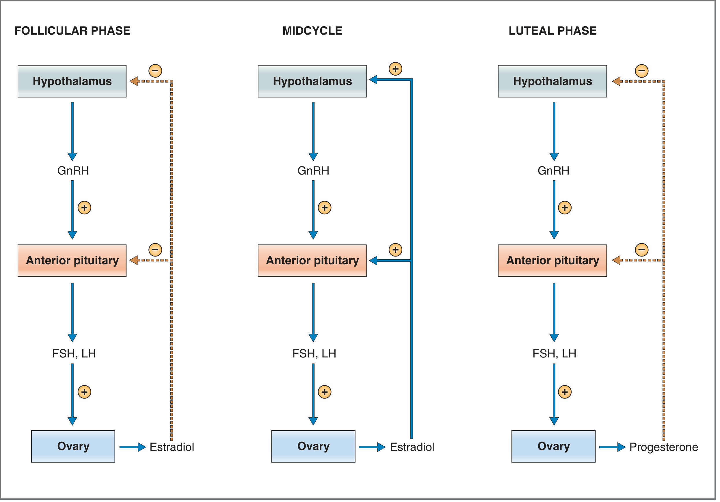
The menstrual cycle uses both negative and positive feedback:
- For most of the cycle, estradiol and progesterone inhibit GnRH, FSH, and LH release by negative feedback.
- Near mid-cycle, sustained high estradiol changes to positive feedback, causing the LH surge that triggers ovulation.
Costanzo Physiology, 7th ed., pp. 477-479.
3. Ovarian and uterine phases
| Ovarian event | Dominant hormone | Endometrial phase | Main event |
|---|---|---|---|
| Follicular phase | Estrogen | Proliferative phase | Follicle development and endometrial growth |
| Ovulation | LH surge | Late proliferative | Release of oocyte |
| Luteal phase | Progesterone | Secretory phase | Endometrium prepared for implantation |
| Menstruation | Falling estrogen and progesterone | Menstrual phase | Functional endometrium is shed |
A. Follicular phase
The follicular phase starts on the first day of menstruation and ends with ovulation.
Hormonal events
At the end of the preceding cycle, the corpus luteum regresses. Therefore, progesterone and estrogen fall. This removes negative feedback on the hypothalamus and pituitary, allowing FSH to rise.
FSH:
- Recruits a group of ovarian follicles
- Stimulates granulosa-cell growth
- Promotes estradiol synthesis
A dominant follicle emerges. Its increasing estradiol and inhibin B suppress FSH, so most of the other follicles undergo atresia.
Endometrial effects of estrogen
Estrogen produces the proliferative phase of the endometrium:
- Regrowth of endometrium after menstruation
- Proliferation of glands and stroma
- Lengthening of spiral arteries
- Progressive thickening of endometrium
It also produces thin, watery, alkaline, stretchable cervical mucus. This facilitates sperm survival and transport. Dried mid-cycle mucus may show a fern-like pattern.
Costanzo Physiology, 7th ed., p. 479.
B. Ovulation
As the dominant follicle matures, estradiol becomes persistently high. Sustained high estradiol changes its effect from negative to positive feedback.
This causes:
- A large LH surge
- A smaller FSH surge
- Completion of oocyte maturation
- Rupture of the dominant follicle
- Release of the oocyte
Ovulation occurs approximately 24-36 hours after the beginning of the LH surge. It is generally about 14 days before the next menstrual period, not necessarily on day 14.
Possible clinical signs include:
- Mid-cycle pelvic discomfort, called mittelschmerz
- Clear, slippery cervical mucus
- Slight rise in basal body temperature after ovulation.
C. Luteal phase
After ovulation, the ruptured follicle becomes the corpus luteum. Under LH stimulation, it secretes:
- Progesterone
- Estrogen
- Inhibin A
Progesterone is the dominant hormone of this phase.
Effects of progesterone
Progesterone changes the proliferative endometrium into secretory endometrium:
- Endometrial glands become tortuous and secretory.
- Glands contain glycogen-rich material.
- Stroma becomes edematous.
- Spiral arteries become longer and more coiled.
- Cervical mucus becomes thick, scanty, and less permeable to sperm.
- Basal body temperature rises slightly.
- Further FSH and LH release is suppressed.
This phase prepares the uterus for implantation.
If fertilization occurs, early embryonic hCG maintains the corpus luteum until placental hormone production is sufficient. If fertilization does not occur, the corpus luteum regresses after about 14 days.
D. Menstruation
Regression of the corpus luteum causes a fall in progesterone and estrogen. This leads to:
- Spiral-arteriole vasoconstriction
- Ischemia of the functional endometrium
- Prostaglandin production
- Tissue breakdown and shedding of the functional layer
The basal layer remains and regenerates the endometrium during the next follicular phase.
Menstrual blood contains tissue debris, prostaglandins, and fibrinolysin, which normally breaks down clots. Therefore, prominent clots may suggest a high flow rate. Ganong's Review of Medical Physiology, 26th ed., p. 406.
4. Dysmenorrhea
Dysmenorrhea means painful menstruation. The pain is commonly cramping and located in the lower abdomen or pelvis. It may radiate to the back or thighs and be accompanied by nausea, diarrhea, fatigue, headache, or dizziness.
There are two main types.
A. Primary dysmenorrhea
Primary dysmenorrhea is painful menstruation without identifiable pelvic disease.
Typical pattern
- Begins 6-24 months after menarche, after ovulatory cycles are established
- Starts just before or within hours of menstrual flow
- Peaks during the first day of heaviest bleeding
- Usually lasts 2-3 days
- Pelvic examination is normal when it is appropriate to perform one
Pathophysiology
At menstruation, endometrial prostaglandins, particularly PGF2 alpha, increase. They cause:
- Strong and frequent uterine contractions
- Increased uterine tone
- Uterine vasoconstriction
- Reduced uterine blood flow and ischemic pain
Swanson's Family Medicine Review, section “Dysmenorrhea,” pp. 411-412.
Management
-
NSAIDs are first-line treatment.
They inhibit cyclooxygenase and reduce prostaglandin synthesis. They are most effective when started just before expected menses or at onset of pain and taken regularly through the first 2-3 days of flow. They may be unsuitable in people with certain gastrointestinal, renal, bleeding, allergy, or cardiovascular risks. -
Hormonal suppression or contraception.
Combined hormonal contraceptives or progestogen-based methods can reduce ovulation, endometrial growth, and prostaglandin production. They are useful when contraception is also desired. -
Non-drug measures.
- Local heat
- Exercise
- Sleep and stress management
- Symptom and cycle diary
-
Reassess non-response.
Persistent or worsening pain despite adequate NSAID and hormonal treatment should prompt assessment for secondary dysmenorrhea, particularly endometriosis.
B. Secondary dysmenorrhea
Secondary dysmenorrhea is menstrual pain caused by an underlying pelvic disorder.
Common causes
- Endometriosis
- Adenomyosis
- Uterine fibroids
- Pelvic inflammatory disease
- Endometrial polyps
- Ovarian cysts or pelvic adhesions
- Cervical stenosis
- Congenital obstructive anomalies
- Copper intrauterine device-related symptoms in some people
Features suggesting secondary dysmenorrhea
- New onset after years of painless periods
- Progressively worsening pain
- Pain beginning days before menstruation and continuing after it
- Heavy menstrual bleeding or intermenstrual bleeding
- Deep dyspareunia
- Infertility
- Chronic pelvic pain
- Dyschezia or painful defecation during periods
- Poor response to first-line treatment
- Abnormal pelvic examination
Assessment
Assessment may include:
- Pregnancy test when applicable
- Detailed pain, bleeding, sexual, contraceptive, and fertility history
- Examination when appropriate
- Pelvic ultrasound for fibroids, adenomyosis, ovarian masses, or endometrioma
- Specialist gynecology assessment if endometriosis or another significant pathology is suspected
Management is directed at the cause, such as treatment of endometriosis, fibroids, infection, or structural obstruction.
5. Other menstrual-cycle disorders
A. Anovulatory cycles
An anovulatory cycle occurs when ovulation does not take place. No corpus luteum forms, so progesterone is absent.
Estrogen may still stimulate endometrial growth. Without progesterone to organize normal secretory transformation and shedding, the endometrium becomes unstable and may shed irregularly.
Consequences
- Irregular bleeding
- Prolonged bleeding
- Heavy bleeding
- Infrequent menses
- Unpredictable timing of periods
Anovulatory cycles can be physiological:
- During the first 12-18 months after menarche
- During the perimenopausal transition
They can also occur with:
- Polycystic ovary syndrome, or PCOS
- Thyroid disease
- Hyperprolactinemia
- Obesity
- Stress, eating disorders, low body weight, or excessive exercise
- Chronic systemic illness
Ganong's Review of Medical Physiology, 26th ed., p. 406.
B. Abnormal uterine bleeding
Abnormal uterine bleeding, or AUB, means bleeding that differs from expected menstrual bleeding in volume, duration, frequency, regularity, or timing.
Current terminology favors descriptive terms rather than older labels such as menorrhagia, metrorrhagia, polymenorrhea, and oligomenorrhea.
AUB includes:
- Heavy menstrual bleeding
- Bleeding lasting longer than expected
- Frequent periods
- Infrequent periods
- Irregular periods
- Intermenstrual bleeding
- Postcoital bleeding
Causes: PALM-COEIN classification
| Structural causes: PALM | Non-structural causes: COEIN |
|---|---|
| P - Polyp | C - Coagulopathy |
| A - Adenomyosis | O - Ovulatory dysfunction |
| L - Leiomyoma or fibroid | E - Endometrial disorder |
| M - Malignancy and hyperplasia | I - Iatrogenic, such as medication or device-related |
| N - Not otherwise classified |
Heavy menstrual bleeding
Heavy menstrual bleeding is bleeding that interferes with physical, social, emotional, or material quality of life. It may cause iron deficiency or anemia.
Possible causes include:
- Fibroids
- Adenomyosis
- Endometrial polyps
- Anovulation and PCOS
- Bleeding disorders, including von Willebrand disease
- Thyroid disease
- Anticoagulant medicines
- Endometrial hyperplasia or malignancy, depending on age and risk factors
Evaluation may include pregnancy testing, complete blood count, iron assessment, pelvic ultrasound, assessment for bleeding disorders, and endometrial sampling in selected higher-risk patients. ACOG's AUB guidance notes that bleeding between periods, after sex, longer than 7 days, or excessively heavy bleeding requires assessment.
C. Oligomenorrhea and infrequent cycles
Oligomenorrhea means infrequent menstrual periods or cycles longer than expected. Common causes include:
- PCOS
- Pregnancy
- Hypothalamic dysfunction
- Hyperprolactinemia
- Thyroid disorder
- Primary ovarian insufficiency
- Perimenopause
Repeated long cycles often indicate infrequent ovulation. In PCOS, chronic anovulation can expose the endometrium to unopposed estrogen and may increase risk of endometrial hyperplasia. The aim of treatment is not only cycle regularity but also endometrial protection and management of metabolic risk.
D. Amenorrhea
Amenorrhea means absence of menstruation.
- Primary amenorrhea: no menstruation by age 15 years with normal secondary sexual development, or by age 13 years without secondary sexual development.
- Secondary amenorrhea: no period for 3 months in someone with previously regular cycles, or 6 months in someone with previously irregular cycles.
Pregnancy must be excluded first.
Causes can be grouped by level of dysfunction:
- Hypothalamic: stress, weight loss, eating disorder, intensive exercise
- Pituitary: hyperprolactinemia, pituitary tumor
- Ovarian: PCOS, primary ovarian insufficiency
- Thyroid or other endocrine disorders
- Uterine/outflow tract: intrauterine adhesions, congenital genital tract anomalies, cervical stenosis
Initial assessment commonly includes pregnancy test, TSH, prolactin, FSH, estradiol, androgen evaluation when indicated, and pelvic ultrasound.
E. Premenstrual syndrome and PMDD
Premenstrual syndrome
Premenstrual syndrome, or PMS, consists of physical and emotional symptoms occurring in the luteal phase and improving soon after menstruation begins.
Symptoms include:
- Irritability and mood change
- Breast tenderness
- Bloating
- Headache
- Fatigue
- Food cravings
- Poor concentration
Premenstrual dysphoric disorder
Premenstrual dysphoric disorder, or PMDD, is a more severe condition with marked mood symptoms and functional impairment.
A prospective symptom diary across at least two cycles helps establish the cyclical pattern. Management can include lifestyle measures, cognitive and behavioral approaches, selected hormonal methods, and SSRIs for clinically significant PMDD.
6. Important warning signs
Prompt clinical assessment is needed for:
- Very heavy bleeding, especially soaking a pad or tampon hourly for more than 2 hours
- Dizziness, fainting, shortness of breath, chest pain, or features of severe anemia
- Possible pregnancy with pain or bleeding, because ectopic pregnancy must be excluded
- Severe new or worsening pelvic pain
- Fever, foul-smelling discharge, vomiting, or severe tenderness
- Intermenstrual, postcoital, postmenopausal bleeding
- Amenorrhea with severe headache, visual change, or nipple discharge
- Rapid onset of hirsutism, voice deepening, or other virilization features
Key points for revision
- GnRH pulses stimulate FSH and LH.
- FSH promotes follicle growth and estradiol synthesis.
- Sustained high estradiol causes positive feedback and the LH surge.
- LH causes ovulation and corpus-luteum formation.
- Estrogen causes the proliferative endometrium.
- Progesterone causes the secretory endometrium.
- Withdrawal of estrogen and progesterone causes menstruation.
- Primary dysmenorrhea is prostaglandin-mediated and usually responds to NSAIDs.
- Secondary dysmenorrhea suggests pelvic pathology, especially endometriosis.
- AUB should be assessed using PALM-COEIN.
- Persistent irregular anovulatory cycles require evaluation and endometrial protection.
This is a shared conversation. Sign in to Orris to start your own chat.