
choledocholithiasis obstructive jaundice pathogenesis
gallstone in common bile duct obstructive jaundice diagram
cholelithiasis gallbladder stones cholecystitis
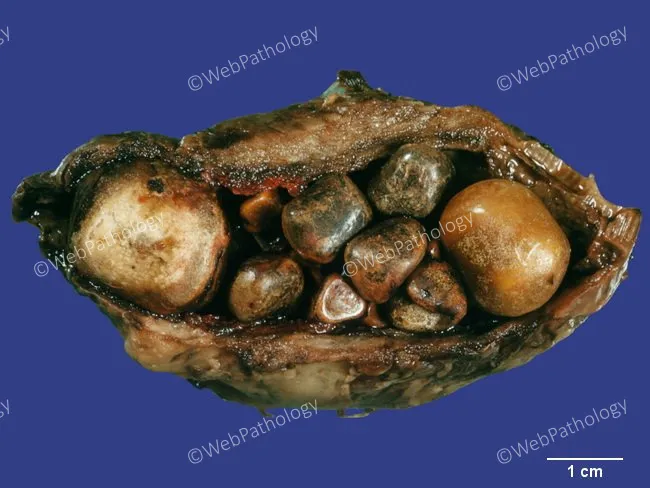
Gross pathology photograph of an opened gallbladder demonstrating chronic calculous cholecystitis with marked cholelithiasis. The lumen is densely filled by numerous mixed cholesterol stones of variable size, ranging from tiny calculi to several millimeters in diameter. Stones exhibit tan to brown coloration with smooth to slightly faceted surfaces, consistent with cholesterol-rich, mixed biliary calculi. The gallbladder wall is focally thickened and fibrotic, reflecting chronic inflammatory change; subtle mucosal irregularities may be present, but no focal ulceration is evident in this view. The specimen is positioned against a color-calibrated background with a 1 cm scale bar for size reference. This gross appearance correlates with long-standing gallstone disease and chronic inflammation, rather than acute cholecystitis. Diagnostic significance lies in confirming cholelithiasis with chronic cholecystitis, guiding surgical pathology submission and influencing prognosis. Clinically, this pattern is associated with episodic biliary colic, risk of cholangitis if obstruction occurs, and potential progression to gallbladder fibrosis or porcelain gallbladder in advanced cases. Differential considerations on gross exam include calcified or pigment stones, but the predominance of cholesterol stones with wall thickening supports chronic calculous cholecystitis. This image serves as an educational reference for gross anatomy, surgical pathology, and hepatobiliary disease assessment, and complements histologic evaluation by providing macroscopic context for stone burden and wall remodeling.
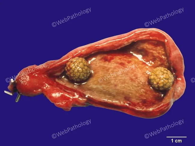
Gross pathology photograph of a cholecystectomy specimen demonstrating cholelithiasis with two cholesterol gallstones within the gallbladder lumen. The specimen is a distended gallbladder with a mucosal surface largely exposed and no obvious perforation. Two yellow-tan, faceted calculi, approximately 0.8-1.2 cm in diameter, are visible protruding into the lumen. The stones are cholesterol stones, common in symptomatic gallstone disease, formed from cholesterol supersaturation with bile and darkened by mucin adherence. The surrounding gallbladder wall shows mild serosal inflammation consistent with chronic cholecystitis rather than acute suppurative inflammation; no porcelain gallbladder is evident. The background tissue is red-brown, with clear gauze and ruler scale indicating 1 cm increments for size estimation. This gross image supports a clinical history of biliary colic and symptomatic cholelithiasis, typically precipitated by fatty meals and nocturnal gallbladder contraction. The image demonstrates classic features of cholesterol stones: pale yellow, radiolucent in typical composition, faceted surfaces, and dense aggregation at the gallbladder neck. Clinically, these findings corroborate preoperative diagnoses of biliary colic, cholecystitis, and cholelithiasis, and are relevant for educational purposes, surgical pathology correlation, and radiologic-pathologic correlation. Useful for teaching on gallbladder disease, stone morphology, and gross correlation with ultrasound findings. This image aids pathology teaching and examination objectively.
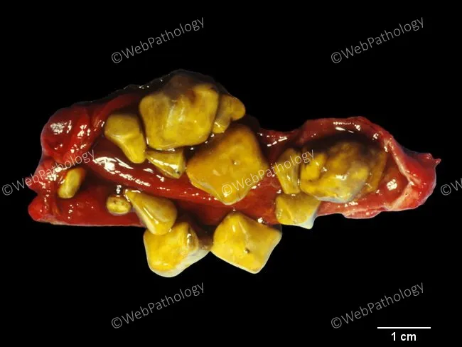
Gross-pathology photograph of gallbladder with cholesterol gallstones. Modality: macroscopic specimen photography under bright-field illumination; scale bar present showing 1 cm for size reference. Location: gallbladder lumen with multiple golden-yellow stones contiguous within a red-brown, flattened gallbladder wall. Stones are irregularly shaped, to molded contours, with smooth, glossy surfaces typical of cholesterol monohydrate composition. Size range spans approximately 2–12 mm for small fragments to larger stones; overall count exceeds a dozen in this specimen. The stones are pale to deep yellow, translucent in appearance, and demonstrate outward molding with faceted edges where they abut the gallbladder mucosa. The surrounding tissue shows minimal inflammatory changes in this specimen, consistent with chronic cholelithiasis without acute cholecystitis. Cholesterol stones arise predominantly in the gallbladder, are the most common calculus type in Western populations, and account for about 75–80% of gallstones. Composition is primarily cholesterol with variable bilirubin content and trace calcium salts. This image highlights a classic presentation: cholelithiasis caused by cholesterol supersaturation of bile; pathogenesis linked to obesity, age, female sex, rapid weight loss, and metabolic syndrome. Clinically, such stones can cause biliary colic, cholecystitis, or pancreatitis if obstructing the cystic or pancreatic duct; definitive management is cholecystectomy. This photograph is suitable for educational, diagnostic, and research contexts.
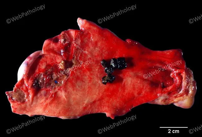
Gross pathology photograph of a gallbladder specimen showing thickened wall with Rokitansky-Aschoff sinuses and mucosal ulceration. Several small black pigment gallstones are embedded within the ulcerated mucosa near the left edge of the tissue. The stones appear faceted with a dense, glossy surface and are surrounded by inflamed, reddened mucosa. Rokitansky-Aschoff sinuses are mucosal outpouchings extending into the muscularis propria, reflecting chronic inflammatory remodeling typical of chronic cholecystitis. The embedded stones indicate mucosal erosion rather than intraluminal lodging. The cut surface reveals fibrous, contracted gallbladder wall with focal firmness and pearling along the serosa. A clear 2 cm scale bar is visible on the image. The overall specimen demonstrates features consistent with long-standing cholelithiasis with secondary inflammation. Pigment stones, often composed of calcium bilirubinate, are associated with hemolysis, infection, biliary stasis, or biliary infections. Clinically, these findings support gallbladder removal (cholecystectomy) and correlate with pain, biliary colic, and potential chronic cholecystitis. Differential considerations include chronic cholecystitis with cholelithiasis; less likely are gallbladder neoplasms arising in chronic inflammatory settings. Histologic confirmation would assess mucosal and muscularis changes and exclude carcinoma. Documentation of orientation, margins, and ancillary testing would enhance diagnostic precision and guide postoperative surveillance planning for at-risk patients and outcomes.
/tmp/workspace
Case Analysis: 55-year-old female with jaundice, dark urine, pale stools, and biliary colic
A. Diagnosis
- Female, over 40, recurrent RUQ pain after fatty meals = classic gallstone risk profile ("Female, Forty, Fertile, Fat, Flatulent")
- Pain radiating to the right shoulder/scapula (referred via phrenic nerve, C3-C5 dermatome, since the diaphragm shares this segmental innervation)
- Jaundice + dark urine + pale (clay-colored) stools = cholestatic/obstructive pattern, distinguishing it from purely hepatocellular jaundice
- Mild right hypochondrial tenderness (Murphy's sign territory)
B. Pathogenesis
- Cholesterol nucleation and crystal formation
- Gallbladder hypomotility/stasis (allowing crystals time to aggregate into stones)
- Excess mucin gel that traps crystals
- Bile (conjugated bilirubin, bile salts, cholesterol) backs up into the biliary radicles and regurgitates into the bloodstream → conjugated hyperbilirubinemia → jaundice (icterus of skin/sclera)
- Bile acids deposited in the skin → pruritus (may or may not be present here)
- No bile reaches the duodenum → loss of stercobilinogen pigment and impaired fat digestion (see C and D below)
- Recurrent stone impaction/dislodgement at the cystic duct or CBD produces episodic biliary colic; when a stone transiently obstructs the cystic duct outlet during gallbladder contraction, pressure rises acutely within a non-distensible viscus, producing pain (biliary colic).
C. Why are stools pale and urine dark?
-
Pale (clay-colored) stools: Normally, conjugated bilirubin reaches the intestine via bile, where gut bacteria convert it to urobilinogen, most of which is oxidized to stercobilin, the pigment that gives stool its normal brown color. When the CBD is obstructed, no (or very little) bile/bilirubin reaches the intestine, so stercobilin is not formed, and stools become pale/clay-colored (acholic), often with a greasy texture from unabsorbed fat.
-
Dark urine: Because the obstruction prevents excretion of conjugated bilirubin into the gut, it regurgitates back into the bloodstream. Conjugated bilirubin is water-soluble (unlike unconjugated bilirubin) and is freely filtered by the kidneys, appearing in urine as bilirubinuria, which darkens the urine (often described as "tea-colored" or "coca-cola colored"). Urobilinogen, which normally contributes some color to urine, is actually reduced in complete obstruction since less bilirubin reaches the gut to be converted to urobilinogen - but the bilirubin itself is what colors the urine dark here.
D. Why does pain occur after fatty meals?
- Fat entering the duodenum stimulates release of cholecystokinin (CCK) from duodenal mucosal I-cells.
- CCK causes forceful gallbladder contraction and relaxation of the sphincter of Oddi, aiming to eject bile to help digest and emulsify the fat.
- If a stone is lodged in the cystic duct or common bile duct (or intermittently obstructs it), this forceful contraction against the obstruction sharply raises intraluminal/intraductal pressure within the gallbladder or bile duct.
- Since the gallbladder and biliary tree are relatively non-distensible, this pressure rise stretches the wall and stimulates visceral pain afferents, producing the colicky right upper quadrant/epigastric pain that radiates to the back or right shoulder (via shared phrenic/diaphragmatic innervation, C3-C5) - classic biliary colic.
- This is why the pain is characteristically post-prandial, particularly after fatty meals, and may be accompanied by nausea/vomiting due to visceral afferent stimulation and reflex activation of the vomiting center.